Báo cáo hóa học: " CdSe Ring- and Tribulus-Shaped Nanocrystals: Controlled Synthesis, Growth Mechanism, and Photoluminescence Properties" potx
Bạn đang xem bản rút gọn của tài liệu. Xem và tải ngay bản đầy đủ của tài liệu tại đây (438.35 KB, 7 trang )
NANO EXPRESS
CdSe Ring- and Tribulus-Shaped Nanocrystals: Controlled
Synthesis, Growth Mechanism, and Photoluminescence Properties
Pengfei Hu Æ Dianzeng Jia Æ Yali Cao Æ
Yudai Huang Æ Lang Liu Æ Jianmin Luo
Received: 24 November 2008 / Accepted: 27 January 2009 / Published online: 18 February 2009
Ó to the authors 2009
Abstract With air-stable and generic reagents, CdSe
nanocrystals with tunable morphologies were prepared by
controlling the temperature in the solution reaction route.
Thereinto, the lower reaction temperature facilitates the
anisotropic growth of crystals to obtain high-yield CdSe
ring- and tribulus-shaped nanocrystals with many branches
on their surfaces. The photoluminescence properties are
sensitive to the nature of particle and its surface. The
products synthesized at room temperature, whose surfaces
have many branches, show higher blue shift and narrower
emission linewidths (FWHM) of photoluminescence than
that of samples prepared at higher temperature, whose
surfaces have no branches. Microstructural studies revealed
that the products formed through self-assembly of primary
crystallites. Nanorings formed through the nonlinear
attachment of primary crystallites, and the branches on the
surfaces grew by linear attachment at room temperature.
And the structure of tribulus-shaped nanoparticle was
realized via two steps of aggregation, i.e., random and
linear oriented aggregation. Along with the elevation of
temperature, the branches on nanocrystal surfaces short-
ened gradually because of the weakened linear attachment.
Keywords CdSe nanostructure Á Tunable morphologies Á
Narrow emission linewidth Á Nonlinear and linear
attachment Á Two-step attachment
Introduction
Cadmium selenide (CdSe), one of the important II–VI
group semiconductors, has received significant interest in
the field of optoelectronic applications due to its broad
range of optical transmissions, excellent nonlinear optical
properties, and quantum size effects [1–6]. For these
applications, the efficiency and line width of the photo-
luminescence (PL) are important factors. And these PL
properties are very sensitive to the nature of the particle
and its surface [3]. Up to now, shape-controlled synthesis
of CdSe nanorods [7–13], nanowires [14–17], nanotubes
[18], nanobelts [15, 19], nanosaws [15], nanobarbells
[20], and many novel nanostructures [21–24] have been
demonstrated. The popular routes to synthesize CdSe,
whether the west coast method (TBP/TOPO) or the east
coast method (TOP/TOPO), generally require rather
complicated procedures including delicate control of
surfactant ratios and inert reaction conditions due to the
toxic and unstable nature of the precursors [25]. So, with
the exploration of the electrical and photoluminescence
properties of the nano-sized CdSe, a convenient and
effective synthetic method is still a focus for researchers.
Concerning the unique structural features and better
properties generated with them, the nonlithographic fab-
rication of free-standing CdSe nanorings (strict circular,
oval, homocentric, or polygonal forms) and other novel
nanostructure objects from small building blocks by self-
organizing means may represent a next challenge of
nanofabrication.
Electronic supplementary material The online version of this
article (doi:10.1007/s11671-009-9265-2) contains supplementary
material, which is available to authorized users.
P. Hu Á D. Jia (&) Á Y. Cao Á Y. Huang Á L. Liu
Institute of Applied Chemistry, Xinjiang University,
Urumqi 830046, People’s Republic of China
e-mail:
J. Luo
Physics and Chemistry Test Centre, Xinjiang University,
Urumqi 830046, People’s Republic of China
123
Nanoscale Res Lett (2009) 4:437–443
DOI 10.1007/s11671-009-9265-2
In view of the advantages of the solution reaction route,
low energy consumption, and facility, we introduce it to
explore a general synthetic method for CdSe nanomaterials
at low temperature. In this work, we successfully devel-
oped a low temperature and convenient solution reaction
approach to fabricate CdSe nanocrystals. This method
does not require complex apparatus, expensive reagents, or
complicated techniques. It can synthesize CdSe nanoma-
terials with branches on surface in high-yield. Shape
control was achieved by varying the reaction temperature
conveniently and it does not need any organic additives.
Based on the self-assembly of nanocrystals, the formation
mechanism of nanorings and tribulus-shaped nanoparticles
were discussed in detail. Furthermore, the photolumines-
cence properties were investigated.
Experimental Section
All the reagents were of analytical-grade and were used
without further purification. The synthesis of sample I was
carried out through solution reaction process. Firstly, the
selenium powder (Se) was put into the hydrazine hydrate
(N
2
H
4
Á H
2
O) in a three-necked flask under magnetic
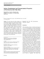
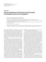
Fig. 1 Typical TEM images of
CdSe nanocrystals synthesized
at a and b room temperature
(sample I), c 60 °C (sample II),
and d 100 °C (sample III)
438 Nanoscale Res Lett (2009) 4:437–443
123
stirring. The process was carried out at room temperature
till a resultant brown solution was got. Subsequently, the
Cd(CH
3
COO)
2
Á 2H
2
O (the molar ratio of Cd
2?
:Se = 2:1)
was added to the above solution and stirred for 2 h. The
products were filtered and washed with distilled water
and ethanol for five times, respectively. Finally, the
orange product powder was dried at 60 °C for 4 h in air
oven and collected for further characterization. Sample II
and III were synthesized at 60 °C and 100 °C, respec-
tively and other experimental parameters are consistent.
Worth the whistle, all processes were carried out in fume
cupboard.
Powder X-ray diffraction (XRD, MXP18AHF, MAC)
using CuKa radiation (k = 0.154056 nm) was adopted to
identify the crystalline phase of the resulting materials.
Transmission electron microscopic analysis (TEM) and high-
resolution transmission electron microscopic analysis
(HRTEM) were performed with HITACHI H-600 (TEM,
HITACHI H-600) microscope operating at 75 kV and a JEOL
JEM-2100 (TEM, JEOL JEM-2100) electron microscope
operating at 200 kV, respectively. The photoluminescence
spectra were obtained by using a HITACHI F-4500 fluores-
cence spectrophotometer at room temperature.
Results and Discussion
The TEM images demonstrated the high-yield of nano-
structure with branches obtained at room temperature
and structural evolution along the temperature. As shown
in Figs. 1a and S1, the products synthesized at room
temperature are dominated by CdSe tribulus-shaped
nanoparticles and nanorings. Figure 1b and the magnified
images of Figs. 1a and S1 clearly display the branched
structure on the surfaces of nanostructure. The ring-like
objects involving circular, hexagonal, and oval forms, with
growing outward radial thorn-like branches were exhibited
in the Figs. 1a, b, 2a and S2a
1
–a
7
. Among them, the hex-
agonal rings are less populated (Fig. S2a
7
). Meanwhile,
there is a portion of CdSe homocentric nanorings (Fig. 2a).
Enlarged images show that some rings are half-penetrated
because of the attachment of the primary particles in the
cavities (Figs. 2a and S2a
2
,a
3
,a
6
). Furthermore, a series of
tribulus-shaped nanocrystals were created at room tem-
perature (Figs. 2c, 5a, c, S1 and S2b). The tribulus-shaped
nanocrystals have about 3–10 cuspidal arms. These arms
are 10–30 nm in length and the cusp of these arms are
3–5 nm in diameters (Figs. 3, 5a and c).
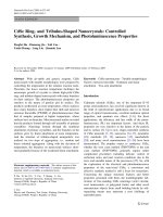
Fig. 2 a–c TEM Images and SAED pattern of CdSe nanocrystals
synthesized at room temperature: a the homocentric ring; b the oval
and irregular rings (inset is the SAED pattern of the ring in the image
b); c the tribulus-shaped nanoparticles (inset is a natural tribulus);
d and e TEM images of CdSe nanorings prepared at 60 °C and
100 °C, respectively; f Room temperature photoluminescence spectra
of products obtained at (I) room temperature, (II)60°C, and (III)
100 °C (Excitation wavelength: 450 nm)
Nanoscale Res Lett (2009) 4:437–443 439
123
The morphology of the products varies upon the reaction
temperature. The thorn-like branches of nanoparticles and
nanorings obviously shortened at 60 °C and completely
disappeared at 100 °C (Fig. 1c and d). In fact, the branches
have become small protuberances at 60 °C (Fig. 2d). The
reason for the shortening of branches on nanocrystals
prepared at higher temperature will be discussed in detail
at the latter paragraph. Furthermore, the yield of rings
decreases while the temperature steps up. According to the
statistic, the average yield of rings is about nine per TEM
image in sample I and three in sample II, and two in
sample III (based on a total of 20 TEM images of sample I,
II, and III, respectively).
As mentioned in the introduction segment, the PL
properties of the materials are very sensitive to the nature
of the particle and its surface. Figure 2f compares the room
temperature PL spectra of three samples in this paper. They
all exhibit a strong fluorescence emission band with the
similar profile centered at 672 nm (I), 676 nm (II), and
677 nm (III), respectively. Each as-prepared CdSe nano-
crystal has a blue shift in the PL spectra, in comparison
with that of bulk CdSe at 730 nm. The Full-Width-at-Half-
Maximum (FWHM) is an important parameter of photo-
luminescence properties, which reveals the crystallinity
and size distribution of nanostructure. It is the distance
between two sides of a peak measured at half the peak
height. The FWHM of the sample I fabricated at room
temperature is about 13 nm, which agrees well with the
higher crystallinity, and that of samples II and III are about
18 nm and 20 nm, respectively. In a word, the PL spectra
demonstrate that the sample I prepared at room tempera-
ture has a stronger blue shift and a narrower FWHM than
the samples II and III synthesized at 60 °C and 100 °C.
The novel surface structure of as-obtained particles formed
at room temperature may be a reason for this result. Fur-
thermore, from the emission spectra, the size of CdSe
nanoparticles which made up the polycrystalline walls of
the rings or tribulus-shaped crystals was estimated to be
around 10 nm in the sample I.
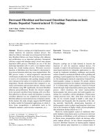
High-resolution TEM (HRTEM) shows that the arms of
tribulus-shaped nanocrystals and radial branches of nan-
orings are well crystalline. Among them, the branches/arms
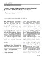
Fig. 4 a XRD patterns of
sample I, II, and III; b EDX
spectrum of CdSe sample
produced at room temperature
(sample I)
Fig. 3 The high-resolution
TEM images of CdSe branches
of nanocrystals synthesized at
room temperature: a the space
between arrowheads
corresponds to the distance
between two (0002) planes;
b the interplanar spacing
corresponds to the distance
between two (10-10) planes;
The inset of (a) and (b) show
the FFT analysis of selective
region in the crystal,
respectively
440 Nanoscale Res Lett (2009) 4:437–443
123
that grow along the [0001] direction are more populated.
The interplanar spacing in the branches/arms is 0.35 nm
which matches well with the (0002) plane of CdSe
(Fig. 3a). Moreover, the (10-10) plane of CdSe can also be
observed in the sample (Fig. 3b). A fast Fourier transform
(FFT) analysis of the branches/arms (see inset of Figs. 3a
and b) confirmed that the thorn-like branches were strictly
oriented. Both the HRTEM result and FFT patterns anal-
yses demonstrate that these thorn-like branches have
\0001[ or \11-20[ preferential growth direction.
The X-ray diffraction (XRD) patterns show that the
CdSe nanocrystals have hexagonal wurtzite structure with
the diffraction peaks which shift to higher angles (Fig. 4a).
The extremely sharp (0002) peak reveals the preferred
\0001[orientation of the CdSe nanocrystallites. The cell
constants were calculated to be a = b = 0.4256 nm and
c = 0.6977 nm from (10-10) and (0002) peaks of (I)
(sample I) after refinement. They revealed that the lattice
contractions of Da = 1.00% and Dc = 0.47% occurred
against the reported data (a = b = 0.4299 nm and
c = 0.7010 nm). A spectrum of energy-dispersive X-ray
spectroscopy (EDX) for the sample I confirms that the
atomic ratio for Cd:Se is approximately 1.13:1. And the
sample contains a little oxygen element (Fig. 4b).
According to the above experimental results, we suggest
the formation mechanism of nanorings and tribulus-shaped
nanoparticles as follows.
For the present synthetic route, N
2
H
4
Á H
2
O is used as
the reducing agent (N
2
/N
2
H
4
,OH
-
, -1.15 V) and the
selenium powder was reduced from zero valence to -2
valence. After this course, the as-reduced Se
2-
reacts with
Cd
2?
to generate orange CdSe.
Our experimental results revealed that the resultant
nanostructures came into being through the self-assembly
of primary particles. It is well-known that the main driv-
ing force for aggregation of nanoparticles can be generally
attributed to the tendency for reducing the high surface
energy through the attachment among the primary nano-
particles and the formation of coherent lattice structure at
grain interfaces [26–29]. The aggregation of nanoparticles
contains the linear alignment which can be achieved by
sharing a common crystallographic orientation among the
primary particles and the nonlinear arrangement which
can be attained with the lateral lattice fusion of the pri-
mary particles [30]. Generally, the linear alignment can
lead to the formation of one-dimensional nanostructures
(nanorods or nanowires) mostly. The nonlinear arrange-
ment which can result in the ring-like structure has been
demonstrated in the literatures [30, 31]. Literature [30]
described three types of CdS nanorings which were based
on the statistical assembly or specific crystallographic
requirement of the tiny CdS hexagons. The rectangular
PbSe nanorings resulted from the dipole-induced
orientational attachment of cubic primary PbSe nanopar-
ticles in the literature [31].
Similar to the formation of CdS nanorings [30], the first
type (i) of organization of CdSe nanorings was achieved by
the attachments among neighboring hexagonal building
units with their {10-10} and {0001} family planes on their
external surfaces. Sixfold symmetry of wurtzite CdSe
{0001} surfaces make hexagonal building units having six
equal chances to attach to their neighboring crystallites,
and a statistical assembly of the tiny hexagons may lead to
the formation of a curvature. In order to ensure a smooth
curvature development, building segments of {10-10} and
{11-20} should be connected in an alternative manner.
Otherwise, the aggregation process will run out of a cir-
cumferential track, and the tortile and incomplete circular
ring structures will be formed (Fig. 2b). The study on
Fig. 5 Top: Schematic illustration depicted the two steps of growth
based on self-assembly from primary particles to a tribulus-shaped
crystal. a TEM image showing attachment of new particles to
elongate the branches (arrowheads in the section A, B and C);
b HRTEM image of section B in (a) indicating the connecting region
(between arrowheads) between a new particle and the arm; c high-
resolution TEM image of a tribulus-shaped nanoparticle; d magni-
fication of the section A in (c) exhibiting three random arrangement
particles A1, A2, A3 and two oriented attachment branches A4
(\11-20[ direction) and A5 (\0001[ direction)
Nanoscale Res Lett (2009) 4:437–443 441
123
selected area electron diffraction (SAED) in the inset of
Fig. 2b reveals the polycrystalline nature for the CdSe
rings.
Apart from the horizontal ring formation with the
{10-10} family facets, some CdSe primary nanocrystallites
can also stack along the \0001[ axis, where the c-plane
terminates either with positively charged (0001)-Cd or
negatively charged (000-1)-Se polar surfaces. This vertical
oriented attachment of building primary nanoparticles lead
to the formation of thorn-like branches on the surface of
rings. As shown in Fig. 5a, this vertical organization can be
further confirmed by attachment of new particles at the end
of a branch (showed by arrowheads in the A, B and C
sections). This behavior is believed to be a consequence of
further development of oriented attachment. The connect-
ing region with coherent lattice structure indicated by
arrowheads in Fig. 5b (high-magnification HRTEM image
of B section in Fig. 5a) powerfully reveal that the building
primary crystallites assemble via the ‘‘oriented attach-
ment’’ mechanism.
Type (ii) and type (iii) of organization, which can bring
on the birth of hexagonal ring, must be formed with
straight segments as a result of the \11-20[and \10-10[
directional alignments [30]. So, the quantity of hexagonal
rings is less. In this paper, the latter two cases will not be
further investigated.
Additionally, lower reaction temperature benefits the
anisotropic growth [13, 27]. When the reaction is conducted
at higher temperature, the anisotropic growth becomes
weaker. Therefore, the branches on the surface of nano-
crystals shorten at higher temperature, and even disappear.
The two steps of self-assembly from primary particles to
a tribulus-shaped crystal are schematically illustrated in
Fig. 5 top. It is generally believed that the synthesis of
some nanoparticles often involves the fast nucleation of
primary particles and the subsequent growth via their
aggregation. The aggregation includes random and oriented
aggregation [27]. Firstly, the ‘‘core’’ of tribulus-shaped
nanoparticle is formed by the random aggregation of
building primary particles. Some facets tending to be the
anisotropic growth of wurtzite CdSe nanocrystals [29],
which is coming from the primary building blocks, are
bared on the surfaces of the ‘‘cores’’. Subsequently, the
nomadic building particles attach to these facets with a
highly oriented fashion and then produce branches.
Figure 5c clearly displays a tribulus-shaped nanoparticle
which generated through two steps of attachment. The
section A in Fig. 5c contained three random arrangement
particles A1, A2, A3, and two oriented attachment bran-
ches A4 and A5. Figure 5d, the magnification of section A
in Fig. 5c, displays them clearly. Thereinto, the branches
A4 and A5 display the \11-20[ and \0001[ directional
attachment, respectively.
Conclusions
In conclusion, a shape-controlled synthesis of CdSe nan-
orings and tribulus-shaped nanoparticles can be developed
without any organic additives at low temperature. The
lower reaction temperature benefits the anisotropic growth
of crystals with oriented attachment mechanism to produce
the high-yield ring- and tribulus-shaped nanocrystals, and
produce many branches/arms of products. In photolumi-
nescence spectra, the emission of CdSe nanocrystals
synthesized at room temperature has higher blue shift and
narrower FWHM than that of products prepared at 60
°C
and 100 °C. The structure of tribulus-shaped nanoparticles
is achieved via random aggregation and succedent linear
oriented aggregation of the building primary particles at
room temperature. The nanorings are constructed through a
nonlinear arrangement of the building primary particles at
lower temperature. This process can provide a new way to
fabricate novel architectures. Further investigations are
currently under way to identify the underlying mechanism
about the hexagonal nanorings growth and to control the
size distribution.
Acknowledgments This work was partially supported by the
National Nature Science Foundation of China (Grant No.20666005
and 20661003), the Nature Science Foundation of Xinjiang Province
(Grant No. 200821121 and 200721102), and The Research Fund for
the Doctoral Program of Higher Education (Grant No. 20070755001).
References
1. W.U. Huynh, J.J. Dittmer, A.P. Alivisatos, Science 295, 2425
(2002). doi:10.1126/science.1069156
2. Y.N. Xia, P.D. Yang, Y.G. Sun, Y.Y. Wu, B. Mayers, B. Gates,
Y.D. Yin, F. Kim, Y.Q. Yan, Adv. Mater. 15, 353 (2003). doi:
10.1002/adma.200390087
3. N. Myung, Y. Bae, A.J. Bard, Nano Lett. 3, 747 (2003). doi:
10.1021/nl034165s
4. B.Q. Sun, E. Marx, N.C. Greenham, Nano Lett. 3, 961 (2003).
doi:10.1021/nl0342895
5. W. Luan, H. Yang, N. Fan, S T. Tu, Nanoscale Res. Lett. 3, 134
(2008). doi:10.1007/s11671-008-9125-5
6. M.A. Hahn, P.C. Keng, T.D. Krauss, Anal. Chem. 80, 864 (2008).
doi:10.1021/ac7018365
7. X.G. Peng, L. Manna, W.D. Yang, J. Wickham, E. Scher, A.
Kadavanich, A.P. Alivisatos, Nature 404, 59 (2000). doi:10.1038/
35003535
8. Q. Peng, Y. Dong, Z. Deng, Y. Li, Inorg. Chem. 41, 5249 (2002).
doi:10.1021/ic0257266
9. R.F. Li, Z.T. Luo, F. Papadimitrakopoulos, J. Am. Chem. Soc.
128, 6280 (2006). doi:10.1021/ja058102i
10. F. Shieh, A.E. Saunders, B.A. Korgel, J. Phys. Chem. B 109,
8538 (2005). doi:10.1021/jp0509008
11. A. Salant, E. Amitay-Sadovsky, U. Banin, J. Am. Chem. Soc.
128, 10006 (2006). doi:10.1021/ja063192s
12. D.H. Son, S.M. Hughes, Y.D. Yin, A.P. Alivisatos, Science 306,
1009 (2004). doi:10.1126/science.1103755
442 Nanoscale Res Lett (2009) 4:437–443
123
13. L. Ouyang, K.N. Maher, C.L. Yu, J. McCarty, H. Park, J. Am.
Chem. Soc. 129, 133 (2007). doi:10.1021/ja066243u
14. Z.Y. Tang, N.A. Kotov, Adv. Mater. 17, 951 (2005). doi:10.1002/
adma.200401593
15. C. Ma, Z.L. Wang, Adv. Mater. 17, 2635 (2005). doi:10.1002/
adma.200500805
16. L.L. Zhao, T.Z. Lu, M. Yosef, M. Steinhart, M. Zacharias, U.
Go
¨
sele, S. Schlecht, Chem. Mater. 18, 6094 (2006). doi:10.1021/
cm062014v
17. N. Pradhan, H.F. Xu, X.G. Peng, Nano Lett. 6, 720 (2006). doi:
10.1021/nl052497m
18. X.C. Jiang, B. Mayers, T. Herricks, Y.N. Xia, Adv. Mater. 15,
1740 (2003). doi:10.1002/adma.200305737
19. J. Joo, J.S. Son, S.G. Kwon, J.H. Yu, T. Hyeon, J. Am. Chem.
Soc. 128, 5632 (2006). doi:10.1021/ja0601686
20. J.E. Halpert, V.J. Porter, J.P. Zimmer, M.G. Bawendi, J. Am.
Chem. Soc. 128, 12590 (2006). doi:10.1021/ja0616534
21. L. Manna, E.C. Scher, A.P. Alivisatos, J. Am. Chem. Soc. 122,
12700 (2000). doi:10.1021/ja003055
22. D. Battaglia, J.J. Li, Y.J. Wang, X.G. Peng, Angew. Chem. Int.
Ed. 42, 5035 (2003). doi:10.1002/anie.200352120
23. G. Zlateva, Z. Zhelev, R. Bakalova, I. Kanno, Inorg. Chem. 46,
6212 (2007). doi:10.1021/ic062045s
24. L. Liu, Q. Peng, Y. Li, Inorg. Chem. 47, 3182 (2008). doi:
10.1021/ic702203c
25. S.J. Rosenthal, J. McBride, S.J. Pennycook, L.C. Feldman, Surf.
Sci. Rep. 62, 111 (2007). doi:10.1016/j.surfrep.2007.02.001
26. R.L. Penn, J.F. Banfield, Science 281, 969 (1998). doi:10.1126/
science.281.5379.969
27. R.L. Penn, J. Phys. Chem. B 108, 12707 (2004). doi:10.1021/
jp036490
28. Y. Cheng, Y. Wang, D. Chen, F. Bao, J. Phys. Chem. B 109, 794
(2005). doi:10.1021/jp0460240
29. L. Manna, L.W. Wang, R. Cingolani, A.P. Alivisatos, J. Phys.
Chem. B 109, 6183 (2005). doi:10.1021/jp0445573
30. B. Liu, H.C. Zeng, J. Am. Chem. Soc. 127, 18262 (2005). doi:
10.1021/ja055734w
31. K S. Cho, D.V. Talapin, W. Gaschler, C.B. Murray, J. Am.
Chem. Soc. 127, 7140 (2005). doi:10.1021/ja050107s
Nanoscale Res Lett (2009) 4:437–443 443
123