Acute Ischemic Stroke Part 3 ppt
Bạn đang xem bản rút gọn của tài liệu. Xem và tải ngay bản đầy đủ của tài liệu tại đây (1.45 MB, 18 trang )
Diaschisis, Degeneration, and Adaptive Plasticity After Focal Ischemic Stroke
25
Nudo R.J. & Milliken G.W. (1996) Reorganization of movement representations in primary
motor cortex following focal ischemic infarcts in adult squirrel monkeys. Journal of
Neurophysiology, Vol. 75, pp. (2144-2149).
O'Brien, M. D., Waltz, A. G., & Jordan, M. M. (1974). Ischemic cerebral edema. distribution
of water in brains of cats after occlusion of the middle cerebral artery. Archives of
Neurology, Vol. 30, No. 6, pp. (456-460).
Pettigrew, L. C., Kindy, M. S., Scheff, S., Springer, J. E., Kryscio, R. J., Li, Y., et al. (2008).
Focal cerebral ischemia in the TNFalpha-transgenic rat. Journal of
Neuroinflammation, Vol. 5, pp. (47).
Pierpaoli, C., Barnett, A., Pajevic, S., Chen, R., Penix, L. R., Virta, A., et al. (2001). Water
diffusion changes in wallerian degeneration and their dependence on white matter
architecture. NeuroImage, Vol. 13, No. 6, pp. (1174-1185).
Platz, T., Denzler, P., Kaden, B., & Mauritz, K. H. (1994). Motor learning after recovery from
hemiparesis. Neuropsychologia, Vol. 32, No. 10, pp. (1209-1223).
Puig, J., Pedraza, S., Blasco, G., Daunis-I-Estadella, J., Prats, A., Prados, F., Boada, I., et al.
(2010). Wallerian degeneration in the corticospinal tract evaluated by diffusion tensor
imaging correlates with motor deficit 30 days after middle cerebral artery ischemic
stroke. AJNR. American journal of neuroradiology, Vol. 31, No. 7, pp. (1324–1330).
Que, M., Schiene, K., Witte, O. W., & Zilles, K. (1999). Widespread up-regulation of N-
methyl-D-aspartate receptors after focal photothrombotic lesion in rat brain.
Neuroscience Letters, Vol. 273, No. 2, pp. (77–80).
Qü, M., Mittmann, T., Luhmann, H. J., Schleicher, A., & Zilles, K. (1998). Long-term changes
of ionotropic glutamate and GABA receptors after unilateral permanent focal
cerebral ischemia in the mouse brain. Neuroscience, Vol.85, No. 1, pp. (29–43).
Reinecke, S., Dinse, H. R., Reinke, H., & Witte, O. W. (2003). Induction of bilateral plasticity
in sensory cortical maps by small unilateral cortical infarcts in rats. The European
Journal of Neuroscience, Vol. 17, No. 3, pp. (623-627).
Reitmeir, R., Kilic, E., Kilic, U., Bacigaluppi, M., ElAli, A., Salani, G., Pluchino, S., et al.
(2011). Post-acute delivery of erythropoietin induces stroke recovery by promoting
perilesional tissue remodelling and contralesional pyramidal tract plasticity. Brain :
a journal of neurology, Vol.134, No. 1, pp. (84–99).
Remple M.S., Bruneau R.M., VandenBerg P.M., Goertzen C., Kleim J.A. (2001) Sensitivity of
cortical movement representations to motor experience: Evidence that skill learning
but not strength training induces cortical reorganization. Behavioral Brain Research,
Vol. 123, pp. (133-141).
Riecker, A., Gröschel, K., Ackermann, H., Schnaudigel, S., Kassubek, J., & Kastrup, A. (2010).
The role of the unaffected hemisphere in motor recovery after stroke. Human brain
mapping, Vol. 31, No. 7, pp. (1017–1029).
Ritter, L. S., Orozco, J. A., Coull, B. M., McDonagh, P. F., & Rosenblum, W. I. (2000).
Leukocyte accumulation and hemodynamic changes in the cerebral
microcirculation during early reperfusion after stroke. Stroke; a Journal of Cerebral
Circulation, Vol. 31, No. 5, pp. (1153-1161).
Rosenzweig, E. S., Courtine, G., Jindrich, D. L., Brock, J. H., Ferguson, A. R., Strand, S. C.,
Nout, Y. S., et al. (2010). Extensive spontaneous plasticity of corticospinal projections
after primate spinal cord injury. Nature Neuroscience, Vol. 13, No. 12, pp. (1505–1510).
Schaechter J.D. (2004) Motor rehabilitation and brain plasticity after hemiparetic stroke.
Progress in Neurobiology, Vol. 73, pp. (61-72).
Acute Ischemic Stroke
26
Schaechter J.D., Moore C.I., Connell B.D., Rosen B.R., Dijkhuizen R.M. (2006) Structural and
functional plasticity in the somatosensory cortex of chronic stroke patients. Brain,
Vol. 129, pp. (2722-2733).
Schiene, K., Bruehl, C., Zilles, K., Qü, M., Hagemann, G., Kraemer, M., & Witte, O. W. (1996).
Neuronal hyperexcitability and reduction of GABAA-receptor expression in the
surround of cerebral photothrombosis. Journal of cerebral blood flow and metabolism :
official journal of the International Society of Cerebral Blood Flow and Metabolism, Vol.
16, No. 5, pp. (906–914).
Seitz R.J., Hoflich P., Binkofski F., Tellmann L., Herzog H., Freund H.J. (1998) Role of the
premotor cortex in recovery from middle cerebral artery infarction. Archives of
Neurology, Vol. 55, pp. (1081-1088).
Sigler, A., Mohajerani, M. H., & Murphy, T. H. (2009). Imaging rapid redistribution of
sensory-evoked depolarization through existing cortical pathways after targeted
stroke in mice. Proceedings of the National Academy of Sciences of the United States of
America, Vol. 106, No. 28, pp. (11759-11764).
Slater, R., Reivich, M., Goldberg, H., Banka, R., & Greenberg, J. (1977). Diaschisis with cerebral
infarction. Stroke; a Journal of Cerebral Circulation, Vol. 8, No. 6, pp. (684-690).
Somjen, G. G. (2001). Mechanisms of spreading depression and hypoxic spreading
depression-like depolarization. Physiological Reviews, Vol. 81, No. 3, pp. (1065-1096).
Steriade, M., & Llinas, R. R. (1988). The functional states of the thalamus and the associated
neuronal interplay. Physiological Reviews, Vol. 68, No. 3, pp. (649-742).
Steward, O., Zheng, B., Tessier-Lavigne, M., Hofstadter, M., Sharp, K., & Yee, K. M. (2008).
Regenerative Growth of Corticospinal Tract Axons via the Ventral Column after
Spinal Cord Injury in Mice. Journal of Neuroscience, Vol. 28, No. 27, pp. (6836–6847).
Stroemer, R. P., Kent, T. A., & Hulsebosch, C. E. (1995). Neocortical neural sprouting,
synaptogenesis, and behavioral recovery after neocortical infarction in rats. Stroke,
Vol. 26, No. 11, pp. (2135–2144).
Takasawa, M., Watanabe, M., Yamamoto, S., Hoshi, T., Sasaki, T., Hashikawa, K., . . .
Kinoshita, N. (2002). Prognostic value of subacute crossed cerebellar diaschisis:
Single-photon emission CT study in patients with middle cerebral artery territory
infarct. AJNR.American Journal of Neuroradiology, Vol. 23, No. 2, pp. (189-193).
Takatsuru, Y., Fukumoto, D., Yoshitomo, M., Nemoto, T., Tsukada, H., & Nabekura, J.
(2009). Neuronal Circuit Remodeling in the Contralateral Cortical Hemisphere
during Functional Recovery from Cerebral Infarction. Journal of Neuroscience, Vol.
29, No. 32, pp. (10081–10086).
Tamura, A., Tahira, Y., Nagashima, H., Kirino, T., Gotoh, O., Hojo, S., & Sano, K. (1991).
Thalamic atrophy following cerebral infarction in the territory of the middle
cerebral artery. Stroke, Vol. 22, No. 5, pp. (615–618).
Thomalla, G., Glauche, V., Koch, M., Beaulieu, C., Weiller, C., & Rother, J. (2004). Diffusion
tensor imaging detects early Wallerian degeneration of the pyramidal tract after
ischemic stroke. NeuroImage, Vol. 22, No. 4, pp. (1767–1774).
Thornton, P., McColl, B. W., Greenhalgh, A., Denes, A., Allan, S. M., & Rothwell, N. J. (2010).
Platelet interleukin-1alpha drives cerebrovascular inflammation. Blood, Vol. 115,
No. 17, pp. (3632-3639).
Traversa R., Cicinelli P., Bassi A., Rossini P.M., Bernardi G. (1997) Mapping of motor cortical
reorganization after stroke. A brain stimulation study with focal magnetic pulses.
Stroke, Vol. 28, pp. (110-117).
Diaschisis, Degeneration, and Adaptive Plasticity After Focal Ischemic Stroke
27
Tsai, S. Y., Markus, T. M., Andrews, E. M., Cheatwood, J. L., Emerick, A. J., Mir, A. K., et al.
(2007). Intrathecal treatment with anti-nogo-A antibody improves functional
recovery in adult rats after stroke. Experimental Brain Research.Experimentelle
Hirnforschung.Experimentation Cerebrale, Vol. 182, No. 2, pp. (261-266).
Umegaki, M. (2005). Peri-Infarct Depolarizations Reveal Penumbra-Like Conditions in
Striatum. Journal of Neuroscience, Vol. 25, No. 6, pp. (1387–1394).
van der Zijden J.P., van der Toorn A., van der Marel K., Dijkhuizen R.M. (2008)
Longitudinal in vivo MRI of alterations in perilesional tissue after transient
ischemic stroke in rats. Experimental Neurology, Vol. 212, pp. (207-212).
van der Zijden J.P., Wu O., van der Toorn A., Roeling T.P., Bleys R.L., Dijkhuizen R.M.
(2007) Changes in neuronal connectivity after stroke in rats as studied by serial
manganese-enhanced MRI. Neuroimage, Vol. 34, pp. (1650-1657).
Vila, Nicolás, Castillo, J., Dávalos, A., Esteve, A., Planas, A. M., & Chamorro, A. (2003).
Levels of anti-inflammatory cytokines and neurological worsening in acute
ischemic stroke. Stroke, Vol. 34, No. 3, pp. (671–675).
Vila, N, Castillo, J., Dávalos, A., & Chamorro, A. (2000). Proinflammatory cytokines and early
neurological worsening in ischemic stroke. Stroke, Vol. 31, No. 10, pp. (2325–2329).
Wang, T., Wang, J., Yin, C., Liu, R., Zhang, J. H., & Qin, X. (2010). Down-regulation of Nogo
receptor promotes functional recovery by enhancing axonal connectivity after
experimental stroke in rats. Brain Research, Vo. 1360, pp. (147–158).
Ward N.S., Brown M.M., Thompson A.J., Frackowiak R.S. (2006) Longitudinal changes in
cerebral response to proprioceptive input in individual patients after stroke: An
FMRI study. Neurorehabilitation and Neural Repair, Vol. 20, pp. (398-405).
Ward, N. S., Brown, M. M., Thompson, A. J., & Frackowiak, R. S. (2003). Neural correlates of
outcome after stroke: A cross-sectional fMRI study. Brain : A Journal of Neurology,
Vol. 126, No. 6, pp. (1430-1448).
Weber R., Ramos-Cabrer P., Justicia C., Wiedermann D., Strecker C., Sprenger C., Hoehn M.
(2008) Early prediction of functional recovery after experimental stroke: Functional
magnetic resonance imaging, electrophysiology, and behavioral testing in rats.
Journal of Neuroscience, Vol. 28, pp. (1022-1029).
Wei L., Erinjeri J.P., Rovainen C.M., Woolsey T.A. (2001) Collateral growth and angiogenesis
around cortical stroke. Stroke, Vol. 32, pp. (2179-2184).
Weiller C., Ramsay S.C., Wise R.J., Friston K.J., Frackowiak R.S. (1993) Individual patterns of
functional reorganization in the human cerebral cortex after capsular infarction.
Annals of Neurology, Vol. 33, pp. (181-189).
Weishaupt, N., Silasi, G., Colbourne, F., Fouad, K. (2010). Secondary damage in the spinal
cord after motor cortex injury in rats. Journal of Neurotrauma, Vol. 27, pp. (1387-
1397).
Werring, D. J., Toosy, A. T., Clark, C. A., Parker, G. J., Barker, G. J., Miller, D. H., et al. (2000).
Diffusion tensor imaging can detect and quantify corticospinal tract degeneration after
stroke. Journal of Neurology, Neurosurgery, and Psychiatry, Vol. 69, No. 2, pp. (269-272).
Wiessner, C., Bareyre, F. M., Allegrini, P. R., Mir, A. K., Frentzel, S., Zurini, M., et al. (2003).
Anti-nogo-A antibody infusion 24 hours after experimental stroke improved
behavioral outcome and corticospinal plasticity in normotensive and
spontaneously hypertensive rats. Journal of Cerebral Blood Flow and Metabolism :
Official Journal of the International Society of Cerebral Blood Flow and Metabolism, Vol.
23, No. 2, pp. (154-165).
Acute Ischemic Stroke
28
Winship, I. R., & Murphy, T H. (2009). Remapping the Somatosensory Cortex after Stroke:
Insight from Imaging the Synapse to Network. The Neuroscientist, Vol. 15, No. 5, pp.
(507–524).
Winship I.R. & Murphy T.H. (2008) In vivo calcium imaging reveals functional rewiring of
single somatosensory neurons after stroke. Journal of Neuroscience, Vol. 28, pp.
(6592-6606).
Witte, O. W., Bidmon, H. J., Schiene, K., Redecker, C., & Hagemann, G. (2000). Functional
differentiation of multiple perilesional zones after focal cerebral ischemia. Journal of
Cerebral Blood Flow and Metabolism : Official Journal of the International Society of
Cerebral Blood Flow and Metabolism, Vol. 20, No. 8, pp. (1149-1165).
Wolf, T., Lindauer, U., Reuter, U., Back, T., Villringer, A., Einhaupl, K., et al. (1997).
Noninvasive near infrared spectroscopy monitoring of regional cerebral blood
oxygenation changes during peri-infarct depolarizations in focal cerebral ischemia in
the rat. Journal of Cerebral Blood Flow and Metabolism : Official Journal of the International
Society of Cerebral Blood Flow and Metabolism, Vol. 17, No. 9, pp. (950-954).
Yu, C., Zhu, C., Zhang, Y., Chen, H., Qin, W., Wang, M., & Li, K. (2009). A longitudinal diffusion
tensor imaging study on Wallerian degeneration of corticospinal tract after motor
pathway stroke. NeuroImage, Vol. 47, No. 2, pp. (451–458). ISSN: 1053-8119
Zai, L., Ferrari, C., Dice, C., Subbaiah, S., Havton, L. A., Coppola, G., et al. (2011). Inosine
augments the effects of a nogo receptor blocker and of environmental enrichment
to restore skilled forelimb use after stroke. The Journal of Neuroscience : The Official
Journal of the Society for Neuroscience, Vol. 31, No. 16, pp. (5977-5988).
Zhang, S., & Murphy, Timothy H. (2007). Imaging the impact of cortical microcirculation on
synaptic structure and sensory-evoked hemodynamic responses in vivo. PLoS
biology, Vol. 5, No. 5, pp. (e119).
Zhang, Y., Xiong, Y., Mahmood, A., Meng, Y., Liu, Z., Qu, C., & Chopp, M. (2010). Sprouting
of corticospinal tract axons from the contralateral hemisphere into the denervated
side of the spinal cord is associated with functional recovery in adult rat after
traumatic brain injury and erythropoietin treatment. Brain Research, Vol. 1353, pp.
(249–257).
Zipfel, G. J., Babcock, D. J., Lee, J. M., & Choi, D. W. (2000). Neuronal apoptosis after CNS
injury: The roles of glutamate and calcium. Journal of Neurotrauma, Vol. 17, No. 10,
pp. (857-869).
2
Excitotoxicity and Oxidative
Stress in Acute Ischemic Stroke
Ramón Rama Bretón
1
and Julio César García Rodríguez
2
1
Department of Physiology & Immunology, University of Barcelona
2
CENPALAB
1
Spain
2
Cuba
1. Introduction
The term “stroke” is applied to a heterogeneous group of diseases caused by decreased
perfusion of the brain due to occlusion of the blood vessels supplying the brain or a
haemorrhage originating in them. Most strokes (~ 85%) are ischemic; that is, they result
from occlusion of a major cerebral artery by a thrombus or embolism. This results in
reduced blood flow and a major decrease in the supply of oxygen and nutrients to the
affected region. The rest of strokes are haemorrhagic: caused by the rupture of a blood
vessel either in the brain or on its surface.
Strokes deprive the brain not only of oxygen but also of glucose and of all other nutrients, as
well as disrupting the nutrient/waste exchange process required to support brain
metabolism. The result is the development of a hypoxic-ischemic state. Ischemia is defined
as a decrease in blood flow to tissues that prevents adequate delivery of oxygen, glucose
and others nutrients. Ischemic stroke is the result of total or partial interruption of cerebral
arterial blood supply, which leads to oxygen and glucose deprivation of the tissue
(ischemia). If cerebral arterial blood flow is not restored within a short period, cerebral
ischemia is the usual result, with subsequent neuron death within the perfusion territory of
the vessels affected. Ischemic stroke is characterized by a complex sequence of events that
evolves over hours or even days [1-3]. Acute ischemic stroke results from acute occlusion of
cerebral arteries. Cerebral ischemia occurs when blood flow to the brain decreases to a level
where the metabolic needs of the tissue are not met. Cerebral ischemia may be either
transient (followed by reperfusion) or essentially permanent. In all cases, a stroke involves
dysfunction and death of brain neurons and neurological damage that reflects the location
and size of the brain area affected [1, 2].
2. Ischemic core and ischemic penumbra
Neuropathological analysis after focal brain ischemia reveals two separate areas: the
ischemic core, and ischemic penumbra. Once onset of a stroke has occurred, within
minutes of focal ischemia occurring, the regions of the brain that suffer the most severe
degrees of blood flow reduction experience irreversible damage: these regions are the
Acute Ischemic Stroke
30
“ischemic core”. This area exhibits a very low cerebral blood flow (CBF) and very low
metabolic rates of oxygen and glucose [2, 3]. Thus, reduced or interrupted CBF has
negative effects on brain structure and function. Neurons in the ischemic core of the
infarction are killed rapidly by total bioenergetic failure and breakdown of ion
homeostasis, lipolysis and proteolysis, as well as cell membrane fragmentation [4]. The
result is cell death within minutes [5]. Tissue in the ischemic core is irreversibly injured
even if blood flow is re-established.
The necrotic core is surrounded by a region of brain tissue which suffers moderate blood
flow reduction, thus becoming functionally impaired but remaining metabolically active;
this is known as the “ischemic penumbra” [6]. This metabolically active border region
remains electrically silent [7]. From experiments in non-human primates, it has been shown
that in this region, the ability of neurons to fire action potentials is lost. However, these
neurons maintain enough energy to sustain their resting membrane potentials and when
collateral blood flow improves, action potentials are restored. The ischemic penumbra may
comprise as much as half the total lesion volume during the initial stages of ischemia, and
represents the region in which there is an opportunity to salvage functionality via post-
stroke therapy [8, 9].
Ischemic penumbra refers to the region of brain tissue that is functionally impaired but
structurally intact; tissue lying between the lethally damaged core and the normal brain,
where blood flow is sufficiently reduced to result in hypoxia that is severe enough to arrest
physiological function, but not so complete as to cause irreversible failure of energy
metabolism and cellular necrosis [8]. The ischemic penumbra has been documented in
laboratory animals as severely hypoperfused, non-functional, but still viable brain tissue
surrounding the irreversibly damaged ischemic core [10]. The penumbra can be identified
by the biochemical and molecular mechanisms of neuron death [11, 12] and by means of
clinical neuroimaging tools [10, 13].
Thus, the ischemic penumbra refers to areas of the brain that are damaged during a stroke
but not killed. The concept therefore emerges that once onset of a stroke has begun, the
necrotic core is surrounded by a zone of less severely reduced blood flow where the
neurons have lost functional activity but remain metabolically active. Tissue injury in the
ischemic penumbra is the outcome of a complex series of genetic, molecular and
biochemical mechanisms, which contribute either to protecting –and then penumbral
tissue is repaired and recovers functional activity– or to damaging –and then the
penumbral area becomes necrotic –brain cells. Tissue damage and functional impairment
after cerebral ischemia result from the interaction between endogenous neuroprotective
mechanisms such as anti-excitotoxicity (GABA, adenosine and K
ATP
activation), anti-
inflammation and anti-apoptosis (IL-10, Epo, Bcl-proteins), and repair and regeneration
(c-Src formation, vasculogenesis, neurogenesis, BM-derived cells) on the one hand, with
neurotoxic events such as excitotoxicity, inflammation and apoptosis that ultimately lead
to cell death, on the other [14]. The penumbra is the battle field where the ischemic
cascade with several deleterious mechanisms is triggered, resulting in ongoing cellular
injury and infarct progression. Ultimately, the ischemic penumbra is consumed by
progressive damage and coalesces with the core, often within hours of the onset of the
stroke. However, the penumbra can be rescued by improving the blood flow and/or
interfering with the ischemic cascade. At the onset of a stroke, the evolution of the
ischemic penumbra is only partially predictable from the clinical, laboratory and imaging
methods currently available [3, 10].
Excitotoxicity and Oxidative Stress in Acute Ischemic Stroke
31
3. Pathophysiological basis of the stroke
In the last 30 years, experimental and clinical results have led to characterizations of the
pathophysiological basis of strokes [1-3]. Cerebral ischemia (ischemic stroke) triggers a
complex series of physiological, biochemical, molecular and genetic mechanisms that impair
neurologic functions through a breakdown of cellular integrity mediated by ionic
imbalance, glutamate-mediated excitotoxicity and also such phenomena as calcium
overload, oxidative stress, mitochondrial dysfunction and apoptosis [1-3, 15, 16]. These
mediate injury to neurons, glia cells and vascular elements by means of disturbing the
function of important cellular organelles such as mitochondria, nuclei, cell membranes,
endoplasmic reticula and lysosomes. The result is cell death via mechanisms that promote
rupture, lysis, phagocytosis or involution and shrinkage [11, 16]. Knowledge of the
molecular mechanisms that underlie neuron death following a stroke is important if we are
to devise effective neuroprotective strategies.
We will examine how ischemic injury occurs, which cell death mechanisms are activated,
especially excitotoxicity and oxidative stress, and how these can be manipulated to induce
neuroprotection. Unfortunately, despite their effectiveness in preclinical studies, a large
number of neuroprotectants have failed to produce the desired effects in clinical trials
involving stroke sufferers, which suggests that we still lack essential knowledge of the
triggers and mediators of ischemic neuron death. We will discuss why, after 30 years or so
of intense basic and clinical research, we still find it extremely difficult to translate
experimental neuroprotective success in the laboratory to the clinical setting [17-20].
3.1 Acute ischemic injury in strokes
Acute ischemic injury is the result of a transient or permanent reduction of CBF in a
restricted vascular territory. Normal CBF is between 45 and 60 ml blood/100 g/min. It is
well documented that time-dependent neuronal events are triggered in response to reduced
CBF [21, 22]. The brain has critical thresholds for CBF and for oxygen tension. Oxygen
supply to the brain below a critical level reduces, and eventually blocks, oxidative
phosphorylation, drastically decreases cellular ATP and leads to the collapse of ion
gradients. Neuron activity ceases and if oxygen is not re-introduced quickly, cells die [22]. A
reduction of cortical blood flow to levels of approximately 20 ml/100 g/min may be
tolerated without functional consequences, but it is associated with the loss of consciousness
and ECG alterations. At values of CBF below 18 ml/100 g/min, the tissue infarction is time
dependent: CBF of 5 ml/100 g/min lasting about 30 minutes cause infarction; CBF of 10
ml/100 g/min needs to last for more than 3 hours to cause infarction; permanent CBF below
18 ml/100 g/min causes irreversible damage [22, 23]. In focal ischemia, complete cessation
of blood flow is uncommon because collateral vessels sustain CBF at 5 to 15 ml/100
g/minute in the ischemic core and at 15 to 25 ml/100 g/minute in the outer areas of the
ischemic zone [5, 21, 24]. Global ischemia results from transient CBF below 0.5 ml/100
g/min or severe hypoxia to the entire brain. When CBF falls to zero within seconds, loss of
consciousness occurs after approximately 10 s, EEG activity ceases after 30–40 s, cellular
damage is initiated after a few minutes, and death occurs within 10 min, at least under
normothermic conditions [25].
The brain is highly vulnerable to ischemia. In part, the vulnerability of brain tissue to
ischemia reflects its high metabolic demands. The brain has a relatively high energy
production demand and depends almost exclusively on oxidative phosphorylation for
Acute Ischemic Stroke
32
energy production. Although the weight of the human brain is only about 2% of the total
bodyweight, it has high metabolic activity and uses 20% of the oxygen and 25% of the
glucose consumed by the entire body [23]. Proper functioning of brain cells depends on an
abundant and continuous supply of oxygen. Even with such high metabolic demands, there
is essentially no oxygen storage in cerebral tissue, and only limited reserves of high-energy
phosphate compounds and carbohydrate substrates are available. More than 90% of the
oxygen consumed by the brain is used by mitochondria to generate ATP. Energy in the
brain is mainly formed when glucose is oxidized to CO
2
and water through mitochondrial
oxidative phosphorylation. At rest, about 40% of cerebral energy is used to maintain and
restore ionic gradients across cell membrane; even more energy is used during activity [23].
The brain requires large amounts of oxygen to generate sufficient ATP to maintain and
restore ionic gradients.
3.2 Basic mechanisms of ischemic cell death
After the onset of a stroke, the disruptions to the blood flow in areas affected by vascular
occlusion limit the delivery of oxygen and metabolic substrates to neurons causing ATP
reduction and energy depletion. The glucose and oxygen deficit that occurs after severe
vascular occlusion is the origin of the mechanisms that lead to cell death and consequently
to cerebral injury. These mechanisms include: ionic imbalance, the release of excess
glutamate in the extracellular space, a dramatic increase in intracellular calcium that in turn
activates multiple intracellular death pathways such as mitochondrial dysfunction, and
oxidative and nitrosative stress that finally cause neuron death.
After ischemic onset, the primary insult that ischemia causes neurons is a loss of oxygen and
glucose substrate energy. While there are potentially large reserves of alternatives substrates
to glucose, such as glycogen, lactate and fatty acids, for both glycolysis and respiration,
oxygen is irreplaceable in mitochondrial oxidative phosphorylation, the main source of ATP
in neurons. Consequently, the lack of oxygen interrupts oxidative phosphorylation by the
mitochondria and drastically reduces cellular ATP production, which results in a rapid
decline in cellular ATP [26, 27]. Although there are potentially large reserves of substrates
such as glycogen, lactate and fatty acids that may be alternatives to glucose, anaerobic
metabolism is insufficient to produce sufficient ATP. Reduced ATP stimulates the glycolytic
metabolism of residual glucose and glycogen, causing an accumulation of protons and
lactate, which leads to rapid intracellular acidification and increases the depletion of ATP
[26]. When the lack of oxygen is severe and glucose is diminished, inhibition of oxidative
phosphorylation leads to ATP-synthase functioning backwards and consuming ATP, thus
contributing to an increase in the loss of ATP [27]. If ATP levels are low, the Na
+
/K
+
-ATPase
function fails [27]. After several minutes, inhibition of the Na
+
/K
+
-ATPase function causes a
profound loss of ionic gradients and the depolarization of neurons and astrocytes [28].
Membrane depolarization and changes in the concentration gradients of Na
+
and K
+
across
the plasma membrane result in activation of voltage-gated calcium channels. This leads to
excessive release of excitatory amino acids –particularly glutamate– to the extracellular
compartment (Fig. 1).
Uncontrolled membrane depolarization by massive changes in the concentration gradients of
Na
+
and K
+
across the plasma membrane results in a large and sustained release of glutamate
and other neurotransmitters to the extracellular compartment [29]. Simultaneously,
neurotransmitter re-uptake from the extracellular space is reduced [30, 31]. The rise in the
extracellular glutamate concentration initiates a positive feedback loop, with further activation
Excitotoxicity and Oxidative Stress in Acute Ischemic Stroke
33
of glutamate receptors in neighbouring neurons and as a result, more Na
+
inflow to neurons
via monovalent ion channels that decrease ionic gradients and consume ATP, both of which
promote further release of glutamate [32, 33]. Simultaneously, glutamate transporters in
neurons and astrocytes can function backwards, releasing glutamate into the extracellular
space [31, 34] and contributing to glutamate overload there. A marked and prolonged rise in
the extracellular glutamate concentration kills central neurons [2, 11, 32]. Excessive glutamate
in the synapses activates the ionotropic glutamate receptors at a pathophysiological level; this
type of neuronal insult is called excitotoxicity [29] and is defined as cell death resulting from
the toxic actions of excitatory amino acids. Because glutamate is the most important excitatory
neurotransmitter in primary perception and constitutes the basis of synaptic transmission in
about 10
14
synapses in the human brain, neuronal excitotoxicity usually refers to the injury and
death of neurons arising from prolonged intense exposure to glutamate and the associated
ionic imbalance in the cell. Excessive activation of glutamate receptors by excitatory amino
acids leads to a number of deleterious consequences, including impairment of calcium
buffering, generation of free radicals, activation of the mitochondrial permeability transition
and secondary excitotoxicity.
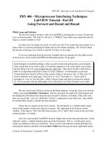
Fig. 1. Excitotoxicity in ischemic stroke. The reduction of blood flow supply to the brain
during ischemic stroke results in oxygen and glucose deprivation and thus a reduction in
energy available to maintain the ionic gradients. This results in excessive neuronal
depolarization and deregulated glutamate release.
3.3 Excitotoxic mechanisms
Excitotoxicity is considered to be the central mechanism underlying neuron death in stroke
[29, 32-35]. Excitotoxicity is considered to trigger tissue damage in both focal experimental
ischemia [34, 36] and clinical ischemia [37]. Glutamate is released at high concentrations in
the penumbral cortex [38], particularly if blood flow is reduced for a long period, and the
amount of glutamate released correlates with early neurological deterioration in patients
Acute Ischemic Stroke
34
with acute ischemic stroke [37]. Glutamate concentrations greater than 200 mmol/l in
plasma and greater than 8.2 mmol/l in CSF are associated with neurological deterioration in
the acute phase of cerebral infarction.
The excitotoxic mechanisms which lead to neuron death are complex, but primarily involve
the generation of free radicals [35; 39, 40], mitochondrial dysfunction [41, 42] and the
participation of various transcription factors as activators of gene expression [43, 44]. All of
these mechanisms acting synergistically can damage cellular proteins [45], lipids [46] and
DNA [47, 48], which leads to the deterioration of cellular architecture and signalling,
resulting in necrosis, apoptosis or both depending on the severity of the insult and of
relative speed of each process [49-51].
3.4 The role of glutamate receptors in excitotoxicity
The excitatory effects of glutamate are mediated through two kinds of glutamate receptors –
ionotropic receptors and metabotropic receptors linked to G-protein [52]– found in the pre-
and post-synaptic neuron membranes of the central nervous system (CNS). Glutamate
ionotropic receptors are ligand-gated cation channels permeable to Ca
2+
. Although virtually
all members of the glutamate receptor family are believed to be involved in mediating
excitotoxicity [90], N-methyl-d-aspartate (NMDA) glutamate receptors are believed to be the
key mediators of death during excitotoxic injury [53].
In recent years, the role of the structure of the NMDA glutamate receptors (NMDARs) in
excitotoxicity has caused great therapeutic interest. NMDARs are complex heterotetramer
combinations of three major subfamilies of subunits: the ubiquitously expressed NR1
subunit together with one of the four possible NR2 (A-D) subunits and, in some cases, two
NR3 (A and B) subunits [54, 55]. Subunit NR1 contains the site where the glutamate is
united to the receptor, whereas subunit NR2 contains the site where the glycine is united
[56]. The NR3 subunit is present predominantly during brain development [57]. The distinct
pharmacological and biophysical properties mediated by NMDARs are largely determined
by the type of NR2 subunits incorporated into the heteromeric NR1/NR2 complex [58, 59].
Specific NR2 subtypes appear to play a pivotal role in strokes [60]. In a four-vessel occlusion
model of transient global ischemia in rats, the blocking of NMDARs that contained NR2A
enhanced neuron death and prevented the induction of ischemic tolerance, whereas
inhibiting NMDARs that contained NR2B attenuated ischemic cell death and enhanced
preconditioning-induced neuroprotection [61]. It has been suggested that excitotoxicity is
triggered by the selective activation of NMDARs containing the NR2B subunit [61, 62] and a
correlation between NR2B expression, a rise in cytosolic calcium and excitotoxicity was
observed in cortical neurons [63]. Because NR2A and NR2B are the predominant NR2
subunits in the adult forebrain, where stroke most frequently occurs, NMDA receptors that
contain NR2A and NR2B may play different roles in supporting neuronal survival and
mediating neuron death, and hence have opposing impacts on excitotoxic brain damage
after acute brain insults such as a stroke or brain trauma [60, 61].
NMDARs are found at synaptic or extrasynaptic sites [64, 65]. These different locations on
cellular membrane have been considered a determining factor in excitotoxicity after a stroke
[65, 66]. Depending on their location on the cell membrane, activation of NMDARs has
dramatically different effects. Evidence suggests that synaptic NMDAR activity is necessary
for neuronal survival while the extrasynaptic NMDARs are involved in cell death [65, 66].
Stimulation of synaptic NMDARs leads to expression of pro-survival proteins, such as
BDNF (brain-derived neurotrophic factor) whereas activation of extrasynaptic NMDARs
Excitotoxicity and Oxidative Stress in Acute Ischemic Stroke
35
leads to expression of pro-apoptotic proteins and suppression of survival pathways [64,65,
67]. However, it has also been postulated that the apparent differences in excitotoxicity
mediated by NMDARs could be due to differences in synaptic/extrasynaptic NMDAR
molecular composition as opposed to the location of the receptors per se. In adults brain,
NMDARs located in synapses predominantly contain the NR2A subtype; while
extrasynaptic NMDARs predominantly contain NR2B [67-69]. Although there is little
evidence that differences in subunit composition explain the differences between the
synaptic and extrasynaptic effects of glutamate, a recent study showed that activation of
NMDARs containing NR2B subunits tends to promote neuron death, irrespective of
location, whereas activation of NMDARs containing NR2A subunits promotes survival [66].
However, have been shown that NR2A-NMDARs are capable of mediating excitotoxicity
[70] and NR2B-NMDARs are capable of mediating both pro-survival and pro-death
signalling, depending on the stimulation paradigm [69].
It has further been proposed that lethal Ca2+ signalling by NMDARs is determined by the
molecules with which they interact [85]. At the synapse, NMDAR receptors are found
localized within electron-dense structures known as the postsynaptic densities (PSDs)
where they form large and dynamic multiprotein signalling complexes [71-73]. NMDARs
interact with multiple intracellular synaptic and cytoskeletal proteins, mainly through the
cytoplasmatic C-termini of the NR1 and NR2 subunits [74, 75]. The PSD is a multiprotein
complex that includes a group of proteins called MAGUKs (membrane-associated
guanylate kinases) [74-76]. These proteins contain several PDZ (post-synaptic density-
95/discs large/zonula occludens-1) protein interaction domains through which they are
connected to other proteins. PDZ is a common structure domain of 80-90 amino acids
found in the signalling proteins. PDZ domains often function as modules in scaffolding
proteins that are involved in assembling large protein complexes in the cell [73]. A
prominent protein component in the PDZ complex is post-synaptic density-95 (PSD-95)
[74, 75], which couples NMDARs to intracellular proteins and signalling enzymes. It also
functions as a scaffolding and organizer protein of PSD [75, 76]. PSD-95 contains three
PDZ domains, of the which the first two (PDZ1 and PDZ2) interact with the C termini of
the NMDAR NR2B subunit. The NMDAR is linked to nNOS through the first and second
PDZ domains of PSD-95 [76, 77]. Activation of the nNOS by NMDARs leads to the
production of excessive levels of nitric oxide (NO) [71]. NO serves as a substrate for the
production of highly reactive free radicals such as peroxynitrites, which promote cellular
damage and ultimately neuron death [78-80]. Thus, during ischemia, Ca2+ influx through
NMDARs promotes cell death more efficiently than through other Ca2+ channels [81],
suggesting that proteins responsible for Ca2+-dependent excitotoxicity reside within the
NMDAR signalling complex. Disrupting the NMDAR-PSD-95 or nNOS-PSD-95 complexes
may reduce the efficiency by which Ca2+ ions activate excitotoxic signalling through
molecules such as nNOS. In cortical neurons, suppression of PSD-95 selectively blocks NO
production by NMDARs without affecting NOS expression [71]. In cultured neurons and
in experimental animals, through the use of small peptides that disrupted the interaction
of NMDARs with PSD-95, neurons were rendered resistant to focal cerebral ischemia [82].
It has been shown that inhibition of the NMDAR/PSD-95 interaction prevents ischemic
brain damage, while the physiological function of the NMDAR remains intact [83]. The
use of small peptides that bind to the PDZ domains of PSD-95 and block protein-protein
interactions protected cultured neurons from excitotoxicity and dramatically reduced
cerebral infarction in rats subjected to transient focal cerebral ischemia, and effectively
Acute Ischemic Stroke
36
improved their neurological function. The treatment was effective when applied either
before, or 1 h after, the onset of excitotoxicity in vitro and cerebral ischemia in vivo [83].
Perturbing NMDAR/PSD-95 interactions with peptides that comprise the nine C-terminal
residues of the NR2B subunit reduces the vulnerability of neurons to excitotoxicity and
ischemia. Proteomic and biochemical analysis of all the known human PDZs with
synaptic signalling proteins that include NR1 or NR2A-NR2D, shows that only neurons
lacking PSD-95 or nNOS exhibited reduced excitotoxic vulnerability. Of all the PDZs
examined, only PSD-p5 and nNOS participated significantly in excitotoxicity signalling.
Thus, despite the ubiquity of proteins that contain the PDZ domain, the importance of the
role of PSD-95 and nNOS over and above that of any other PDZ proteins in mediating
NMDAR-dependent excitotoxicity was recently demonstrated [70]. Deletion of the PSD-95
dissociates NMDAR activity from NO production and suppresses excitotoxicity [84].
It remains an open question whether the death of neurons is mediated by different types of
NMDAR subunits [66, 68] or only by distinct locations of the receptors [64, 65]. It is possible
that Ca
2+
toxicity is linked to the route of Ca
2+
entry and the different second messenger
pathways activated by Ca
2+
entry [85].
Perhaps consideration of the NMDARs as the route to excitotoxicity is over-simplistic, since
others mechanisms may be involved [86]. AMPA receptors are not normally calcium
permeable due to their GluR2 subunit, nevertheless, after ischemia this subunit is reduced
and the permeability of these receptors by calcium increases 18-fold, allowing AMPARs to
contribute to increased intracellular calcium [87]. As just mentioned, injury during stroke
may result from Ca
2+
-overload due to overstimulation of AMPA receptors together with
indirect Ca
2+
entry through gated voltage-channels, Ca
2+
-permeable acid-sensing ion
channels [88], activation of metabotropic glutamate receptors via the release of Ca
2+
from
endoplasmic reticulum and via a cleavage of Na
+
/ Ca
2+
exchangers [89]. Consequently, it
seems that in relation to the mechanisms that mediate cell death in stroke, the more
important factor is the amount of cytosol Ca
2+
free to accumulate and not the route of entry.
3.5 Ca
2+
cytoplasmic overload, mitochondria dysfunction and oxidative stress
After a stroke, as a consequence of excessive extracellular glutamates, NMDARs are
excessively activated resulting in increased Ca
2+
influx [35, 81, 84]. Calcium plays a critical
role in the excitotoxic cascade, because either removing Ca
2+
from extracellular medium
[90] or preventing Ca
2+
from entering mitochondria by uncouplers [91] protects neurons
against excitotoxic injury. There is strong evidence that perturbed cellular Ca
2+
homeostasis is pivotal in the death of neurons
following a stroke [35, 81, 84, 92]. It is now
well established that a strong relationship exists between excessive Ca
2+
influx and
glutamate-triggered neuronal injury during stroke [2, 43, 93]. The earliest studies of the
mechanisms resulting in neuron death as a consequence of glutamate excitotoxicity
established the essential role of calcium in neuron cell death resulting from excessive
NMDAR activation [93-95]. Sustained overstimulation of NMDARs leads to Ca
2+
and Na
+
overload in postsynaptic neurons [92, 94, 95]. After ischemia, cytoplasmic Ca
2+
levels rise
to 50-100 µM. Such excessive Ca
2+
levels can trigger many downstream neurotoxic
cascades [35, 92, 94, 95], including the activation and overstimulation of proteases, lipases,
phosphatases and endonucleases (Fig. 2). The results include the activation of several
signalling pathways, mainly causing an overproduction of free radicals, dysfunction of
mitochondria, cell membrane disruption, and DNA fragmentation, which acting
synergistically cause neuron death [1, 2, 11, 84, 96].
Excitotoxicity and Oxidative Stress in Acute Ischemic Stroke
37
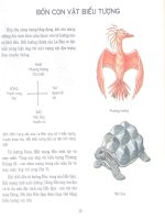
Fig. 2. Effects of very high Ca
2+
accumulation in neurons after ischemia. Excitotoxicity
causes a sudden increase in cytoplasmic Ca
2+
concentrations in neurons after ischemia,
which induces activation of several signaling pathways, leading to apoptototic or necrotic
neuronal death. Activation of calpains, caspases, other proteases, kinases and
endonucleases, cause mitochondrial disturbance, overproduction of free radicals and DNA
fragmentation, that synergistically lead to neuronal death.
Major excitotoxic events promoted by cytoplasmic Ca
2+
overload due to massively activated
glutamate receptors include mitochondrial dysfunction, oxidative/nitrosative stress and
calpain activation (Fig. 3).
The excitotoxicity can contribute to neuron death by altering the functions of mitochondria.
Mitochondrial disturbance is the result of both oxidative-nitrosative stress and a direct effect of
excessive Ca
2+
intracellular levels. Mitochondrial dysfunction is caused by free radicals and the
mitochondrial disturbance, in turn, increases the production of free radicals. Mitochondria
play an important role in calcium homeostasis [97, 98]. Under conditions of cytoplasmic excess
of Ca
2+
, mitochondria are very important for cell survival, as they have the ability to sequester
large amounts of Ca
2+
. From in vitro studies [98] it can be inferred that mitochondria within
intact neurons will act as temporary reversible stores of Ca
2+
, accumulating the cation when
cytoplasmic Ca
2+
is above a set point, and releasing the cation back to the cytoplasm when the
plasma membrane Ca
2+
-ATPase succeeds in pumping down cytoplasmic Ca
2+
to below the set
point [96, 99]. For this cytoplasmic buffering to occur with no deleterious effects for the
mitochondria and hence the cell, the time during which cytoplasmic Ca
2+
is above the set point
must be brief, thus avoiding mitochondrial Ca
2+
overload [96]. During stroke, electron
microscope analyses show that Ca
2+
accumulates in mitochondria very soon after global
ischemia and this state persists for several hours [100]. Excessive and prolonged uptake of Ca
2+
in mitochondria causes mitochondrial dysfunction [41, 96, 101], which is considered the
primary event in neuron death due to excitotoxicity [41].
Acute Ischemic Stroke
38
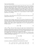
Fig. 3. Excitotoxic signaling by overstimulation of the NMDA receptors. Major excitotoxic
events promoted by extrasynaptic NMDAR activation. Cerebral ischemia elevates cytosolic
Ca
2+
levels through the stimulation of NMDARs. The calcium overload: a) activates calpains
that inactivate of the Na
+
/Ca
2+
exchanger (NCX3), b) induces mitochondrial disturbance
that activate intrinsic apoptotic pathway and c) activates NOS that increases the NO
production. Higher concentrations of nitric oxide can produce irreversible modifications of
proteins, lipids and impairment of mitochondrial respiration. All of these processes trigger
pathological mechanisms leading to neuronal death.
Mitochondrial dysfunction as a consequence of prolonged accumulation of Ca
2+
is
considered a major source of free radicals that are generated after ischemia-reperfusion [102,
103]. As a result of the mitochondrial dysfunction induced by the free Ca
2+
cytosol
accumulation, two events seem to play an important role in the death of neurons: the
increase in the production of free radicals associated with a diminution of the antioxidant
defences [102, 103], and the induction of the apoptotic cascade (Fig. 4) [104, 105].
Under physiological conditions, free radicals are generated at low levels and play
important roles in signalling and metabolic pathways (106-108]. However, free radicals
avidly interact with a large number of molecules including other small inorganic
molecules as well as proteins, lipids, carbohydrates, and nucleic acids. Through such
interactions, free radicals may irreversibly destroy or alter the function of the target
molecule. Consequently, free radicals have been increasingly identified as major
contributors to damage in biological organisms. The significance of free radicals as
aggravating or primary factors in numerous pathologies is firmly established [109, 110].
Excitotoxicity and Oxidative Stress in Acute Ischemic Stroke
39
Importantly, free radicals are produced continually during normal oxidative metabolism,
but there are counteracted by a sophisticated system of enzymes and non-enzymatic
antioxidants which maintains physiological homeostasis [111](Fig. 5). Enzymatic
components mainly comprise superoxide dismutases (SOD) [112], catalases [111],
glutathione [113] glutathione reductase/glutathione peroxidases (GR/GPX) [114], and
peroxiredoxins [115]. Also, small molecular non-enzymatic antioxidants are important in
scavenging free radicals. These include ascorbic acid, pyruvate, α-tocopherol and
glutathione, which are also involved in the detoxification of free radicals, provision of
antioxidant defence and prevention of tissue damage [111].
Fig. 4. Mitochondrial dysfuntion by disruption of calcium homeostasis leads to oxidative
stress and apoptosis. Mitochondria are involved in both, the necrosis and the apoptotic
pathways, which depend on the severity of the insult or nature of the signaling pathways.
When cytosolic Ca
2+
reaches non-physiological levels, the mitochondrial membrane may
become more permeable, which causes release of cytochrome c and activation of the
apoptotic pathway. Ca
2+
-induced mitochondria disturbance involves dysfunction of ETC,
increased ROS and oxidative stress.
When an imbalance occurs, either by increasing free radical formation or decreased anti-
oxidant defences, and the formation of free radicals exceeds the protective capacity of
antioxidant systems, the accumulation of free radicals is known as a state of “oxidative
stress” [116]. Oxidative stress is generally defined as an imbalance that favours the
production of free radicals over their inactivation by antioxidant defence systems [117].
Acute Ischemic Stroke
40
Fig. 5. Cellular reactions lead to oxidative damage of lipids, proteins and DNA via the
Fenton reaction and their protection by main endogenous antioxidant enzymes (SOD,
catalases and proxidaxes). The deleterious effects of ROS and RNS are controlled by
antioxidant defences. Neurons are particularly vulnerable to oxidative stress owing to their
high metabolic activity and oxygen consumption, which leads to high levels of ROS
production, together with relatively low levels of endogenous antioxidant enzymes,
particularly catalase. Moreover, the high lipid content of the brain can react with ROS to
generate peroxyl radicals, leading to lipid oxidation of the neuronal membrane. The
combination of these factors makes the CNS particularly vulnerable to oxidative damage.
Oxidative stress induced by excitotoxicity is considered the main event leading to brain
damage after cerebral ischemia [35, 103, 109]. The most important free radicals induced by
excitotoxicity are molecular derivates of oxygen and oxide nitric. Owing to their high
oxidizing power, the intermediate reduction states of oxygen are called reactive oxygen species
(ROS) and nitrogen-containing oxidants are called reactive nitrogen species (RNS). ROS are
small oxygen-derived molecules, including the superoxide anion radical (O
2
•-
), hydroxyl
radical (OH·), and certain non-radicals that are either oxidizing agents or easily converted
into radicals, such as hydrogen peroxide (H
2
O
2
) and the oxygen singlet (
1
O
2
). RNS are
nitrogen-derived molecules, such as nitric oxide (NO
•
), which has a relatively long half-life
(approx. 1 s) and whose reactions with biological molecules are slow due to its very rapid
diffusion into the blood and consequent inactivation by haemoglobin. NO
•
is an important
free radical because it combines with H
2
O
2
and O
2
•-
to form OH
•
and peroxynitrite
(ONOO
−
), which is stable at an alkaline pH and fairly non-reactive, but it is readily
protonated at cellular pH to peroxynitrous acid (ONOOH), which is very cytotoxic.
Following early suggestions [118], free radicals and other small reactive molecules have
emerged as important players in the cell mechanisms involved in the pathophysiology of
strokes [35, 119-121]. Several lines of research indicate that oxidative stress is a primary
mediator of neurologic injury following cerebral ischemia [103, 120, 121]. After cerebral
ischemia and particularly reperfusion, robust oxidants are generated including superoxide
and hydroxyl radicals, which overwhelm endogenous scavenging mechanisms [122, 123]
Excitotoxicity and Oxidative Stress in Acute Ischemic Stroke
41
and are directly involved in the damage to cellular macromolecules, such as lipids, proteins,
and nucleic acids, eventually leading to cell death [1,2] (Fig. 6). Re-oxygenation during
reperfusion provides
oxygen to sustain neuronal viability and also provides oxygen
as a
substrate for numerous enzymatic oxidation reactions that
produce reactive oxidants. In
addition, reflow after occlusion
often causes an increase in oxygen to levels that cannot be
utilized by mitochondria under normal physiological flow conditions. During reperfusion,
perturbation of the antioxidative defence
mechanisms is a result of the overproduction of
oxygen radicals,
inactivation of detoxification systems, consumption of antioxidants,
and
failure to adequately replenish antioxidants in the ischemic
brain tissue [122-123].
Fig. 6. ROS-mediated damage of cellular macromolecules may lead to neuron death.
Excessive release of glutamate can trigger ROS increase. Antioxidant defences include
several enzymes. In the healthy subjects, there is a balance between the production of
antioxidants defences and of reactive species. When an imbalance occurs, either by
increasing free radical formation and/or decreased anti-oxidant defences, and the formation
of free radicals exceeds the protective capacity of antioxidant systems, the accumulation of
free radicals leads to oxidative stress.
The important role of free radicals in cell damage during stroke is emphasized by the fact
that even delayed treatment with the use of antioxidants and inhibitors of free radical
producing enzymes can be effective in experimental focal cerebral ischemia [124, 125]. In
addition, the overproduction of radical-scavenging enzymes protects against stroke [126]
and animals that are deficient in radical-scavenging enzymes are more susceptible to
cerebral ischemic damage [127]. In addition, neuroprotection is evident in animal models
where genes coding for enzymes that promote oxidative stress are knocked down or out,
and where genes coding for antioxidant enzymes, e.g., superoxide dismutase (SOD) are
over-expressed [44, 126].
Increased levels of ROS and RNS generated extra- and intra-cellularly can, by various
processes, initiate and promote neuron death during ischemic stroke. ROS and RNS can
directly oxidize and damage macromolecules such as DNA, proteins, and lipids,
culminating in neuron death[1, 2, 45-47]. ROS and RNS can also indirectly contribute to
tissue damage by activating a number of cellular pathways resulting in the expression of
stress-sensitive genes and proteins that cause oxidative injury [43].
Intracellular sources of ROS include the mitochondrial electron transport chain (ETC),
xanthine oxidase, arachidonic acid, and NADPH oxidases. It is generally thought that
Acute Ischemic Stroke
42
mitochondria are the primary source of ROS involved in oxidative stress induced after
cerebral ischemia. Free radicals are produced in the mitochondria as by-products of
respiratory chain reactions. While passing through the mitochondrial ETC, some electrons
escape from the mitochondrial ETC, especially from complexes I and III, and react with O
2
to
form superoxide anion radicals (O
2
•
−
) (Figure 7), which rapidly dismutate to H
2
O
2
either
spontaneously, particularly at low pH, or catalyzed by superoxide dismutase [128, 129].
Approximately 1%–2% of the molecular oxygen consumed during normal physiological
respiration is converted into superoxide radicals [130].
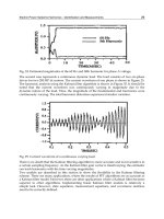
Fig. 7. Cerebral ischemia and reperfusion generated reactive oxygen species by
mitochondria and reactive nitrogen species by nitric oxide synthase. The generation of
peroxynitrite (ONOO
-
), formed by the reaction of nitric oxide with superoxide anion, and
subsequent hydroxil radical (OH
•
) production can directly damage lipids, proteins, and
DNA and lead to neuron death.
4. Oxidative stress in acute ischemic stroke
Neurons are particularly vulnerable to oxidative stress owing to their high metabolic
activity and oxygen consumption which lead to high levels of ROS production, together
with relatively low levels of endogenous antioxidant enzymes, particularly catalase [131].
Moreover, the high lipid content of the brain can react with ROS to generate peroxyl radicals
that lead to neuron membrane lipid oxidation [132]. The combination of these factors makes
the CNS particularly vulnerable to oxidative damage [113].
The primary source of free radical generation in cells during cerebral ischemia has been
reported to be due to a decrease in mitochondria redox potential causing ROS production from
the ETC, mainly at the level of cytochrome III [102, 103, 118, 130]. After ischemia, an excess of
cytosolic free Ca
2+
due to excitotoxicity may overload the mitochondrial proton circuit, which
leads to failure in oxidation together with increased ROS production [102, 109].
Overproduction of ROS by mitochondria causes the impairment of the ETC, which in turn,
leads to decreased ATP production, increased formation of free radicals, altered calcium
homeostasis and mitochondrial dysfunction [130]. In the rat, transient middle cerebral artery
occlusion (MCAO) induces ROS production and mitochondrial dysfunction, including the
inactivity of ETC enzymes. The mitochondrial dysfunction is attenuated by treatment with an