Cell Metabolism Cell Homeostasis and Stress Response Part 8 pptx
Bạn đang xem bản rút gọn của tài liệu. Xem và tải ngay bản đầy đủ của tài liệu tại đây (393.44 KB, 15 trang )
Cell Metabolism – Cell Homeostasis and Stress Response
96
Lee, Y.Y., Iyer, P. & Torget, R.W. (1999). Dilute-acid hydrolysis of lignocellulosic biomass.
Adv Biochem Eng Biotechnol, Vol. 65, pp. 93–115
Lemasters, J.J. (2005). Selective mitochondrial autophagy, or mitophagy, as a targeted
defense against oxidative stress, mitochondrial dysfunction, and aging.
Rejuvenation Res, Vol. 8, pp. 3-5
Li, B Z. & Yuan, Y J. (2010). Transcriptome shifts in response to furfural and acetic acid in
Saccharomyces cerevisiae. Appl Microbiol Biotechnol, Vol. 86, pp. 1915–1924
Ligr, M., Madeo, F., Frohlich, E., Hilt, W., Frohlich, K.U., Wolf, D.H. (1998). Mammalian Bax
triggers apoptotic changes in yeast. FEBS Lett, Vol. 438, pp 61-65
Ludovico, P. (1999). Efeitos do ácido acético no potencial de membrana mitocondrial e sua
relação com a perda de integridade e viabilidade celular em Zygosaccharomyces bailii
e Saccharomyces cerevisiae. Estudos por citometria de fluxo e espectrofluorimetria.
Tese de Mestrado, Universidade do Minho
Ludovico, P., Sansonetty, F., Silva, M.T. & Côrte-Real, M (2003). Acetic acid induces a
programmed cell death process in the food spoilage yeast Zygosaccharomyces bailii,
FEMS Yeast Res, Vol. 3, pp. 91–96
Ludovico, P., Rodrigues, F., Almeida, A., Silva, M.T., Barrientos, A. & Côrte-Real, M. (2002).
Cytochrome c release and mitochondria involvement in programmed cell death
induced by acetic acid in Saccharomyces cerevisiae. Mol Biol Cell,Vol. 13, pp. 2598-
2606
Ludovico, P., Sousa, M.J., Silva M.T., Leão, C. & Côrte-Real, M. (2001). Saccharomyces
cerevisiae commits to a programmed cell death process in response to acetic acid.
Microbiology, Vol. 147, pp. 2409-2415
Madeo, F., Frohlich, E. & Frohlich, K.U. (1997). A yeast mutant showing diagnostic markers
of early and late apoptosis. J Cell Biol, Vol. 139, pp. 729-734
Madeo, F., Frohlich, E., Ligr, M., Grey, M., Sigrist, S.J., Wolf, D.H. & Frohlich, K.U. (1999).
Oxygen stress: a regulator of apoptosis in yeast. J Cell Biol, Vol. 145, pp. 757-767
Madeo, F., Herker, E., Maldener, C., Wissing, S., Lächelt, S., Herlan, M., Fehr, M., Lauber, K.,
Sigrist, S.J., Wesselborg, S. & Fröhlich, K.U. (2002). A caspase related protease
regulates apoptosis in yeast. Mol Cell, Vol. 9, pp. 911–917
Maiorella, B., Blanch, H.W. & Wilke, C.R. (1983). By-product inhibition effects on ethanolic
fermentation by Saccharomyces cerevisiae. Biotechnol Bioeng, Vol. 25, pp. 103–121
McInerny, C.J. (2011), Cell cycle regulated gene expression in yeasts. Adv Genet, Vol. 73, pp.
51-85
Mason, D.A., Shulga, N., Undavai, S., Ferrando-May, E., Rexach, M.F. & Goldfarb, D.S.
(2005). Increased nuclear envelope permeability and Pep4p-dependent degradation
of nucleoporins during hydrogen peroxide-induced cell death. FEMS Yeast Res,
Vol. 5, pp. 1237-1251
Masson, O., Bach A.S., Derocq, D., Prébois, C., Laurent-Matha, V., Pattingre, S. & Liaudet-
Coopman, E. (2010). Pathophysiological functions of cathepsin D: targeting its
catalytic activity versus its protein binding activity? Biochemie, Vol. 92, pp. 1635-
1643
Matsui, M., Yamamoto, A., Kuma, A., Ohsumi, Y. & Mizushima, N. (2006). Organelle
degradation during the lens and erythroid differentiation is independent of
autophagy. Biochem Biophys Res Commun, Vol. 339, pp. 485–489
Stress and Cell Death in Yeast Induced by Acetic Acid
97
Matsuyama, S., Llopis, J., Deveraux, Q.L., Tsien, R. & Reed, J.C. (2000). Changes in
mitochondrial and cytosolic pH: early events that modulate caspase activation
during apoptosis. Nat Cell Biol, Vol. 2, pp. 318–325
Mira, N.,P., Lourenço, A.B., Fernandes, A.R., Becker, J.D. & Sá-Correia, I. (2009). The RIM101
pathway has a role in Saccharomyces cerevisiae adaptive response and resistance to
propionic acid and other weak acids. FEMS Yeast Res, Vol. 9, No. 2, pp. 202-216
Mira, N.P., Palmam, M., Guerreiro, J.F. & Sá-Correia, I. (2010). Genome-wide identification
of Saccharomyces cerevisiae genes required for tolerance to acetic acid. Microb Cell
Fact, Vol. 9, pp. 79
Mollapour, M. & Piper, P.W. (2006). Hog1p mitogen-activated protein kinase determines
acetic acid resistance in Saccharomyces cerevisiae. FEMS Yeast Res, Vol. 6, No. 8, pp.
1274-1280
Mollapour, M. & Piper, P.W. (2007). Hog1 mitogen-activated protein kinase
phosphorylation targets the yeast Fps1 aquaglyceroporin for endocytosis, thereby
rendering cells resistant to acetic acid. Mol Cell Biol, Vol. 27, pp. 6446–6456
Mollapour, M., Fong, D., Balakrishnan, K., Harris, N., Thompson, S., Schuller, C., Kuchler,
K. & Piper, P.W. (2004). Screening the yeast deletant mutant collection for
hypersensitivity and hyperresistance to sorbate, a weak organic acid food
preservative. Yeast, Vol. 21, pp. 927–946
Mollapour, M., Shepherd, A. & Piper, P.W. (2009). Presence of the Fps1p aquaglyceroporin
channel is essential for Hog1p activation, but suppresses Slt2(Mpk1)p activation,
with acetic acid stress of yeast. Microbiology, Vol. 155, pp. 3304–3311
Mozdy, A.D., McCaffery, J.M. & Shaw, J.M. (2000). Dnm1p GTPase-mediated mitochondrial
fission is a multi-step process requiring the novel integral membrane component
Fis1p. J Cell Biol, Vol. 151, pp. 367-380
Nowikovsky, K., Reipert, S., Devenish, R.J. & Schweyen, R.J. (2007). Mdm38 protein
depletion causes loss of mitochondrial K+/H+ exchange activity, osmotic swelling
and mitophagy. Cell Death Differ, Vol. 14, pp. 1647-1656
Palmqvist, E. & Hahn-Hägerdal, B. (2000). Fermentation of lignocellulosic hydrolysates. I:
inhibition and detoxification. Bioresour Technol, Vol. 74, pp. 17-24
Pampulha, M.A. & Loureiro-Dias, M.C. (1989). Combined effect of acetic acid, pH and
ethanol on intracellular pH of fermenting yeast. Appl Microbiol Biotechnol, Vol. 31,
pp. 547–550
Pampulha, M.A. & Loureiro-Dias, M.C. (1990). Activity of glycolytic enzymes of
Saccharomyces cerevisiae in the presence of acetic acid. Appl Microbiol Biotechnol, Vol.
34, pp. 375–380
Pampulha, M.A. & Loureiro-Dias, M.C. (2000). Energetics of the effect of acetic acid on
growth of Saccharomyces cerevisiae. FEMS Microbiol Lett, Vol. 184, pp. 69–72
Parone, P.A. & Martinou, J.C. (2006). Mitochondrial fission and apoptosis: an ongoing trial,
Biochim Biophys Acta, Vol. 1763, pp. 522-530
Pereira, C., Camougrand, N., Manon, S., Sousa, M.J. & Côrte-Real, M. (2007). ADP/ATP
carrier is required for mitochondrial outer membrane permeabilization and
cytochrome c release in yeast apoptosis. Mol Microbiol, Vol. 66, pp. 571-582
Pereira, C., Silva, R.D., Saraiva, L., Johansson, B., Sousa, M.J. & Côrte-Real, M. (2008).
Mitochondria dependent apoptosis in yeast. Biochim Biophys Acta, Vol. 1783, 1286-
1302
Cell Metabolism – Cell Homeostasis and Stress Response
98
Pereira, C., Chaves, S., Alves, S., Salin, B., Camougrand, N., Manon, S., Sousa, M.J. & Côrte-
Real, M. (2010). Mitochondrial degradation in acetic acid-induced yeast apoptosis:
The role of Pep4 and the ADP/ATP carrier. Mol Microbiol, Vol. 76, pp. 1398-1410
Petranovic, D. & Nielsen, J. (2008). Can yeast systems biology contribute to the
understanding of human disease? Trends Biotechnol, Vol. 26, pp. 584-590
Phillips, A.J., Crowe, J.D. & Ramsdale, M. (2006). Ras pathway signaling accelerates
programmed cell death in the pathogenic fungus Candida albicans. Proc Natl Acad
Sci USA, Vol. 103, pp. 726–731
Phillips, A.J., Sudbery, I. & Ramsdale, M. (2003). Apoptosis induced by environmental
stresses and amphotericin B in Candida albicans. Proc Natl Acad Sci U S A, Vol. 100,
25 pp. 14327-14332
Phowchinda, O., Délia-Dupuy, M.L. & Strehaiano, P. (1995). Effects of acetic acid on growth
and fermentative activity of Saccharomyces cerevisiae. Biotechnol Lett, Vol. 17, pp.
237–242
Pinto, I., Cardoso, H. & Leão, C. (1989). High enthalpy and low enthalpy death in
Saccharomyces cerevisiae induced by acetic acid. Biotechnol Bioeng, Vol. 33, pp. 1350-
1352
Piper, P., Mahé, Y., Thompson, S., Pandjaitan, R., Holyoak, C., Egner, R., Mühlbauer, M.,
Coote, P. & Kuchler, K. (1998). The Pdr12 ABC transporter is required for the
development of weak organic acid resistance in yeast. EMBO J, Vol. 17, pp. 4257-
4265
Pozniakovsky, A.I., Knorre, D.A., Markova, O.V., Hyman, A.A, Skulachev, V.P. & Severin,
F.F. (2005). Role of mitochondria in the pheromone- and amiodarone-induced
programmed death of yeast. J Cell Biol, Vol. 168, pp. 257-269
Priault, M., Salin, B., Schaeffer, J., Vallette, F.M., di Rago, J.P. & Martinou, J.C. (2005).
Impairing the bioenergetic status and the biogenesis of mitochondria triggers
mitophagy in yeast. Cell Death Differ, Vol. 12, pp. 1613–1621
Prudêncio, C., Sansonetty, F. & Côrte-Real, M. (1998). Flow cytometric assessment of cell
structural and functional changes induced by acetic acid in the yeasts
Zygosaccharomyces bailii and Saccharomyces cerevisiae. Cytometry, Vol. 31, pp. 307-313
Ramsdale, M. (2006). Programmed Cell Death and Apoptosis in Fungi, In: The Mycota XIII,
Fungal Genomics, Alistair J.P. Brown, pp. 113-146, Springer-Verlag, Berlin
Heidelberg
Repnik, U., & Turk, B. (2010). Lysosomal-mitochondrial cross-talk during cell death.
Mitochondrion, Vol. 10, pp. 662-669
Ribeiro, G.F., Côrte-Real, M. & Johansson, B. (2006). Characterization of DNA damage in
yeast apoptosis induced by hydrogen peroxide, acetic acid, and hyperosmotic
shock. Mol Biol Cell, Vol. 17, pp. 4584-4591
Rodrigues, F., Côrte-Real, M., Leão, C., van Dijken, J.P. & Pronk, J.T. (2001). Oxygen
requirements of the food spoilage yeast Zygosaccharomyces bailii in synthetic and
complex media. Appl Environ Microbiol
, Vol. 67, pp. 2123-2128
Rodrigues, F., Ludovico, P. & Leão, C. (2005). Sugar Metabolism in Yeasts: an Overview of
Aerobic and Anaerobic Glucose Catabolism. In: Biodiversity and Ecophysiology of
Yeasts, Carlos A. Rosa & Gábor Péter, pp. 101-121, Springer Springer Lab Manuals,
Germany
Stress and Cell Death in Yeast Induced by Acetic Acid
99
Rodriguez-Enriquez, S., Kim, I., Currin, R.T., & Lemasters, J.J. (2006) Tracker dyes to probe
mitochondrial autophagy (mitophagy) in rat hepatocytes. Autophagy Vol. 2, pp. 39–
46
Roset, R., Ortet, L. & Gil-Gomez, G. (2007). Role of Bcl-2 family members on apoptosis: what
we have learned from knock-out mice. Front Biosci, Vol. 12, pp. 4722-4730
Sagulenko, V., Muth, D., Sagulenko, E., Paffhausen, T., Schwab, M. & Westermann, F. (2008).
Cathepsin D protects human neuroblastoma cells from doxorubicin-induced cell
death. Carcinogenesis, Vol. 29, pp. 1869-1877
Santos, J., Sousa, M.J., Cardoso, H., Inácio, J., Silva, S., Spencer-Martins, I. & Leão, C. (2008).
Ethanol tolerance of sugar transport and the rectification of stuck wine
fermentations. Microbiology, Vol. 154, pp. 422-430
Saraiva L., Silva R.D., Pereira G., Gonçalves J. & Côrte-Real M. (2006). Specific modulation of
apoptosis and Bcl-xL phosphorylation in yeast by distinct mammalian protein
kinase C isoforms. J Cell Sci, Vol. 119, pp. 3171-3181
Schauer, A., Knauer, H., Ruckenstuhl, C., Fussi, H., Durchschlag, M., Potocnik, U. &
Frohlich, K.U. (2009). Vacuolar functions determine the mode of cell death. Biochim
Biophys Acta, Vol. 1793, pp. 540-545
Schuller, C., Mamnun, Y.M., Mollapour, M., Krapf, G., Schuster, M., Bauer, B.E., Piper, P.W.
& Kuchler, K. (2004). Global phenotypic analysis and transcriptional profiling
defines the weak acid stress response regulon in Saccharomyces cerevisiae. Mol Biol
Cell, Vol. 15, pp. 706–720
Scorrano, L. (2005). Proteins that fuse and fragment mitochondria in apoptosis: con-fissing a
deadly con-fusion? J Bioenerg Biomembr, Vol. 37, pp. 165-170
Severin, F.F. & Hyman, A.A. (2002). Pheromone induces programmed cell death in S.
cerevisiae. Curr Biol, Vol. 12, pp. 233-235
Shinohara, K., Tomioka, M., Nakano, H., Tone, S., Ito, H. & Kawashima, S. (1996). Apoptosis
induction resulting from proteasome inhibition. Biochem J, Vol. 317, pp. 385–388
Silva, R.D., Sotoca, R., Johansson, B., Ludovico, P., Sansonetty, F., Silva, M.T., Peinado, J.M.
& Côrte-Real, M. (2005). Hyperosmotic stress induces metacaspase- and
mitochondria-dependent apoptosis in Saccharomyces cerevisiae. Mol Microbiol, Vol.
58, pp. 824–834
Sokolov, S., Knorre, D., Smirnova, E., Markova, O., Pozniakovsky, A., Skulachev, V. &
Severin, F. (2006). Ysp2 mediates death of yeast induced by amiodarone or
intracellular acidification, Biochim Biophys Acta, Vol. 1757, pp. 1366-1370
Sousa, M.J., Miranda, L., Côrte-Real, M. & Leão, C. (1996). Transport of acetic acid in
Zygosaccharomyces bailii: effects of ethanol and their implications on the resistance of
the yeast to acid environments. Appl Environ Microbiol, Vol. 62, pp. 3152-3157
Sousa, M.J., Rodrigues, F., Côrte-Real, M. & Leão, C. (1998). Mechanisms underlying the
transport and intracellular metabolism of acetic acid in the presence of glucose by
the yeast Zygosaccharomyces bailii. Microbiology, Vol. 144, pp. 665-670
Susin, S.A., Lorenzo, H.K., Zamzami, N., Marzo, I., Snow, B.E., Brothers, G.M., Mangion, J.,
Jacotot, E., Costantini, P., Loeffler, M., Larochette, N., Goodlett, D.R., Aebersold, R.,
Siderovski, D.P., Penninger, J.M. & Kroemer, G. (1999). Molecular characterization
of mitochondrial apoptosis-inducing factor. Nature, Vol. 397, pp. 441–446
Cell Metabolism – Cell Homeostasis and Stress Response
100
Tal, R., Winter, G., Ecker, N., Klionsky, D.J. & Abeliovich, H. (2007). Aup1p, a yeast
mitochondrial protein phosphatase homolog, is required for efficient stationary
phase mitophagy and cell survival. J Biol Chem, Vol. 282, pp. 5617-5624
Thomas, S. & Davenport, R.R. (1985). Zygosaccharomyces bailii, a profile of characteristics and
spoilage activities. Food Microbiol, Vol. 2, pp. 157–169
Tolkovsky, A.M., Xue, L., Fletcher, G.C. & Borutaite, V. (2002). Mitochondrial disappearance
from cells: A clue to the role of autophagy in programmed cell death and disease?
Biochimie, Vol. 84, pp. 233-240
Vahsen, N., Cande, C., Briere, J.J., Benit, P., Joza, N., Larochette, N., Mastroberardino, P.G.,
Pequignot, M.O., Casares, N., Lazar, V., Feraud, O., Debili, N., Wissing, S.,
Engelhardt, S., Madeo, F., Piacentini, M., Penninger, J.M., Schagger, H., Rustin, P. &
Kroemer, G. (2004). AIF deficiency compromises oxidative phosphorylation. EMBO
J, Vol. 23, pp. 4679–4689
Valenti, D., Vacca, R.A., Guaragnella, N., Passarella, S., Marra, E. & Giannattasio, S. (2008).
A transient proteasome activation is needed for acetic acid-induced programmed
cell death to occur in Saccharomyces cerevisiae. FEMS Yeast Res, Vol. 8, pp. 400-404
Valle, E., Bergillos, L., Gascon, S., Parra, F. & Ramos, S. (1986). Trehalase activation in yeasts
is mediated by an internal acidification. Eur J Biochem, Vol. 154, pp. 247–251
van Uden, N. (1984). Temperature profiles of yeasts. Adv Microb Physiol, Vol. 25, 195-251
Vilela-Moura, A., Schuller, D., Mendes-Faia, A., Silva, R.D., Chaves, S.R., Sousa, M.J. &
Côrte-Real, M. (2011). The impact of acetate metabolism on yeast fermentative
performance and wine quality: reduction of volatile acidity of grape musts and
wines. Appl Microbiol Biotechnol, Vol. 89, pp. 271–280
Wei, M.C., Zong, W.X., Cheng, EH., Lindsten, T., Panoutsakopoulou, V., Ross, A.J., Roth,
K.A., MacGregor, G.R., Thompson, C.B. & Korsmeyer, S.J. (2001). Proapoptotic
BAX and BAK: a requisite gateway to mitochondrial dysfunction and death.
Science, Vol. 292, pp. 727-730
Wissing, S., Ludovico, P., Herker, E., Buttner, S., Engelhardt, S.M., Decker, T., Link, A.,
Proksch, A., Rodrigues, F., Côrte-Real, M., Frohlich, K.U., Manns, J., Cande, C.,
Sigrist, S.J., Kroemer, G. & Madeo, F. (2004). An AIF orthologue regulates apoptosis
in yeast. J Cell Biol, Vol. 166, pp. 969-974
Youle, R.J. & Karbowski, M. (2005). Mitochondrial fission in apoptosis, Nat Rev Mol Cell Biol,
Vol. 6, pp. 657-663
Zhang, J.G., Liu, X.Y., He, X.P., Guo, X.N., Lu, Y. & Zhang, B.R. (2011). Improvement of
acetic acid tolerance and fermentation performance of Saccharomyces cerevisiae by
disruption of the FPS1 aquaglyceroporin gene. Biotechnol Lett, Vol. 33, pp. 277-284
5
Metabolic Optimization by Enzyme-Enzyme and
Enzyme-Cytoskeleton Associations
Daniela Araiza-Olivera et al.
*
Instituto de Fisiología Celular, Universidad Nacional Autónoma de México,
Mexico
1. Introduction
Probably enzymes are not dispersed in the cytoplasm, but are bound to each other and to
specific cytoskeleton proteins. Associations result in substrate channeling from one enzyme
to another. Multienzymatic complexes, or metabolons have been detected in glycolysis, the
Krebs cycle and oxidative phosphorylation. Also, some glycolytic enzymes interact with
mitochondria. Metabolons may associate with actin or tubulin, gaining stability. Metabolons
resist inhibition mediated by the accumulation of compatible solutes observed during the
stress response. Compatible solutes protect membranes and proteins against stress.
However, when stress is over, compatible solutes inhibit growth, probably due to the high
viscosity they promote. Viscosity inhibits protein movements. Enzymes that undergo large
conformational changes during catalysis are more sensitive to viscosity. Enzyme association
seems to protect the more sensitive enzymes from viscosity-mediated inhibition. The
association-mediated protection of the enzymes in a given metabolic pathway would
constitute a new property of metabolons: that is, to enhance survival during stress. It is
proposed that resistance to inhibition is due to elimination of non-productive conformations
in highly motile enzymes.
2. Metabolons: Enzyme complexes that channel substrates
The cytoplasm should not be regarded as a liquid phase containing a large number of
soluble enzymes and particles. Instead, it has become evident that there is a high degree of
organization where different lipid and protein structures associate among themselves and
with other molecules. The high molecule concentration found in the cytoplasm promotes
macromolecule associations such as protein-protein, protein-membrane, protein-nucleic
acid, protein-polysaccharide and thus is a control factor for all biological processes (Srere &
Ovadi, 1990). Indeed, the classical studies by Green (Green et al., 1965), Clegg (Clegg, 1964)
*
Salvador Uribe-Carvajal
1,**
, Natalia Chiquete-Félix
1
, Mónica Rosas-Lemus
1
, Gisela Ruíz- Granados
1
,
José G. Sampedro
2
, Adela Mújica
3
and Antonio Peña
1
1
Instituto de Fisiología Celular, Universidad Nacional Autónoma de México,
2
Instituto de Física, Universidad Autónoma de San Luís Potosí and
3
CINVESTAV, Instituto Politécnico Nacional
Mexico
**
Corresponding Author
Cell Metabolism – Cell Homeostasis and Stress Response
102
and Fulton (Fulton, 1982) have suggested that enzymes are not dispersed in the cytoplasm.
Instead, enzymes are localized at specific sites where they are associated between them and
with the cytoskeleton. The cytoskeleton is a trabecular network of fibrous proteins that
micro-compartmentalizes the cytoplasm (Porter et al., 1983). Associated enzymes channel
substrates from one to another preventing their diffusion to the aqueous phase (Gaertner et
al., 1978; Minton & Wilf, 1981; Ovadi et al., 1996).
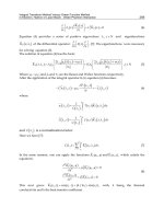
In a multienzyme complex, intermediaries can be channeled more than once from the active
site of an enzyme to the next to obtain the final product (Al-Habori, 2000; Robinson et al.,
1987). Channeling requires stable interactions of the multienzymatic metabolons (Al-Habori,
2000; Cascante et al., 1994; Ovadi & Srere, 1996; Ovadi & Saks, 2004; Srere & Ovadi, 1990;
Srere, 1987). The metabolon stability is facilitated by the compartmentalization of the cell in
different organelles and structures (Jorgensen et al., 2005).
There are many advantages inherent to metabolons (Jorgensen et al., 2005) (Fig. 1):
I) Improved catalytic efficiency of the enzymes. This is obtained by channeling an
intermediary from the active site of an enzyme directly to the active site of the next.
II) Channeling optimizes kinetic constants. III) Labile or toxic intermediates are retained
within the metabolon. IV) Inhibitors are excluded from the active site of enzymes.
V) Control and coordination of the enzymes in a given pathway is enhanced. VI) Finally,
alternative metabolons may favor different pathways (Fig. 1). Most metabolons seem to be
transient, opening the possibility for a quick change in some elements that allows them to
redirect metabolism (Jorgensen et al., 2005).
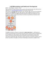
Fig. 1. Advantages of Metabolons. (A) In isolated enzymes the substrate (green),
intermediaries (red and yellow) and product (orange) diffuse into the aqueous phase (little
arrows). Toxic intermediaries and inhibitors (grey) are free to exit/enter the active site in
each enzyme. (B) In metabolons (we show filamentous actin in red and white) channeling
allows transfer of the substrate (green) from the active site of an enzyme direct to the next to
obtain a final product (orange) without diffusion to the cytoplasm of intermediaries (not-
depicted) are prevented, while inhibitors (grey) are excluded from the active sites.
The enzymes from the Krebs cycle are attached to the mitochondrial membrane in an
enzymatic complex; this was the first “metabolon” described (Srere, 1987). In oxidative
A
B
Metabolic Optimization by Enzyme-Enzyme and Enzyme-Cytoskeleton Associations
103
phosphorylation, multiprotein complexes seem to associate in supercomplexes and
eventually in respiratory chains resulting in controlled electron channeling and proton-
pumping stoichiometry (Guerrero-Castillo et al., 2011). It has been proposed that these
supercomplexes constitute an exquisite mechanism to regulate the yield of ATP (Guerrero-
Castillo et al., 2009; 2011; Schägger et al., 2001). In addition, in some organisms such as
trypanosomes, glycolytic enzymes are contained in small organelles called glycosomes,
where channeling is highly efficient (Aman et al., 1985). Tumor cells also produce aggregates
containing glycolytic enzymes (Coe & Greenhouse, 1973). Interactions between organelles
such as the endoplasmic reticulum and mitochondria have been described (Dorn &
Scorrano, 2010; Kornmann et al., 2009; Lebiedzinska et al., 2009). Mitochondria are both, the
main source of ATP and inducers of cellular death (Anesti & Scorrano, 2006). Mitochondrial
functions are regulated by interactions with other organelles and cytoplasmic proteins
(Kostal & Arriaga, 2011). Cytoskeletal proteins such as actin and tubulin, direct
mitochondria to specific sites in the cell (Senning & Marcus, 2010) and control coupling of
phosphorylation by interacting with mitochondrial porin (Xu et al., 2001; Lemasters &
Holmuhamedov, 2006; Rostovtseva et al., 2008; Rostovtseva et al., 2004; Xu et al., 2001). In
addition to cytoeskeletal proteins, hexokinase, a glycolytic enzyme binds mitochondria in
mammalians (Pastorino & Hoek, 2008), yeast and plants (Balasubramanian et al., 2008)
regulatin the energy yield of mitochondria as well as the induction of programmed cell
death (Kroemer et al., 2005; Pastorino & Hoek, 2008; Xie & Wilson, 1988). All the above data
suggest that enzymes are highly organized (Clegg & Jackson, 1989) and the cytoskeleton
plays an important role (Minaschek et al., 1992; Keleti et al., 1989; Porter et al., 1983).
3. The cytoskeleton: A scaffold where metabolons are bound
The eukaryotic cytoplasm is supported by the cytoskeleton, a network of structural proteins
that shapes the cell and has binding sites for different enzymes. Such sites have been
identified in filamentous actin (F-actin), in microtubules and in the cytoplasmic domain of
the erythrocyte band 3, which is also an anion exchanger. Glycolytic enzyme binding to
actin usually results in stimulation, whereas binding to microtubules or to band 3 inhibits
activity (Real-Hohn et al., 2010). Actin is involved in a variety of cell functions that include
contractility, cytokinesis, maintenance of cell shape, cell locomotion and organelle
localization. In addition, glycolytic enzymes and F-actin co-localize in muscle cells, probably
reflecting compartmentation of the glycolytic pathway (Waingeh et al., 2006).
Actin is highly conserved in eukaryotic cells. It may be found as a monomer (G-actin) or as a
polymeric filament (F-actin) that is interconverted in an extremely dynamic, highly
controlled process. The polar actin monomers polymerize head-to-tail to yield a polar
filament. Actin filaments are constituted by 8 nm diameter, double-helical structures formed
by assemblies of monomeric actin with a barbed end (or plus end) and a pointed end (or
minus end). The spontaneous polymerization of actin monomers occurs in three phases:
nucleation, elongation and maintenance. Nucleation consists in the formation of a dimer,
followed by the addition of a third monomer to yield a trimer; this process is very slow.
Further monomer addition becomes thermodynamically favorable and the filament
elongates rapidly: much faster at the plus end than at the minus end. In the maintenance
phase, there is no net filament growth and the concentration of ATP-G-actin is kept
stationary (Fig. 2).
Cell Metabolism – Cell Homeostasis and Stress Response
104
Upon incorporation to a filament, G-actin-bound ATP is hydrolyzed. ADP and Pi remain
non-covalently bound. Then Pi is released slowly. Thus, the elongating filaments contain:
the barbed end, rich in ATP-actin, the center, rich in ADP-Pi-actin and the pointed end
containing ADP-actin. Many actin-binding proteins regulate actin polymerization. Profilin is
an actin monomer-binding protein; Arp 2/3 complex are nucleation proteins; CapZ and
gelsolin regulate the length of the actin filament and the cofilin/ADP family cuts F-actin and
accelerates depolymerization (Kustermans et al., 2008). However, protein functions may
vary; in Dictyostelium, CapZ prevents filament elongation and increases the concentration of
unpolymerized actin; in contrast, in yeast this same protein prevents depolymerization
increasing F-actin concentration (Welch et al., 1997). The cytoskeleton can be rapidly
remodeled by the small RhoGTPases (Rho, Rac and Cdc42), which act in response to
extracellular stimuli (Kustermans et al., 2008). There are exogenous natural compounds that
can disturb actin dynamics (Kustermans et al., 2008).
4. The glycolytic metabolon
The association of enzymes with the cytoskeleton probably stabilizes metabolons. In this
regard, glycolytic enzymes such as fructose 1,6-bisphosphate aldolase (aldolase),
glyceraldehyde 3-phosphate dehydrogenase (GAPDH), piruvate kinase (PK), glucose
phosphate isomerase (GPI), and lactate dehydrogenase (LDH) associate with actin. Other
glycolytic enzymes such as triose phosphate isomerase and phosphoglycerate mutase bind
indirectly through interactions with other enzymes. Enzyme-enzyme-actin complexes are
called piggy-back interactions. Also, aldolase and GAPDH compete for binding sites (Knull
& Walsh, 1992; Waingeh et al., 2006).
Fig. 2. Actin polymerization. During nucleation, actin monomers aggregate to form a trimer.
Then during elongation actin filaments grow actively at both ends. Growth stops in the
maintenance phase, also known as stationary phase. (Modified from Kustermans et al., 2008)
ATP-actin
ADP-Pi-actin
ADP-actin
Barbed end (+)
Pointed end (-)
Nucleation
Elongation
Stationary state
ATP-actin
ADP-Pi-actin
ADP-actin
Barbed end (+)
Pointed end (-)
Nucleation
Elongation
Stationary state
Metabolic Optimization by Enzyme-Enzyme and Enzyme-Cytoskeleton Associations
105
Enzyme/actin interaction is regulated by ionic strength (Waingeh et al., 2006). In
homogenates of muscle tissue suspended in isosmotic sucrose, proteins such as F-actin,
myosin, troponin and tropomyosin associate with glycolytic enzymes (Brooks & Storey,
1991). Glycolytic enzyme association to actin is not accepted universally, for instance, the F-
actin/glycolytic enzyme interaction has been modeled mathematically at physiological ionic
strength and protein concentrations. The results suggest that under cellular conditions only
a small percentage of TPI, GAPDH, PGK and LDH would be associated with F-actin (Brooks
& Storey, 1991).
Protein dynamics seem important for their interactions. Brownian dynamics (BD)
simulations detect that rabbit F-actin has different binding modes/affinities for aldolase and
GAPDH (Forlemu et al., 2006). Some metabolites such as ATP and ADP modulate enzyme
interactions and the resulting substrate channeling (Forlemu et al., 2006).
A barely explored effect of the association of enzymes with the cytoskeleton is the
modulation of the dynamics of actin polymerization. Such an effect has been reported for
aldolase (Chiquete-Felix et al., 2009; Schindler et al., 2001). An interesting finding is that
some growth factors, such as PGF and EGF enhance the GAPDH/cytoskeleton interaction,
possibly increasing keratinocyte migration (Tochio et al., 2010). Indeed, GAPDH seems to
participate in cytoskeleton dynamics processes such as endocytosis, membrane fusion,
vesicular transport and nuclear tRNA transport (Cueille et al., 2007).
In red blood cell membranes, GAPDH, aldolase and PFK interact with an acidic sequence at
the amino-terminal extreme of band 3 with high affinity (Campanella et al., 2005). Under
physiological conditions, the binding of glycolytic enzymes to band 3 results in inhibition of
the glycolytic flux (Real-Hohn et al., 2010).
Association to microtubules regulates the energetic metabolism (Keleti et al., 1989; Keller et
al., 2007; Walsh et al., 1989) at the level of some glycolytic enzymes such as pyruvate kinase,
phosphofructokinase (Kovács et al., 2003) and enolase (Keller et al., 2007). When the
glycolytic enzymes are associated and anchored to the sarcomere, ATP is produced more
efficiently (Keller et al., 2007). The interaction of enzymes with themselves and with the
cytoskeleton confers more stability to the enzyme activity and to the whole network
(Keleti et al., 1989; Volker et al., 1995; Walsh et al., 1989). F-actin stabilizes some glycolytic
enzymes of muscle and sperm (Walsh & Knull, 1988; Ovadi & Saks, 2004). That is the case of
the phosphofructokinase (PFK) and aldolase where the dilution-mediated inactivation of
PFK is stopped upon aldolase addition. If PFK is associated with microtubules, it still loses
activity when diluted, however, in these conditions it recovers the lost activity upon
aldolase addition (Raïs et al., 2000; Vértessy et al., 1997). All this evidence supports the
existence of a cytoskeleton-bound glycolytic metabolon.
5. Compatible solutes protect cellular structures during stress
Compatible solutes are defined as molecules that reach high concentrations in the cell without
interfering with metabolic functions (Brown & Simpson, 1972). These are mostly amino acids
and amino acid derivatives, polyols, sugars and methylamines. Compatible solutes are
typically small and harbor chemical groups that interact with protein surfaces. Indeed, some
authors have proposed to call them “chemical or pharmacological chaperones” as they
stabilize native structures (Loo & Clarke, 2007; Romisch, 2004). Some compatible solutes are:
Cell Metabolism – Cell Homeostasis and Stress Response
106
glycine betaine, a thermoprotectant in B. subtilis (Chen & Murata, 2011; Holtmann & Bremer,
2004). Ectoine, that in halophile microorganisms confers resistance to salt and temperature
stress (Pastor et al., 2010). Glycerol is accumulated in yeast under high osmotic pressure
(Blomberg, 2000). Glycerol stabilizes thermolabile enzymes preventing their inactivation
(Zancan & Sola-Penna, 2005). The disaccharide trehalose protects against environmental
injuries (heat, cold, desiccation, and anoxia) and nutritional limitations (Argüelles, 2000;
Crowe et al., 1984) in bacteria, yeast, fungi, plants and invertebrates. In biotechnology,
trehalose is one of the best protein stabilizing known (Jain & Roy, 2008; Sampedro et al., 2001).
6. Effect of compatible solutes on the activity of enzymes
Compatible solute synthesis and accumulation is triggered by harsh conditions and results
in protein stabilization and enhanced survival. Proteins may be unfolded, partially unfolded
or native (Chilson & Chilson, 2003). In the absence of stress, high compatible solute
concentrations inhibit cellular growth, metabolism and division (Wera et al., 1999), e.g. a
trehalase-deficient mutant of S. cerevisiae subjected to heat or saline stress accumulated high
amounts of trehalose and survived. However, when these mutants were returned to normal
conditions they are unable to grow or sustain metabolic activity (Garre & Matallana, 2009;
Wera et al., 1999).
6.1 Inhibition of isolated enzymes; possible role of viscosity
Under stress, high compatible solutes change the physicochemical properties of the
cytoplasm. However, the effect of the high viscosity generated by molar concentrations of
compatible solutes on enzyme activity has drawn little attention. Trehalose and other
polyols protect proteins from thermal unfolding via indirect interactions (Liu et al., 2010).
Therefore the stabilizing mechanism must rely in the modified physicochemical properties
of aqueous media.
Large-scale conformational changes in proteins involve the physical displacement of
associated solvent molecules and solutes. The resistance to the movement or displacement
of solvent molecules is a frictional process. Kramers theory provides the mathematical basis
to understand and analyze reactions at high viscosity (Kramers, 1940). The application of
Kramer´s theory to proteins indicates that the movements involved in folding or in enzyme-
substrate association and processing must be highly sensitive to viscosity (Jacob and
Schmid, 1999; Jacob et al., 1999; Sampedro and Uribe, 2004).
Studies on cellular viscosity in yeast cytoplasm showed a value of 2 cP at 30°C (Williams et
al., 1997). Also, in vitro determinations for 0.6 M trehalose solutions showed a viscosity of
1.5 cP at 30°C (Table 1). Therefore, one may infer that yeast cytoplasm viscosity with 0.6 M
trehalose should be in the vicinity of 2.5-3 cP.
The plasma membrane H
+
-ATPase from yeast depends on large domain motion for catalysis
(Kulbrandt, 2004), was inhibited at all trehalose concentrations tested (Sampedro et al.,
2002). The rate constant for the ATPase reaction (V
max
= k
cat
[E
t
]) was inversely dependent on
solution viscosity; as higher the viscosity lower the reaction rate of catalysis (Sampedro et al.,
2002). Notably, when temperature was raised inhibition disappeared, in agreement with the
fact that viscosity decreases when temperature increases (Table 1). Similar results have been
obtained with Na
+
/K
+
-ATPase and Na
+
-ATPase in the presence of polyethylene glycol and
Metabolic Optimization by Enzyme-Enzyme and Enzyme-Cytoskeleton Associations
107
glycerol (Esmann et al., 2008). In glucose oxidase, activity inhibition by varying
concentrations of trehalose was due to the promotion of a highly compact state, which
correlated with the increased viscosity of the medium (Paz-Alfaro et al., 2009).
TREHALOSE (M) 0.2 0.4 0.5 0.6 0.8
TEMP (°C) VISCOSITY (cP)
20
1.35 1.59 1.81 2.04 2.58
25
1.20 1.37 1.51 1.74 2.20
30
1.08 1.18 1.33 1.50 1.91
35
0.94 1.03 1.18 1.31 1.67
40
0.86 0.94 1.04 1.13 1.49
45
0.75 0.81 0.90 1.04 1.31
Data modified from Sampedro et al., 2002.
Table 1. Viscosity of trehalose solutions at different concentrations and temperatures.
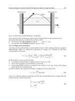
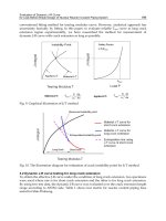
Fig. 3. Reaction coordinate diagram, comparing an enzyme reaction at normal viscosity
(blue) and at high viscosity (h; red). When a diffusive protein domain process is present in
the catalytic cycle, it becomes rate limiting when viscosity is high. Therefore the overall
activation energy (E
a
) increases.
Many enzymes are inhibited by viscosity. Glutathione reductase is inhibited at 25°C, by
trehalose (70% inhibition at 1.5 M trehalose) although inhibition disappears at 40°C
(Sebollela et al., 2004). Also pyrophosphatase and glucose 6-phosphate dehydrogenase show
temperature dependence of trehalose-mediated inhibition (Sebollela et al., 2004).
Cell Metabolism – Cell Homeostasis and Stress Response
108
Aminoglycoside nucleotidyltransferase 2''-I is inhibited by glycerol in a temperature-
dependent way (Gates & Northrop, 1988). The hyaluronan-synthase from Streptococcus
equisimilisis is inhibited by of PEG, ethylene glycol, glycerol or sucrose (Tlapak-Simmons et
al., 2004). At high viscosities (greater than 4 mPa s-1) different carbohydrates inhibit egg-
white lysozyme (Lamy et al., 1990; Monkos, 1997).
Detailed studies on diffusive protein-structural components demonstrated that for -lactam
synthase a conformational change is rate-limiting on k
cat
. Therefore, the rate for catalysis
shows a high inhibition by medium viscosity (Raber et al., 2009). Crystallographic analysis of
adhesion kinase-1 shows a large conformational motion of the activation loop upon ATP
binding. This is an essential step during catalysis and explains the viscosity inhibitory effect
(Schneck et al., 2010). In the plasma membrane H
+
-ATPase, the enzyme fluctuates between
two structural conformations (E1E2) during catalysis. The N-domain (nucleotide binding)
rotates 73° towards the phosphorylation site to deliver ATP to the phosphorylation site
(Kuhlbrandt, 2004). In all cases, the rate-limiting step is a conformational change that seems
to be the one inhibited by viscosity (Fig. 3).
6.2 Enzyme association results in protection against inhibition
Compatible solute-mediated inhibition does not seem to uniformly affect all enzymes.
Furthermore, in the face of both the stress condition and the compatible solute, catabolic
pathways seem to resist inhibition, thus providing the energy needed for survival
(Hoffmann & Holzhütter, 2009; Hounsa et al., 1998). In our hands, in a yeast cytoplasmic
extract, compatible solutes inhibit the whole glycolytic pathway much less than many of its
individual, isolated enzymes (Araiza-Olivera et al., 2010). In contrast, anabolism seems to be
shot both during the stress situation and later (Attfield, 1987). Inhibition of anabolism would
explain the inability of cells to reproduce (Wera et al., 1999). The mechanism for resistance
to inhibition, exhibited by the catabolic enzymes is a matter of study (Marcondes et al., 2011;
Raïs et al., 2000).
The effect of a compatible solute (trehalose) on the activity of some yeast glycolytic enzymes
such as GAPDH, HXK, ALD and PGK has been analyzed. These enzymes were tested
individually or in mixtures (Araiza-Olivera et al., 2010). When isolated, GAPDH and HXK
were inhibited by trehalose while others, such as ALD and PGK were resistant. Probably
GAPDH and HXK are more motile than ALD and PGK. Remarkably, when the sensitive
enzymes were mixed with the resistant enzymes a protection effect was observed. This led
to analyze the whole glycolytic pathway and again, inhibition was minimal in comparison
with the individual, isolated enzymes (Araiza-Olivera et al., 2010). Thus, it was decided to
explore the possible mechanisms underlying this effect, i.e, why some metabolic pathways,
such as glycolysis resist the viscosity-mediated inhibition promoted by compatible solutes,
even if they contain several viscosity-sensitive enzymes.
The protection effect was specific for each protein couple, as GAPDH was not protected by
neither HXK, albumin or lactate-dehydrogenase. Also, the pentose pathway enzyme glucose
6-phosphate dehydrogenase (G6PDH) was not protected by ALD against inhibition by
trehalose. Once in the complexes, probably the more flexible enzymes that are more
sensitive to viscosity (Sampedro & Uribe 2004) are stabilized by the more resistant, more
rigid enzymes forming a less motile, more resistant complex.
Metabolic Optimization by Enzyme-Enzyme and Enzyme-Cytoskeleton Associations
109
The proposal that enzyme association favors a more stable folded state would require the
motile enzymes to eliminate some non-productive conformations (Villali & Kern, 2010).
These associations are probably further stabilized by some elements of the cytoskeleton,
such as tubulin (Raïs et al., 2000; Walsh et al., 1989) or F-actin (Minaschek et al., 1992;
Waingeh et al., 2006). Thus, it is proposed that another function of enzyme association into
metabolons, in addition to substrate channeling and metabolic control might be to resist
compatible solute-mediated inhibition.
7. Concluding remarks
Under stress, compatible solutes accumulate to very high levels in the cytoplasm. This
results in enhanced viscosity. As revised in section 6.1, viscosity is known to inhibit diverse
enzymes. Indeed, high viscosity may be the mechanism by which diverse cell functions are
inhibited in the presence of high compatible solute concentrations, e.g. cells are unable to. In
contrast, catabolism remains active even in the presence of compatible solutes. One possible
mechanism for this resistance to inhibition is probably the specific association of glyolytic
enzymes among themselves and probably with the cytoskeleton. Resistance to viscosity-
mediated inhibition is proposed as a novel, important property of enzyme association into
metabolons. The mechanism of protection that association confers against viscosity still has
to be defined. Protection of activity is needed for survival during stress.
8. References
Al-Habori M. Microcompartmentation, metabolic channelling and carbohydrate
metabolism. Int. J. Biochem. Cell. Biol. 1995; 27(2):123-32.
Aman RA & Wang CC. An improved purification of glycosomes from the procyclic
trypomastigotes of Trypanosoma brucei. Mol. Biochem. Parasitol. 1986; 21(3):211-20.
Anesti V & Scorrano L. The relationship between mitochondrial shape and function and the
cytoskeleton. Biochim Biophys Acta. 2006; 1757(5-6): 692-9.
Araiza-Olivera D, Sampedro JG, Mújica A, Peña A & Uribe-Carvajal S. The association of
glycolytic enzymes from yeast confers resistance against inhibition by trehalose.
FEMS Yeast Res. 2010; 10 (3):282-9.
Argüelles JC. Physiological roles of trehalose in bacteria and yeasts: a comparative analysis.
Arch Microbiol. 2000; 174(4):217-24.
Attfield PV. Trehalose accumulates in Saccharomyces cerevisiae during exposure to agents
that induce heat shock response. FEBS Lett. 1987; 225(1-2):259-63.
Balasubramanian R, Karve A & Moore BD. Actin-based cellular framework for glucose
signaling by Arabidopsis hexokinase1. Plant Signal Behav. 2008; 3(5):322-4.
Blomberg A. Metabolic surprises in Saccharomyces cerevisiae during adaptation to saline
conditions: questions, some answers and a model. FEMS Microbiol Lett. 2000;
82(1):1-8.
Brooks SP & Storey KB. Where is the glycolytic complex? A critical evaluation of present
data from muscle tissue. FEBS Lett.1991; 278(2):135-8.
Brown AD & Simpson JR. Water relations on sugar-tolerant yeast: the role of intracellular
polyols. J. Gen Microbiol. 1972; 72:589-591.
Cell Metabolism – Cell Homeostasis and Stress Response
110
Campanella ME, Chu H & Low PS. Assembly and regulation of a glycolytic enzyme
complex on the human erythrocyte membrane. Proc Natl Acad Sci. 2005; 102(7):2402-
7.
Cascante M, Sorribas A & Canela EI. Enzyme-enzyme interactions and metabolite
channelling: alternative mechanisms and their evolutionary significance. Biochem. J.
1994; 298 ( Pt 2):313-20
Chen TH & Murata N. Glycinebetaine protects plants against abiotic stress: mechanisms and
biotechnological applications. Plant Cell Environ. 2011; 34(1):1-20.
Chilson OP & Chilson AE. Perturbation of folding and reassociation of lactate
dehydrogenase by proline and trimethyl amine oxide. Eur.J.Biochem. 2003;
270,4823–4834.
Chiquete-Felix N, Hernández JM, Méndez JA, Zepeda-Bastida A, Chagolla-López A &
Mújica A. In guinea pig sperm, aldolase A forms a complex with actin, WAS, and
Arp2/3 that plays a role in actin polymerization. Reproduction. 2009; 137(4):669-78.
Clegg JS. The control of emergence and metabolism by external osmotic pressure and the
role of free glycerol in developing cysts of Artemia salina. J. Exp. Biol. 1964; 41:879-
92.
Clegg JS & Jackson SA. Evidence for intermediate channelling in the glycolytic pathway of
permeabilized L-929 cells. Biochem. Biophys. Res. Commun. 1989; 160(3):1409-14.
Coe EL & Greenhouse WV. Possible regulatory interactions between compartmentalized
glycolytic systems during initiation of glycolysis in ascites tumor cells. Biochim.
Biophys. Acta. 1973; 329(2):171-82.
Crowe JH & Crowe LM, Chapman D. Preservation of Membranes in Anhydrobiotic
Organisms: The Role of Trehalose. Science. 1984; 223(4637):701-703.
Cueille N, Blanc CT, Riederer IM & Riederer BM. Microtubule-associated protein 1B binds
glyceraldehyde-3-phosphate dehydrogenase. J Proteome Res. 2007; 6(7):2640-7.
Dorn GW 2
nd
&, Scorrano L. Two close, too close: sarcoplasmic reticulum-mitochondrial
crosstalk and cardiomyocyte fate. Circ Res. 2010; 107(6):689-99. Review.
Esmann M, Fedosova NU & Marsh D. Osmotic Stress and Viscous Retardation of the Na,K-
ATPase Ion Pump. Biophysical J. 2008; 94:2767-2776.
Forlemu NY, Waingeh VF, Ouporov IV, Lowe SL & Thomasson KA. Theoretical study of
interactions between muscle aldolase and F-actin: insight into different species.
Biopolymers. 2007; 85(1):60-71.
Fulton AB. How crowded is the cytoplasm? Cell. 1982; 30(2):345-7.
Gaertner FH. Unique catalytic properties of enzyme clusters. Trends Biochem. Sci. 1978; 3, 63.
Garre E & Matallana E. The three trehalases Nth1p, Nth2p and Ath1p participate in the
mobilization of intracellular trehalose required for recovery from saline stress in
Saccharomyces cerevisiae. Microbiology. 2009; 155:3092–3099.
Gates CA & Northrop DB. Determination of the rate-limiting segment of aminoglycoside
nucleotidyltransferase 2''-I by pH and viscosity-dependent kinetics. Biochemistry.
1988; 27(10):3834-3842.
Green DE, Murer E, Hultin HO, Richardson SH, Salmon B, Brierley GP & Baum H.
Association of integrated metabolic pathways with membranes. I. Glycolytic
enzymes of the red blood corpuscle and yeast. Arch. Biochem. Biophys. 1965;
112(3):635-47.