Fungicides for Plant and Animal Diseases Part 7 pot
Bạn đang xem bản rút gọn của tài liệu. Xem và tải ngay bản đầy đủ của tài liệu tại đây (2.06 MB, 20 trang )
Screening Methods in the Study of Fungicidal Property of Medicinal Plants
111
Soxhlet
extraction
Sonification
Maceration
Supercritical
Fluid
extraction
(SFE)
Microwave
assisted
extraction
(MAE)
Pressurized
Liquid
Extraction,
(PLE)
Common
solvents used
Methanol,
ethanol,
or mixture
of
alcohol and
water
Methanol,
ethanol,
or mixture of
alcohol and
water
Methanol,
ethanol,
or mixture of
alcohol and
water
Carbon
dioxide or
carbon
dioxide
with
modifiers,
such as
methanol
Methanol,
ethanol,
or mixture of
alcohol and
water
Methanol
Temperature
(
o
C)
Depending
on
solvent
used
Can be
heated
Room
temperature
40–100 80–150 80–200
Pressure
applied
Not
applicable
Not
applicable
Not applicable 250–450 atm
Depending
on if it
is closed or
opened
vessel
extraction
10–20 bar
Time required
3–18 hr 1 hr 3-4 days 30–100 min 10–40 min 20–40 min
Volume of
solvent
required (ml)
150–200 50–100
Depending on
the
sample size
Not
applicable
20–50 20–30
References
Zygmunt &
Namiesnik,
2003;
Huie, 2002
Zygmunt &
Namiesnik,
2003;
Huie, 2002
Phrompittayarat
et al.,2007;
Sasidharan et
al.,2008a;
Cunha et al.,
2004;
Woisky et al.,
1998
Zygmunt &
Namiesnik,
2003;
Huie, 2002;
Luque de
Castro et al.,
2000; Liu &
Wai, 2001
Zygmunt &
Namiesnik,
2003;
Huie, 2002;
Camel, 2000;
Pan et al.,
2001; Pan et
al., 2002
Fang et al.,
2000
Ong et al.,
2000; Ong &
Apandi, 2001;
Lee et al.,
2002; Ong,
2002; Ong &
Len, 2003a;
2003b; Choi
et al., 2003;
Table 1. A brief summary of the experimental conditions for various methods of extraction
for plants material
As the target compounds may be non-polar to polar and thermally labile, the suitability of the
methods of extraction must be considered. The choice of solvent depends on several factors
including the characteristics of the constituents being extracted, cost and environmental issues.
SFE has been used for many years for the extraction of volatile components on an industrial
scale. An important advantage of applying SFE to the extraction of active compounds from
medicinal plants is that degradation as a result of lengthy exposure to elevated temperatures
and atmospheric oxygen are avoided. Using MAE, the microwave energy is used for solution
heating and results in significant reduction of extraction time (usually in less than 30 min)
compared with conventional liquid–solid extraction methods in which a relatively long
extraction time (typically 3–48 h) is required. Another advantage of MAE is that it enables a
significant reduction in the consumption of organic solvents, typically less than 40 mL,
compared with the 100–500 mL required in Soxhlet extraction (Huie, 2002).
Fungicides for Plant and Animal Diseases
112
3. In vitro antifungal testing
3.1 Microbial strain
Potato dextrose agar (PDA) medium normally used by the scientist to maintained fungal
isolates and consist of extract of boiled potatoes, 200 mL; dextrose, 20 g; agar, 20 g; deionized
water, 800 mL at 28
C. Spore suspensions can be prepared and diluted in sterile potato dextrose
broth (PDB) to a concentration of 10
7
spores per mL. The spore population needed can be
counted using a haemocytometer. Subsequent dilutions can be made from the aforementioned
suspension to adjust to the required concentration, which can be used in the antifungal test.
3.2 Screening for the antifungal effect
Screening for antifungal effect can be carried out by using the disc diffusion method. The
plate containing 25 mL of PDA medium will be seeded with 1 mL of fungal conidial spore
suspension containing 10
5
spores per mL from a 120-h-old culture. Three Whatman filter
paper No. 1 discs of 6-mm diameter can be used to screen the antifungal activity. Each
sterile disk will be impregnated with 20 mL of the extract corresponding with 100 mg/mL of
crude extract, myconazole 30 g/mL, as positive control, or vehicle as negative control. The
plates will be refrigerated for 2 h to allow the compounds presents in the extract diffused
and then will be incubated at 28
C for 5 days. Diameter of the inhibition zone will be
measured, and the mean of the three replicates are taken (Bauer et al., 1966). The disc
diffusion method is a qualitative test which could provide the information whether the
crude extract possessed antifungal properties
3.3 Determination of the minimal inhibitory concentration
The minimal inhibitory concentration (MIC) can be determined as the lowest concentration
at which no growth occurs and is determined as follows: PDB medium will be prepared and
sterilized in universal bottles, each containing 10 mL medium. Different amounts of the
tested extract will be added to the broth medium to give the following concentration: 0.3125
to 100 mg/mL. To each flask 0.5 mL of Tween-80 will be added as emulsifying agent. The
flasks will be inoculated with 0.5 mL fungal conidial spore suspension containing 10
5
spores
per mL from a 120-h-old culture and will be incubated at 28
C for 5 days. The MIC value is
determined as the lowest concentration of plant extract in the broth medium that inhibited
visible growth of the test fungal strains. Each assay should be carried out in triplicate. The
MIC test will be quantified the antifungal activity of plant extract.
3.4 Determination of minimum fungicidal concentration
The hyphal growth inhibition test can be used to determine the antifungal activity of the plant
extract against fungal strains as previously described Picman et al. (1990). Briefly, dilutions of
the test solutions dissolved in vehicle will be added to sterile melted PDA at 45
C to give final
concentrations of 100, 10, 1, 0.8, 0.6, 0.4, 0.2, and 0.1 mg/mL of plants extracts. The resultant
solution will be thoroughly mixed and approximately 15 mL will be poured onto the petri
plate. Plugs of 1 mm of fungal mycelium cut from the edge of actively growing colonies will be
inoculated in the center of the agar plate and then incubated in a humid chamber at 25
C.
Control cultures will be received an equivalent amount of vehicle. Three replicates will be
used for each concentration. Radial growth is measured when the control colonies almost
Screening Methods in the Study of Fungicidal Property of Medicinal Plants
113
reached 1.5 cm. Results will be expressed as the percentage of hyphal growth inhibited
(Gamliel et al., 1989). Concentration response curves will be prepared in which the percentage
of hyphal growth inhibition is plotted against concentration mg/mL. The concentration
required to give 50% inhibition of hyphal growth IC
50
will be calculated from the regression
equation. Miconazole can be used as a positive control.
4. In situ antifungal activity
Electron microscopy (EM) is one of the many methods available for visual inspection of
fungal strains. The effects of potential antifungal extracts from natural sources can also be
evaluated by using the EM methods. Hence in this section the microscopical techniques such
as Scanning (SEM) and Transmission (TEM) electron microscopy on the in situ antifungal
activity by plant extract will be discussed.
4.1 Scanning electron microscopy
After treatment with plant extract, scanning electron microscopy SEM observation will be
carried out on fungal strains. First of all, the plate containing 25 mL PDA medium will be
seeded with 1 mL of the fungal conidial spore suspension containing 10
5
spores per mL
from a 120-h-old culture. The extract 1mL, at the concentration of IC
50
(obtained from the
hyphal growth inhibition test), is then dropped onto the inoculated agar and will be further
incubated for another 7 days at 28
C. A vehicle-treated culture can be used as a control. Five
to ten mm segments will be cut from cultures growing on potato dextrose plates at various
time intervals 1, 2, 3, 4, 5, 6, and 7 days for SEM examination (Sasidharan et al., 2008b). The
specimen then placed on double-stick adhesive tabs on a planchette and the planchette
placed in a petri plate. In a fume hood, a vial cap containing 2% osmium tetroxide in water
will be placed in an unoccupied quadrant of the plate. After being covered, the plate will be
sealed with parafilm, and vapor fixation of the sample proceeded for 1 h. Once the sample is
vapor fixed, the planchette will be plunged into slushy nitrogen -210
C and then transferred
on to the “peltier-cooled” stage of the freeze dryer, and freeze drying of the specimen will be
proceeded for 10 h. Finally, the freeze dried specimen will be sputter coated with 5–10 nm
gold before viewing in the SEM. The SEM is advantageous over several other microscopy
methods as it is three-dimensional and almost the whole of the specimen is sharply focused.
Furthermore, besides having a combination of higher magnification, larger depth of focus
and greater resolution, the preparation of samples is also relatively easier, compared to the
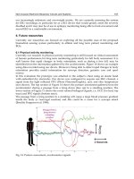
TEM method (Sasidharan et al., 2010). From the SEM micrograph (Fig. 4) we can observe the
changes caused by the plant extract on fungal surface.
4.2 Transmission electron microscopy (TEM)
Further confirmation of SEM finding can be obtained from TEM study. To study the antifungal
activity through TEM method the hyphal specimens (1×3 mm
2
, with approximately 1 mm
thickness of underlying agar blocks) of test fungal strains will be excised from the margin of
actively growing SDA culture treated with plant extract using a sterilized razor blade. The
specimens are then fixed with modified Karnovsky’s fixative (Karnivsky, 1965) consisting of
2% (v/v) glutaraldehyde and 2% (v/v) paraformaldehyde in 0.05 M sodium cacodylate buffer
solution (pH 7.2) at 4°C overnight. Subsequently, the fixed specimens are washed with the
solution three times for 10 min each. The specimens were then will be post-fixed in the
solution with 1% (w/v) osmium tetroxide at 4°C for 2 h and then will be washed briefly with
Fungicides for Plant and Animal Diseases
114
distilled water twice each. The postfixed specimens will be en bloc stained with 0.5% (w/v)
uranyl acetate at 4°C overnight and then will be dehydrated once in a graded ethanol series of
30, 50, 70, 80, and 95% and three times in 100% ethanol for 10 min each. The specimens will be
further treated with propylene oxide twice for 30 min each as a transitional fluid and then will
be embedded in Spurr’s resin. Ultra-thin sections (approximately 50 nm in thickness) will be
cut with a diamond/ glass knife using an ultra-microtome. The sections will be mounted on
copper grids and will be stained with 2% uranyl acetate and Reynolds’ lead citrate (Reynolds,
1963) for 7 min each. Finally the sections will be observed with a transmission electron
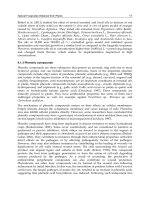
microscope. From the TEM micrograph we can observe the changes caused by the plant
extract on fungal cytoplasm (Fig. 5).
Fig. 4. SEM micrographs of Aspergillus niger
Fig. 5. TEM micrographs of Candida albicans
Screening Methods in the Study of Fungicidal Property of Medicinal Plants
115
4.3 Confocal laser scanning microscopy (CLSM)
Further verification of SEM and TEM finding can be obtained from CLSM study. To study
the antifungal activity through CLSM method the plant extract with MIC concentration will
be prepared. 48 h fungal culture will be developed by culturing the fungal strains on SDA
agar for 48 h. Controls without the plant extract or antimicrobials also will be included as
control groups. The 48 h fungal culture will be gently transferred into a 12-well microtitre
plate and rinsed with PBS for 15 s. The discs will be then immersed in 1 ml of the plant
extract or antimicrobial agents and incubated at 37
o
C in an aerobic incubator for 24 h.
Subsequently, the extract or antimicrobial will be removed and the viability of the fungal
cells will be assessed by Molecular Probes LIVE/DEAD BacLight Bacterial viability kit
which comprise SYTO-9 and propidium iodide (PI) (Molecular Probes, Eugene, OR). After
incubation with the dyes, the polymethylmethacrylate discs with biofilms will be placed on
glass slides and live/dead ratio of cells will be quantified using the CSLM system (Thein et
al., 2007). CLSM has become a precious tool for a wide range of studies in the biological and
medical sciences for imaging thin optical sections in living and fixed specimens ranging in
thickness up to 100 micrometers.
5. Conclusion
The above mentions methods demonstrated the great potential in the development of
antifungal testing to study the fungicidal properties of medicinal plants to develop
fungicide. The main advantages of the presented methods are the following: easy; rapid;
cheap and accurate. Our discussion demonstrates that the use electron microscopy is vital
to reveal the cell injury caused by plants extract on fungal strains. The cell changes
occurring in surface and cytoplasm of fungal cells following exposure to the plant extract
could be visible using a combination of SEM and TEM studies.
6. Acknowledgment
This project was partly supported by USM Short Term Grants (304/CIPPM/639040) from
Universiti Sains Malaysia. Kwan Yuet Ping was supported by MyPhD fellowship from
Ministry of Higher Education, Government of Malaysia, Malaysia.
7. References
Bauer, R.W.; Kirby, M.D.K.; Sherris, J.C. & Turck, M. (1966). Antibiotic susceptibility testing
by standard single disc diffusion method. American Journal of Clinical Pathology, 45,
493–496.
Camel, V. (2000). Microwave-assisted solvent extraction of environmental samples. Trends in
Analytical Chemistry, 19, 229-248.
Choi, M.P.K.; Chan, K.K.C.; Leung, H.W. & Huie, C.W. (2003). Pressurized liquid extraction
of active ingredients (ginsenosides) from medicinal plants using non-ionic
surfactant solutions. Journal of Chromatography A, 983, 153-162.
Fungicides for Plant and Animal Diseases
116
Cunha, I.B.S.; Sawaya, A.C.H.F.; Caetano, F.M.; Shimizu, M.T.; Marcucci, M.C.; Drezza, F.T.;
Povia, G.S. & Carvalho, P.O. (2004). Factors that influence the yield and
composition of Brazilian propolis extracts. Journal of Brazilian Chemical Society, 15,
964–970.
Ebana, R.U.B., Madunagu, B. E. & Etok, C.A. (1993). Anti-microbial effect of Strophantus
hipides Secamone afzeli on some pathogenic bacteria and their drug Research strain.
Nigeria Journal of Botany, 6, 27-31.
Fang, Q.; Teung, H.W.; Leung, H.W. & Huie, C.W. (2000) Micelle-mediated extraction and
preconcentration of ginsenosides from Chinese herbal medicine. Journal of
Chromatography A, 904, 47-55.
Gamliel, A.; Katan, J. & Cohen, E. (1989). Toxicity of chloronitrobezenes to Fusarium
oxysporum and Rhizoctonia solani as related to their structure. Phytoparas, 17, 101–
105.
Handa, S.S.; Khanuja, S.P.S.; Longo, G. & Rakesh, D.D. (2008). Extraction Technologies for
Medicinal and Aromatic Plants. International centre for science and high
technology, Trieste, 21-25.
Huie, C.W. (2002). A review of modern sample-preparation techniques for the
extraction and analysis of medicinal plants. Analytical and Bioanalytical Chemistry,
373, 23-30.
Khaing, T.A. (2011). Evaluation of the antifungal and antioxidant activities of the leaf extract
of aloe Vera (Aloe barbadensis Miller). Proceedings of World Academy of Science,
Engineering and Technology, 75, 610-612.
Karnovsky, M.J. (1965). A formaldehyde-glutaraldehyde fixative of high osmolality for use
in electron microscopy. Journal of Cell Biology, 27, 137A-138A.
Lang, Q. & Wai, C.M. (2001). Supercritical fluid extraction in herbal and natural product
studies-a practical review. Talanta, 53, 771-782.
Lee, H.K.; Koh, H.L.; Ong, E.S. & Woo, S.O. (2002). Determination of ginsenosides in
medicinal plants and health supplements by pressurized liquid extraction (PLE)
with reversed phase high performance liquid chromatography. Journal of Separation
Science, 25,160-166.
Luque de Castro, M.D. & Jiménez-Carmona, M.M. (2000). Where Is Supercritical Fluid
Extraction Going? Trends in Analytical Chemistry, 19, 223-228.
Ong, E. S. (2002). Chemical assay of glycyrrhizin in medicinal plants by pressurized liquid
extraction (PLE) with capillary zone electrophoresis (CZE). Journal of Separation
Science, 25, 825-831.
Ong, E.S. & Binte Apandi, S.N. (2000). Determination of Berberine and Strychnine in
Medicinal Plants and Herbal Preparations by Pressurized Liquid Extraction with
Capillary Zone Electrophoresis. Electrophoresis, 22, 2723-2729.
Ong, E.S. & Len, S.M. (2003a). Pressurized hot water extraction of berberine, baicalein, and
glycyrrhizin in medicinal plants. Analytica Chimica Acta, 482, 81-89.
Ong, E.S. & Len, S.M. (2003b).
Evaluation of surfactant assisted pressurized hot water
extraction for marker compounds in Radix Codonopsis pilosula using liquid
Screening Methods in the Study of Fungicidal Property of Medicinal Plants
117
chromatography and liquid chromatography/electrospray ionization mass
spectrometry. Journal of Separation Science, 26, 1533-1540.
Ong, E.S. & Len, S.M. (2004). Evaluation of pressurized liquid extraction and pressurized hot
water extraction for tanshinone I and IIA in Salvia miltiorrhiza using LC and LC-ESI-
MS. Journal of Chromatographic Science, 42, 211-216.
Ong, E.S. (2004). Extraction methods and chemical standardization of botanicals and herbal
preparations. Journal of Chromatography B, 812, 23-33.
Ong, E.S.; Woo, S.O. & Yong, Y.L. (2000). Pressurized liquid extraction of berberine
and aristolochic acids in medicinal plants. Journal of Chromatography A, 904,
57-64.
Mann, A., Banso, A. & Clifford, L.C. (2008). An antifungal property of crude plant extracts
from Anogeissus leiocarpus and Terminalia avicennioides. Tanzania Journal of Health
Research, 10, 34-38.
Pan, X.; Niu, G. & Liu, H. (2001). Microwave assisted extraction of tanshinones from Salvia
miltiorrhiza bunge with analysis by high performance liquid chromatography.
Journal of Chromatography A, 922, 371-375.
Pan, X.J.; Niu, G.G. & Liu, H.Z. (2002). Comparison of microwave-assisted extraction and
conventional extraction techniques for the extraction of tanshinones from Salvia
miltiorrhiza Bunge. Biochemical Engineering Journal, 12, 71-77.
Phrompittayarat, W.; Putalun, W.; Tanaka, H.; Jetiyanon, K.; Wittaya-areekul, S. &
Ingkaninan, K. (2007). Comparison of various extraction methods of Bacopa
monnier. Naresuan University Journal, 15, 29-34.
Picman, A.K.; Schneider, E.F. & Gershenzon, J. (1990). Antifungal activities of sunflower
terpenoids. Biochemical Systematics and Ecology, 18, 325–328.
Raaman, N. (2006). Phytochemical Techniques, p. 306, New India Publishing, India.
Reynolds, E.S. (1963). The use of lead citrate at high pH as an electron-opaque stain in
electron microscopy. Journal of Cell Biology, 17, 208-212.
Sasidharan, S.; Darah, I. & Jain K. (2008a). In Vivo and In Vitro toxicity study of Gracilaria
changii. Pharmaceutical Biology, 46, 413–417.
Sasidharan, S.; Yoga Latha, L. & Angeline, T. (2010). Imaging In vitro Anti-biofilm Activity to
Visualize the Ultrastructural Changes, In: Microscopy: Science, Technology,
Applications and Education A. Méndez-Vilas & J. Díaz, (Eds.), 622-626, Formatex,
Spain.
Sasidharan, S.; Zuraini, Z.; Yoga Latha, L. & Suryani, S. (2008b). Fungicidal effect and oral
acute toxicity of Psophocarpus tetragonolobus root extract. Pharmaceutical Biology, 46,
261–265.
Thein, Z.M., Samaranayake, Y.H. & Samaranayake, L.P. (2007). Dietary sugars, serum and
the biocide chlorhexidine digluconate modify the population and structural
dynamics of mixed Candida albicans and Escherichia coli biofilms. Acta Pathologica,
Microbiologica et Immunologica Scandinavica, 115: 1241–1251.
Woisky, R.G. & Salatino, A. (1998). Analysis of propolis: some parameters and procedures
for chemical quality control. Journal of Apicultural Research, 37, 99–105.
Fungicides for Plant and Animal Diseases
118
Zygmunt, J.B. & Namiesnik, J. (2003). Preparation of samples of plant material for
chromatographic analysis. Journal of Chromatographic Science, 41, 109–116.
6
In Vitro Multiplication of Aromatic
and Medicinal Plants and Fungicide Activity
Fernanda Leal
1
, Manuela Matos
1
,
Ana Cláudia Coelho
2
and Olinda Pinto-Carnide
1
1
IBB-Institute for Biotechnology and Bioengineering,
Centre of Genomic and Biotechnology, University of Trás-os-Montes and Alto-Douro,
Department of Genetics and Biotechnology
2
CECAV- Center for Animal Science and Veterinary,
University of Trás-os-Montes and Alto-Douro, Department of Veterinary Sciences,
Portugal
1. Introduction
Aromatic and medicinal plants, widely used as folk medicine are, beyond fruits, vegetables
grains and spices, the principal source of antioxidant compounds. Several studies
demonstrated that antioxidants have also antifungal activity (Jayashree & Subramanyam,
2000; Rasooli & Abyaneh, 2004). More and more, humanity try to replace synthetic
metabolites by natural metabolites. Therefore, studies in aromatic and medicinal plants with
the capacity to produce a different range of secondary metabolites extremely increase in late
years. On the other hand, chemical products, like pesticides, fungicides or bactericides are
widely used in agriculture. However, they have disadvantages to the environment, due to
contamination of the soils, the final consumers or the producers. Still, the indiscriminate and
recurrent use of synthetic fungicides has been found to induce resistance in several fungi,
the residual toxicity of these compounds result in human health hazards and requires
caution in their use for plant disease control (Singh et al., 2009). Thus, some aromatic and
medicinal plants, with antifungal capacity (Soliman & Badeaa, 2002; Goun et al. 2003;
Sucharita & Padma, 2010), like genus Thymus, Mentha, Calendula and Catharanthus were
micropropagated for antifungal activity evaluation.
Medicinal and aromatic plants are important sources for plant secondary metabolites that
are involved in many other aspects of a plant’s interaction with its immediate
environment. The genetic manipulation of plants together with the establishment of in
vitro plant regeneration systems facilitates efforts to engineer secondary product
metabolic pathways (Kumar & Gupta, 2007). Improvement of the yield and quality of
these natural plant products through conventional breeding is still a challenge. However,
recent advances in plant genomics research has generated knowledge leading to a better
understanding of the complex genetics and biochemistry involved in biosynthesis of these
plant secondary metabolites (Gómez-Galer et al., 2008). Advances in the cloning of genes
involved in relevant pathways, the development of high throughput screening systems
for chemical and biological activity, genomics tools and resources, and the recognition of
a higher order of regulation of secondary plant metabolism operating at the whole plant
Fungicides for Plant and Animal Diseases
120
level facilitate strategies for the effective manipulation of secondary products in plants
(Kumar & Gupta, 2007).
To overcome the problem of antifungal resistance in human pathogens, plants with
antimicrobial properties have been extensively studied for a possible application in food
microbiology and as alternative treatments for diseases or to prevent bacterial and fungal
growth. Many studies have proven very good fungicide effect of plants (Zabka et al., 2011).
The details of plants screened, their families, vernacular names and their therapeutic uses
are given in Table 1.
Plant species Family
Common
name
Therapeutic use
Thymus mastichina
and Thymus zygis
Lamiaceae Thyme
Antiseptic, antispasmodic, antitussive
(Pina-Vaz et al., 2004)
Mentha rotundifolia
Lamiaceae Applemint
Antiseptic, antispasmodic,
expectorant, vasoconstrictor
(Edris et al., 2003)
Calendula sp.
Asteraceae Marigold
Skin problems, fevers, anti-inflammatory,
anti-viral, anti-bacterial and fungicide
(Hänsel et al., 1992)
Catharanthus roseus
Apocynaceae
Madagascar
periwinkle
Anticancer, antidiabetic, laryngitis,
rheumatism, dysmenorrhea
(Jaleel et al., 2009)
Table 1. Ethnomedical information of the studied species.
2. In vitro multiplication of plants from genus Thymus, Mentha, Calendula
and Catharanthus
The multiplication by in vitro culture, means micropropagation, is a very important
methodology to obtain a great number of homogeneous plants in a short period of time. In
vitro culture is an important system in order to optimize and increase the secondary
metabolites production. With this technique, explants from different species could be
micropropagated under optimized condition of culture media, temperature and photoperiod.
In this study, different species of plants with antifungal activity were micropropagated, and
the fungicide activity of in vitro plants compared with the field plants.
In all the studies presented, the culture media were solidified with 0.7% of agar, and pH was
adjusted to 5.6-5.8. Culture media were autoclaved at 121ºC for 15 min. The cultures were
maintained in a growth chamber at 24 ± 1 ºC on a 16/8-h photoperiod (73 mol m
-2
s
-1
).
Data were subjected to analysis of variance (ANOVA) using STATVIEW 5.0 program,
treatment means separated using Fisher´s Least Significant Difference (LSD) test at P = 0.05.
2.1 Micropropagation of Thymus
Thyme is a perennial herb, a 20 to 50 cm shrub, of the Lamiaceae family, an aromatic plant
native to the Mediterranean region (Miguel et al., 1999). The genus Thymus is exceptionally
rich in species, and due to the diversity and plasticity of these plants, their geographical
range is very wide. In Portugal are known, at least, 11 Thymus species (Afonso & McMurtrie,
In Vitro Multiplication of Aromatic and Medicinal Plants and Fungicide Activity
121
1991). Thymus plants are of much interest owing to their use in different applications, in
medicine because of their antiseptic properties, in the cosmetic industry or as a food
additive for their organoleptic properties (Duke et al., 2002; Torras et al., 2007). Thymus
species differ with regard to their morphological features and metabolism, which influences
their chemical constitution. Within individual species there are chemical variations that are
characterized by different plant oil compositions, usually without any morphological
differences (Smolik et al., 2009). Increasingly, plant breeding has taken advantage of
developments in molecular biology in order to genotype the species of interest in a way that
considerably accelerates their selection. These types of approaches consist of choosing
desired genotypes on the basis of molecular markers, or having prior knowledge of the
genes that determine the formation of a particular trait in a plant (Pradeep-Reedy et al.,
2002). There are many publications related to the antibacterial and antifungal activities of
thyme essential oil (Urbanczyk et al., 2002; Priestley et al., 2003; Rasooli & Mirmostafa.,
2003). It has been used as weed germination inhibitor (Angelini et al., 2003), and the
different extracts from thyme leaves have shown the presence of a large number of
flavonoids and vitamin E, compounds of great interest in the food industry due to their
antioxidant activities (Sotomayor et al., 2004). There is also interest in using thyme essential
oil for delaying the autoxidation of food lipids (Youdim et al., 2002).
2.1.1 Methodology
Branches of Thymus plants, species Thymus mastichina L. and Thymus zygis L., with 10 to 20 cm
lenght were collected in University of Trás-os-Montes e Alto Douro (UTAD) Botanical Garden,
in Vila Real, Portugal. The explants disinfected with commercial bleach 60% (v/v) and washed
three times with sterile water, were fragmented into nodal segments and placed in different
culture media. Basal culture media MS (Murashige & Skoog, 1962) were evaluated without
growth regulators or supplemented with a cytokinin, 1 mg/L BAP (6-benzylaminopurine),
alone or combined with an auxin, 0.2 mg/L NAA (α-naphthalene-acetic acid). Two carbon
sources, sucrose and glucose, at three concentrations (2, 3 or 4 % w/v) were also tested.
Number and length of shoots have been evaluated after four weeks in culture.
2.1.2 Results and discussion
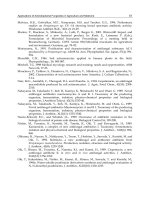
The two Thymus species studied showed a good response to in vitro culture conditions (Fig.
1). After four weeks in culture, T. mastichina L. presented higher length of shoots (11.2 mm
vs 6.0 mm), while T. zygis L. showed bigger shoot number per explant (1.9 vs 1.8). The
culture media revealed a statistical significant effect (P<0.05) for the parameters evaluated,
shoot number and length.
In what concern to carbon source, sucrose seems to be the best carbon source to thyme
micropropagation. For both species, MS medium containing 3 % sucrose produced the highest
shoots length (Fig. 2), similar results were obtained by Bandeira et al. (2007) in T. vulgaris L
With 3 % sucrose concentration the highest shoots length (15.9 mm) was observed in T.
mastichina L., in MS medium supplied with 1 mg/L BAP. One of the explanations for this is the
presence of endogen auxin that releases the addition NAA. The biggest shoot number (9.7)
appeared in T. zygis L. in MS medium with 2 % sucrose and 1 mg/L BAP. However,
considering all the media, the ones with glucose revealed higher values in shoot number when
compare with the sucrose ones, 3.6 vs 3.3 (though, the difference was not statistically
significant P>0.05). Cunha and Fernandes-Ferreira (1999) and Harada e Murai (1996) obtain
similar results with Linum usitatissium L. and Prunus mume, respectively.
Fungicides for Plant and Animal Diseases
122
Fig. 1. Micropropagation of Thymus plants. A – Explant in the establishment medium
culture; B – In vitro plants of T. zygis; C – In vitro plants of T. mastichina.
Fig. 2. Effect of culture media (MS) in shoot number and length in T. zygis (A and B) and
T. mastichina (C and D). GLU- glucose, SUC- sucrose, 20- 2%, 30- 3% 40- 4%. Error bars show
the 95% confidence interval.
A
B
C
In Vitro Multiplication of Aromatic and Medicinal Plants and Fungicide Activity
123
2.2 Micropropagation of Mentha
The genus Mentha belongs to the family Lamiaceae (Labiatae) consisting of about 25 to 30
species mainly found in temperate regions of Eurasia, Australia , South Africa and North
America (Shinwari et al., 2011). Mints grow 10 - 120 centimetres tall and can spread over an
indeterminate sized area. Mints are used either in the herb form or as an essential oil for
flavouring, perfume production and medicinal purposes (Edris et al., 2003). The Mentha
plant produces secondary metabolites such as alkaloids, flavanoids, phenols, gummy
polysaccharides. Terpens and quinines are used in food and pharmaceutical, cosmetics and
pesticide industries (Khanuja et al., 2000). Some members of this genus are also used as
herbal teas and condiments both in fresh and dried form due to their distinct aroma.
Morphological markers (such as plant height, leaf shape, colour, etc.) are among the oldest
markers used in the evaluation of genetic variability, namely in Mentha. However, they are
not sufficiently specific and informative because different gene expression in different
environments causes wide variability of phenotypic characters in individuals. In some cases
congruence between morphology and molecular phylogenetics were reported (Shinwari et
al., 2011 as cited in Shinwari, 1995). Genetic diversity refers to the variation at the level of
individual gene and provides a mechanism for the plants to adapt in ever changing
environment.
The existence of different chemotypes, based on qualitative differences within a taxon, is a
common feature in most Mentha species and hybrids. As a result, the mint plants produce a
number of commercially valuable essential oils, viz. spearmint oil, peppermint oil,
pennyroyal oil, etc. (Edris et al., 2003).
2.2.1 Methodology
Nodal segments of Mentha rotundifolia plants were collected in the greenhouse of Botanical
Garden of UTAD (Vila Real, Portugal). The explants were disinfected first with ethanol 70%
(v/v) then with commercial bleach 40% (v/v) and washed three times with sterile water,
then were fragmented into nodal segments and placed in different culture media. MS
culture media with different concentrations of BAP (1.0 or 2.0 mg/L) and NAA (0.1 or 0.2
mg/L) were used.
The number and length of shoots and roots, and the presence and diameter of calli were
evaluated during the eight weeks of in vitro culture. After that acclimatization was done in a
mixture of peat and perlite (1:1).
2.2.2 Results and discussion
The micropropagation of Menthe had, among others, the purpose to evaluate the effect of
cytokinin and auxin concentrations in the plant development. After four weeks of in vitro
culture there was the development of plants in all culture media (Fig. 3). The results
obtained in MS + 1 mg/L BAP medium with different concentrations of auxin (0.1 or 0.2
mg/L) revealed that the lowest concentration of auxin induces higher values of shoots
length (27.0 mm), and the highest concentration causes higher values of the roots length
(16.9 mm)(Table 2). These results are due to the fact that a low ratio of auxin / cytokinin
stimulates the development of stem sprouts, and a higher ratio to promote root
development (Torres et al., 1998). Pascal et al. (1991) found that the addition of a synthetic
Fungicides for Plant and Animal Diseases
124
auxin (IBA) induces an increase in mulberry shoots. Analyzing the effect of BAP
concentration, culture media MS + 1 mg/L BAP + 0.2 mg/L NAA and MS + 2 mg/L BAP +
0.2 mg/L NAA, it is clear that the medium with the lowest BAP concentration led to higher
values for all parameters, except for calli induction that are similar in both medium (0.7 mm
diameter). Similar results were obtained by Erig et al. (2002) with black mulberry tree by
checking that an increase in BAP concentration resulted in a decrease in length of shoots.
The results showed that the composition of the culture medium influences the response of
Menthe to micropropagation. Overall, MS medium without growth regulators showed
better results in terms of the number of shoots (2.0 / explant), although shoots length was
higher in MS medium with 1 mg/L BAP and 0.1 mg/L NAA (27.0 mm). The plants were
successfully acclimatized with a rate of 100%.
Fig. 3. Micropropagation of M. rotundifolia, plants after four weeks in different culture
media. A – MS; B – MS + 1 mg/L BAP + 0.1 mg/L NAA; C – MS + 1 mg/L BAP + 0.2 mg/L
NAA; D – MS + 2 mg/L BAP + 0.2 mg/L NAA.
Culture
media
MS
MS +
1BAP+0.1NAA
MS +
1BAP+0.2NAA
MS +
2BAP+0.2NAA
Shoot number 2.0 1.9 1.8 1.5
Shoot length
(mm)
18.0 27.0 22.2 11.8
Root number 3.2 2.6 2.5 0.4
Root length
(mm)
18.9 14.5 16.9 4.3
Explants with
calli (%)
0.0 15 15 6
Calli diameter
(mm)
0.0 0.7 0.8 0.7
Table 2. Number and length of shoots and roots and percentage and diameter of calli per
explant, during eight weeks of in vitro culture.
2.3 Micropropagation of Calendula
The genus Calendula belongs to Asteraceae family, also known as Compositae, and includes
several species, namely Calendula arvensis and Calendula officinalis, which are commonly used
AB C D
In Vitro Multiplication of Aromatic and Medicinal Plants and Fungicide Activity
125
as ornamental and medicinal plants. These plants are known to contain saponins, triterpenic
alcohols and their fatty acid esters, carotenoids, flavonoids, coumarins, essential oils,
hydrocarbons and fatty acids (Hänsel et al., 1992). Calendula is known by its medicinal
properties - mainly anti-inflammatory, anti-viral, anti-bacterian and fungicide. Considering
secondary metabolites, C. arvensis is very similar to C. officinalis and therefore, the majority
of the traditional or folk medicinal uses are similar for both species, including for the cure of
skin problems, fevers, chronic infections, wounds, bites and stings (Grieve, 1984; Chevallier,
1996). Calendula sp. micropropagation was achieved for the first time using several types of
explants (Çoçu et al., 2004). Callogenesis can be useful to obtain suspension cell cultures and
regeneration of plants via indirect organogenesis. Calli induction can be obtained with the
addition of growth regulators, namely auxins. However, some genotypes are able to induce
callus formation even in its absence. In C. officinalis, 2,4-D and IAA associated with
cytokinins promoted an efficient callus development (Grzelak & Janiszowska, 2002). As
secondary metabolites are accumulated in cell cultures from numerous species, much work
is focused on their production in shoot or root cultures formed by dedifferentiation of callus
(Banthorpe, 1994). Metabolites extracts can be obtained from in vitro material, namely calli
(Grzelak & Janiszowska, 2002), plantlets (Schmeda-Hirschmann, 2005) and flowers
(Hamburger et al., 2003).
2.3.1 Methodology
Nodal segments obtained from plants growing in greenhouse conditions were washed in
running water, surface-sterilized with a 25% (v/v) sodium hypochlorite solution for 15 min.
and finally rinsed 3 times with sterile distilled water (10 min./wash). The explants were
inoculated in MS media supplemented with BAP (1 or 2 mg/L) alone or combined with 0.1
mg/L of NAA. All the media were supplemented with 30 g/L sucrose. After 4 weeks, the
results were analyzed by several parameters like the number and length of shoots and
induction and development of calli. The calli obtained were transferred to MS culture
medium added with 3 mg/L 2,4-D, for organogenic development.
2.3.2 Results and discussion
In general calli induction was detected in all the media. Callogenesis occurred
simultaneously with organogenesis and, after 4 weeks of in vitro culture, flower induction
was obtained (Fig. 4). It was not observed a significant effect of the culture medium
on the number and length of shoots. The highest number of shoots was obtained in the
medium with 2 mg/L BAP and the larger length was obtained in the medium with 1
mg/L BAP.
Considering all the media tested, the average rate of multiplication of C. arvensis was 4.21
shoots/explant. ÇoÇu et al. (2004), using different culture media to micropropagate C.
officinalis obtained similar values. The best results were obtained in the culture medium
with 2.0 mg/L of BAP (7.8 shoots/explant) (Fig. 5). The presence of the auxin NAA (in
combination with the cytokinin BAP), did not induce a positive effect in the number of
shoots (Fig. 5). The media with 2.0 mg/L BAP and 0.1 mg/L NAA registered 4.75
shoots/explant. The medium with 1 mg/L BAP produced a higher number of
shoots/explant in the presence of NAA, but not statistically different (P>0.05) of the
medium without it.
Fungicides for Plant and Animal Diseases
126
Fig. 4. C. arvensis plants after four weeks of in vitro culture showing flower development.
The weak response of NAA was also observed by ÇoÇu et al. (2004) with hypocotyl and
cotyledon explants of C. officinalis concluding that the addition of KIN and NAA to culture
media reduced the frequency of shoot organogenesis. Considering the length of the shoots,
they reached an average of 23.44 mm at the 4
th
week of in vitro culture.
0
2
4
6
8
10
12
MS
MS+1mg/L BAP
MS+2mg/L BAP
MS+1mg/L
BAP+0.1mg/L NAA
MS+2mg/L
BAP+0.1mg/L NAA
Shoot number
Shoot length (cm)
Fig. 5. Effect of different culture media on the number and length of shoots of C. arvensis.
2.4 Micropropagation of Catharanthus
Catharanthus roseus, an important medicinal plant of Apocynaceae family, is an herbaceous
sub-shrub, also known as Madagascar periwinkle and Vinca rosea (synonym). Worldwide
has been extensively studied by different groups and has been identified to be a source of
numerous active principles of therapeutic importance. It is cultivated mainly for its
alkaloids (Taylor et al., 1975), with anticancer activities (Jaleel et al., 2009). Catharanthus
Culture medium
In Vitro Multiplication of Aromatic and Medicinal Plants and Fungicide Activity
127
roseus (L.) G. Don is one of the most important medicinal plants due the accumulation in the
leaves of two indolalcaloids vinblastine and vincristine, which were the first natural
anticancer agents to be clinically used. The high pharmaceutical value of these secondary
metabolites made of C. roseus one of the most studied medicinal species. A few protocols for
micropropagation of C. roseus were reported in the last years (Junaid et al., 2007;
Dhandapani et al., 2008; Ilah et al., 2009).
2.4.1 Methodology
Nodal segments of Catharanthus roseus were collected from plants that were potted. Explants
were disinfected with a 40% (v/v) sodium hypochlorite solution for 15 min. and finally
rinsed 3 times with sterile distilled water (10 min./wash). After that, explants were
inoculated in MS media alone or supplemented with BAP (1 or 1.5 mg/L) alone or
combined with 0.2 mg/L of IBA (indole butyric acid) or NAA. All the media were
supplemented with 30 g/L sucrose. To evaluate the effect of culture medium in the
development of explants was recorded, over eight weeks, the number and length of their
shoots and roots, the number of internodes and the presence and diameter of calli.
2.4.2 Results and discussion
In this study a protocol was established for the micropropagation of Catharantus roseus (Fig.
6), MS culture medium was used because Pietrosiuk et al (2007) and Dhandapani et al (2008)
described good results with this media.
Fig. 6. Different phases of the micropropagation of C. roseus. A – Explants after two weeks of
in vitro culture; B - Plantlet of C. roseus, at six weeks of in vitro culture; C – Plantlet of C.
roseus with flower, at eight weeks of in vitro culture.
A
B
C
Fungicides for Plant and Animal Diseases
128
Regarding the addition of growth regulators and their effect on the development of the
explants, it was found that, in general, the number of shoots was favored in medium
supplemented with BAP (MS medium + 1 mg/L BAP) and without auxins (2.23
shoots/explant) (Table 3). The length of shoots was higher in the media with the highest
concentration of BAP but with the addition of the auxin NAA, MS + 1.5 mg/L BAP + 0.2
mg/L NAA (16.07 mm). It is explained, among other things, by the presence of auxin which
promotes cell elongation.
Culture media MS
MS +
1BAP
MS +
1BAP+0.2
IBA
MS +
1BAP+0.2
NAA
MS +
1.5BAP+0.2
NAA
Shoot number 1.33 2.23 2.08 2.16 2.15
Shoot length (mm) 9.45 13.58 15.16 11.62 16.07
Root number 3.13 0 5.94 5.79 11.26
Root length (mm) 3.50 0 8.86 12.88 35.40
Number of internodes 0.78 0.78 1.0 0.89 0.91
Explants with calli (%) 0 0 0.27 0.44 0.45
Calli diameter (mm) 0 0 1.23 3.35 3.41
Table 3. Number and length of shoots and roots, number of internodes and percentage and
diameter of calli per explant, during eight weeks of in vitro culture.
In rooting, the results were not as expected because, as has been said, the auxin IBA, is
related to the root development, so was expect that MS medium + BAP + 0.2 IBA had had a
higher rate of rooting, as results obtained by Dhandapani et al. (2008). However, in our
study, MS medium supplied with 1.5 BAP + 0.2 NAA revealed the largest number and
length of roots, 11.3 roots/explant with 35.4 mm. Similar results were verified by
Echeverrigaray and colleagues (2005) for thyme cultivars. With regard to the number of
internodes, the medium with the addition of IBA (MS + 1 mg/L BAP + 0.2 mg/L IBA)
originated the highest values.
This result was not expected since the auxin IBA is more related with rooting, as reported by
Dhandapani et al. (2008) also in Catharanthus roseus. The culture media with the lowest
values for the internodes was MS medium supplemented with NAA (MS + 1 mg/L BAP +
0.2 mg/L NAA). The culture medium MS + 1.5 mg/L BAP + 0.2 mg/L NAA proved to be
the best in the number and diameter of calli (3.41 mm), this result was expected, since
Paramageetham et al. (2007) in Centella asiática L. had found that the formation of calli
occurred in response to an interaction between auxin, its concentration and type of explant.
3. Fungicide activity
3.1 Aspergillus fumigatus
Aspergillus genus is a famous taxonomic group of molds. Some of its species are very
important animal and human pathogens. The disease in humans is caused mainly by
Aspergillus fumigatus, Aspergillus flavus and Aspergillus niger. Other species, for example,
In Vitro Multiplication of Aromatic and Medicinal Plants and Fungicide Activity
129
Aspergillus terreus or Aspergillus nidulans are quantitatively less prevalent (Karthaus, 2010).
A. fumigatus is one of the most ubiquitous of the airborne saprophytic fungi that is
pathogenic to plants, animals and humans. Inhalation of conidia by immunocompetent
individuals rarely has any adverse effect (Latgé, 1999). However, apart from the production
of mycotoxins, A. fumigatus is a dangerous human pathogenic, which is able to cause very
serious human and animal mycoses with a high frequency of resistance to chemical
antifungal drugs (Verweij et al., 2009; Lass-Florl et al., 2010; Xu et al., 2010; Zabka et al.,
2011). Dramatic increases in the incidence of aspergillosis caused primarily by A. fumigatus
have occurred in recent years. A high infection-associated death rate of up to and over 50%
is attributed even today to these fungi (Karthaus, 2010). A. fumigatus has become the most
important airborne pathogen in developed countries, causing a significant mortality in
invasive aspergillosis (Latgé, 1999; Chamilos & Kontoyiannis, 2005). Patients who have been
treated with steroid therapy or those with chronic obstructive pulmonary disease or severe
hepatic failure are at high risk for developing pulmonary aspergillosis (Meersseman et al.,
2007; Morace & Bhorgi, 2010).
The treatment of aspergillosis is most problematic and questionable owing to toxicity and
side effects of the used medicines on the base of synthetic fungicides (Karthaus, 2010).
Fungal infections remain a significant cause of morbidity and mortality despite advances in
medicine and the emergence of new antifungal agents. Drug resistance in fungi is increasing
and is becoming a serious concern and the high use and misuse of antifungal as probably
the main cause of this situation. Therefore, there is an urgent need to search for effective
new antifungal agents in treatment of infectious diseases at present (Xing et al., 2011).
Currently, there are four classes of antifungal agents with activity against Aspergillus: the
polyenes, such as amphotericin B its lipid formulations, and nystatin (including liposomal
nystatin); the triazoles, including itraconazole, the voriconazole and the investigational
posaconazole, the echinocandins, such as caspofungin, the micafungin, and the
anidulafungin; and the allylamines such as terbinafine (Groll & Kolve, 2004; Chamilos &
Kontoyiannis, 2005; Meersseman et al., 2007; Shi et al., 2010). Clinical resistance of invasive
aspergillosis to amphotericin B based therapy is observed frequently in clinical practice
(Chamilos & Kontoyiannis, 2005). However, they are intrinsically resistant to fluconazole
(Moghaddam et al., 2010).
In other way, pathogenic and toxinogenic fungi are one of the major economic problems of
crop and food production (Zabka et al., 2011). In terms of food safety, species of Fusarium,
Aspergillus and Penicillium genera are considered the most significant because they produce
the great majority of known mycotoxins (Palumbo et al., 2008; Zabka et al., 2011). There has
been increasing concern of the consumers about foods free or with lower level of chemical
preservatives because these could be toxic for humans (Bedin et al., 1999; Souza et al., 2005).
This resulted in increasing search for new technologies for use in food conservation systems
which include alternatives antimicrobials (Brull & Coote, 1999; Souza et al., 2005).
3.2 Aromatic plants and antifungal activity
The spread of multidrug-resistant strains of fungus and the reduced number of drugs
available led to a search for therapeutic alternatives, namely among medicinal plants and
compounds isolated from them used for their antifungal properties. In these natural sources, a
series of molecules with antifungal activity have been found, which are of great importance to
Fungicides for Plant and Animal Diseases
130
humans and plants. Several molecules obtained from the natural environment are studied and
described in bibliography with antimycotic activity. Several extracts are tested for antifungal
activities like crude extracts or isolated constituents like, essential oils, terpenoids, saponins,
phenolic compounds, alkaloids, peptides and proteins (Abad et al., 2007).
Aromatic plants have been widely used in folk medicine. About three quarter of the world’s
population relies on plants and plant extracts for healthcare (Parekh & Chanda, 2007).
Several plants have been used in folklore medicine in Portugal (Pina-Vaz et al., 2004;
Figueiredo et al., 2008). Spices have been used with primary purpose of enhancing the flavor
of foods rather than their medicinal and antioxidant properties (Souza et al., 2005).
Plants generally produce many secondary metabolites with antifungal and microbicide
activity (Bobbarala et al., 2009). According to the WHO (World Health Organization), plants
would be the best source for obtaining a variety of drugs and a possible way to treat
diseases caused by multidrug resistant microorganisms (Bhattacharjee et al., 2006). The
medicinal value of plants lies in some chemical substances that produce a definite
physiological action on the human body. The most important of these bioactive compounds
of plants are alkaloids, flavanoids, tannins and phenolic compounds (Edeoga et al., 2005;
Panghal et al., 2011). Some medicinal plants exert strong antifungal properties and could be
conveniently used as a promising alternative source for presently problematic antifungal
treatment in many areas with respect to their natural origin (Zabka et al., 2011).
Many commercially drugs used in modern medicine was initially used in crude form in
traditional or folk healing practices. Benefits of using plant extracts are that they are
relatively safer than synthetic alternatives, offering profound therapeutic benefits and more
affordable treatment (Panghal et al., 2011).
The genus Thymus (Lamiaceae), widely distributed on the Iberian Peninsula, is a
taxonomically complex group of aromatic plants traditionally used for medicinal purposes
because of their antiseptic, antispasmodic and antitussive properties (Pina-Vaz et al., 2004).
Previous studies on the antimicrobial activity of the essential oils of some Thymus spp., most
of them possessing a large amount of phenolic monoterpenes, showed activity against fungi
(Pina-Vaz et al., 2004).
Screening of medicinal plants for antimicrobial activities and phytochemicals is important
for finding potential new compounds for therapeutic use (Duraipandiyan et al., 2006).
The main objective of this study was to investigate the inhibitory effects of Thymus and
Mentha extracts against Aspergillus fumigatus.
3.3 Methodology
Fungal strain was obtained from the collection of pathogenic fungi maintained in the
University of Trás-os-Montes and Alto Douro, Vila Real, Portugal. Subcultivations on Petri
dishes and other manipulations with the strain were carried out in the Bio Security Level 2
(BSL 2) laboratory. The evaluation of antifungal capacity was done by the method of
mycelial growth (Zhang et al., 2006). The fungus used in the assays was the fungi Aspergillus
fumigatus. The mold was grown on potato dextrose agar (PDA). The solution was mixed
with PDA culture media respectively to give a series of 5, 10, 20, and 25 mg/mL
concentrations of culture media containing the compounds described above. The media