Proteomics Human Diseases and Protein Functions Part 12 pdf
Bạn đang xem bản rút gọn của tài liệu. Xem và tải ngay bản đầy đủ của tài liệu tại đây (1.12 MB, 25 trang )
Proteomics – Human Diseases and Protein Functions
264
Protein name T/N ratio Functions References
Heat shock protein 27 kDa
↓ or ↑
[Du et al., 2007; Fu et al., 2007; Liu et
al., 2011; Zhou et al., 2005]
Similar to heat shock congnate 71-kDa
protein
↑ [Du et al., 2007]
Heat shock 70kDa protein 8 ↓ [Nishimori et al., 2006]
Heat shock protein 70 kDa ↑ [Jazii et al., 2006]
gp96 ↑ [Zhou et al., 2005]
GRP78 ↑ [Du et al., 2007]
Alpha-B-Crystalline ↓ [Qi et al., 2005; Zhu et al., 2010]
Fibrin beta ↓ [Liu et al., 2011]
Crystal structure of huma recombinant
procathepsin B
↑ NA [Du et al., 2007]
M2-type pyruvate kinase
↑or ↑
Energy metabolism
[Du et al., 2007; Fu et al., 2007; Liu et
al., 2011]
Mutant beta-actin(Q6F5I1) ↑ [Du et al., 2007]
Phosphoglycerate kinase 1 ↑ [Du et al., 2007; Nishimori et al., 2006]
Alpha enolase ↑
[Du et al., 2007; Fu et al., 2007;
Nishimori et al., 2006; Qi et al., 2005]
Beat-enolase ↑ [Fu et al., 2007]
Triosephosphate isomerase ↑ [Zhu et al., 2010]
GAPDH ↑ [Qi et al., 2005]
Aldolase A ↓ [Nishimori et al., 2006]
Fructose-bisphosphate aldolase A ↓ [Zhu et al., 2010]
RNA binding motif protein 8A ↑
mRNA/nucleotide/protei
n binding
[Zhou et al., 2005]
Translation initiation factor Eif-1A ↑ Translation [Zhou et al., 2005]
Transmembrane protein 4 ↑
Protein binding
[Zhou et al., 2005]
Transgelin
↓or ↑
[Liu et al., 2011; Qi et al., 2005; Zhou
et al., 2005; Zhu et al., 2010]
COMT protein ↑ [Liu et al., 2011]
Early endosome antigen 1 ↓
Protein binding
[Liu et al., 2011]
Cr
y
stal structure of recombinant human
fibrinogen fragment
↑ [Nishimori et al., 2006]
Similar to ubiquitin -conjugating
enzyme E2 variant 1 isoform
↓
Protein degradation
[Du et al., 2007]
Ubiquitin C-terminal esterase ↑ [Zhou et al., 2005]
Ubiquinol-cytochrome C reductase
complex core protein2
↑ [Nishimori et al., 2006]
Proteosome ↑ [Liu et al., 2011]
Galectin-7 ↓
Interactionof cells and
cell-matrix
[Zhou et al., 2005; Zhu et al., 2010]
Fatty acid-binding protein ↓ Lipid metabolism [Zhou et al., 2005]
TGase ↓ Protein modification [Zhou et al., 2005]
Fascin ↑ actin cross-lining [Zhou et al., 2005]
SCCA1 ↓
Cysteine proteinase
inhibitor
[Qi et al., 2005; Zhou et al., 2005]
Proteinase inhibitor, Clade B ↓
Neutrophil elastase
inhibitor
[Zhou et al., 2005]
Thioredoxin perosidase ↑
Redox homeostasis
[Zhou et al., 2005; Zhu et al., 2010]
Peroxiredoxin 1
↑or ↓ [Fu et al., 2007; Qi et al., 2005]
Peroxiredoxin 2 ↓ [Jazii et al., 2006; Qi et al., 2005]
ARK family 1 ↑ Carcinogen metabolism [Zhou et al., 2005]
GST M
2
↑
glutathione transferase
activity
[Zhou et al., 2005]
Proteasome subunit βtype 4 ↑
Protein degradation
[Zhou et al., 2005]
Proteasome subunit βtype 9 ↓ [Zhou et al., 2005]
Prosomal protein p30-33k ↑ [Zhou et al., 2005]
Elongation factor Tu ↑ Translation [Qi et al., 2005]
(NADP) cytoplasmic ↑ NAD binding [Qi et al., 2005]
Proteomic Study of Esophageal Squamous Cell Carcinoma
265
Protein name T/N ratio Functions References
Prohibitin
↑or ↓
Transcription regulation
[Fu et al., 2007; Qi et al., 2005]
Neuronal protein ↑ Neuronal growth [Qi et al., 2005]
Nuclear autoantigenic sperm protein
isoform 1
↑ Hsp90 protein binding [Nishimori et al., 2006]
Myosin heavy chain nonmuscle form A ↓ Actin binding or
calmodulin binding
[Nishimori et al., 2006]
Caldesmon 1 isoform 1 ↓ [Nishimori et al., 2006]
Myosin regulatory light chain 2 ↓
Ventricular/cardiac
muscle isoform
[Jazii et al., 2006; Zhu et al., 2010]
Myosin light chain 2 ↓ Regulatory light chain of
myosin
[Jazii et al., 2006]
Myosin light chain 1 ↓ [Zhu et al., 2010]
Heterogeneous nuclear
ribonucleoprotein A2/B1:B1
↑
RNA binding and
processing
[Nishimori et al., 2006]
Heterogeneous nuclear
ribonucleoprotein A2/B1:A2
↑ [Nishimori et al., 2006]
Myosin light chain 3 ↓
Regulatory light chain
[Zhu et al., 2010]
Myosin light polypeptide 6 ↑ [Jazii et al., 2006]
Myosin light chain 6B ↓ Regulatory light chain [Zhu et al., 2010]
Similar to alpha-fetoprotein ↓ NA [Nishimori et al., 2006]
Trnasferrin ↓ ferric iron binding [Nishimori et al., 2006]
Alpha-1-antitrypsin precursor ↓
Proteinase inhibitor
[Nishimori et al., 2006]
Alpha-1-antitrypsin ↑ [Fu et al., 2007]
Procollagen-proline ↓ Oxidoreductase activity [Nishimori et al., 2006]
Calponin 1, basic ↓
actin binding ;
calmodulin binding
[Nishimori et al., 2006]
DNA directed RNA polymerase B
(ropB)
↑ Transcription [Jazii et al., 2006]
GH16431P ↑ NA [Jazii et al., 2006]
OPTN protein ↓
Protein C-terminus
binding
[Fu et al., 2007]
67 kDa laminin receptor ↑
Signal transduction
[Fu et al., 2007]
TNF receptor associated factor 7 ↑ [Liu et al., 2011]
Stratifin ↓ [Du et al., 2007; Qi et al., 2005]
Cathepsin D ↑
Aspartyl proteinase
activity
[Liu
et al., 2011]
Chromosome1 open reading frame 8 ↑ NA [Liu et al., 2011]
Cdc42 ↑ GTPase activator activity [Liu et al., 2011]
LLDBP ↑ NA [Liu et al., 2011]
Adenylate kinase 1 ↓ Adenylate kinase activity [Liu et al., 2011]
General transcription factor IIH ↓ Transcription [Liu et al., 2011]
Serpin B5 precursor ↑
serine proteinase inhibitor
[Zhu et al., 2010]
Serpin B3 ↑ [Zhu et al., 2010]
Transthyretin [Precursor] ↑
Thyroid hormone-binding
protein
[Zhu et al., 2010]
Apolipoprotein A-I [Precursor] ↑ lipid metabolism [Zhu et al., 2010]
Peptidyl-prolyl cis-trans isomerase A ↑
Peptidyl-prolyl cis-trans
isomerase activity
[Zhu et al., 2010]
Cystatin-B ↑
Cysteine-type
endopeptidase inhibitor
activity
[Zhu et al., 2010]
Serum amyloid P-component
[Precursor]
↓ Protein binding [Zhu et al., 2010]
Phosphatidylethanolamine-binding
protein1
↓
Serine-type
endopeptidase inhibitor
[Zhu et al., 2010]
Carbonic anhydrase 1 ↓ Carbonate dehydratase
activity
[Zhu et al., 2010]
Carbonic anhydrase 3 ↓ [Zhu et al., 2010]
Creatine kinase M-type ↓ Creatine kinase activity [Zhu et al., 2010]
Table 1. Reported differential proteins in esophageal cancer tissues
Proteomics – Human Diseases and Protein Functions
266
proteomics methods have been developed, which include extracted ion current (XIC)-based
label-free quantification and stable isotope labeling quantification. Stable isotope labeling by
amino acids in cell culture (SILAC) is an in vivo metabolic labeling method in which stable
isotope-labeled amino acids (Heavy vs. Light amino acids) replace the natural amino acids
of preexisting proteome[Ong & Mann, 2006]. We used SILAC medium to label immortalized
cells (NE3 and NE6) with heavy stable isotope [U-
13
C
6
]-H-Lysine and [U-
13
C
6
]-H-Arginine
and cancer cells (EC1, EC109, EC9706) with light stable isotope [
12
C
6
]-L-Lysine and [
12
C
6
]-L-
Arginine, respectively. After complete labeling of the cellular proteome, equal quantity of
proteins from immortalized cells and cancer cells were mixed and then subjected to SDS-
PAGE separation, in-gel trypsin digestion and high performance liquid chromatography on-
line with electrospray ionization-MS/MS analysis (HPLC-ESI-MS/MS). Forty-seven
candidate proteins with differential expression were identified with our arbitrary criteria,
which contains ratio change > 1.5 folds, ≥ 2 peptides for quantification and coefficient of
variation < 50%. Then, we characterized the cellular protein expression pattern and
secretome derived from cisplatin-resistant sub-cell line EC9706 and its parental sensitive cell
line EC9706. By SILAC labeling and MS-based quantification, we successfully identified 74
proteins of cellular origin and 57 proteins of secretome with altered expression levels.
Similar to our approach, Kashyap et al. used a SILAC-based quantitative proteomic
approach to compare the secretome of ESCC cells with that of non-neoplastic esophageal
squamous epithelial cells and identified 120 up-regulated proteins with >2-fold difference in
the ESCC secretome[Kashyap et al., 2010]. In addition of previously known increased ESCC
biomarkers, i.e. matrix metalloproteinase 1, transferrin receptor, and transforming growth
factor beta-induced 68 kDa, a number of novel proteins showed distinct expression pattern,
among which protein disulfide isomerase family a member 3 (PDIA3), GDP dissociation
inhibitor 2 (GDI2), and lectin galactoside binding soluble 3 binding protein (LGALS3BP)
were further validated by immunoblot analysis and immunohistochemical labeling using
tissue microarrays. These identified proteins participate in multiple biological functions,
including molecular chaperones, cytoskeletal proteins, and members of protein inhibitors
family, reducing protein, etc., suggesting multiple dysregulated pathways involving in
ESCC.
2.4 Clinical relevance of potential protein biomarkers in ESCC
To answer clinical questions, the protein biomarkers identified by proteomic techniques
with potential diagnosis and therapeutic targets for ESCC need to be translated into clinical
scenario, which is realized by using clinical samples, such as biopsy samples, resected tissue
samples, plasma or serum samples, urine samples, saliva samples, etc. The methods used for
validation generally comprise Western blot, IHC and ELISA at protein level, and RT-PCR at
transcription level. Using 2DE- and SILAC-based quantitative proteomic approaches, we
have identified a total of 78 non-redundant proteins with aberrant expression associated
with ESCC, suggesting that these proteins may play functional roles in carcinogenesis of
ESCC and may have clinical values. Afterwards, Western blot analysis verified the
decreased expressions of three proteins, i.e. SCCA1, TPM1 and αB-Cryst in cancer, in
accordance with 2DE quantitative results. At transcription level, SCCA1 mRNA was down-
regulated in tumor as well. More importantly, the expression of SCCA1 decreased step by
step as a function of precancer lesions progression, which suggests that SCCA1 may take
part in the multi-stage transformation of ESCC, even in the earliest stages[Qi et al., 2005]. In
the 2DE-based comparative proteomic study using immortalized and cancer cell model, we
Proteomic Study of Esophageal Squamous Cell Carcinoma
267
Accession no. Protein name MW/PI Scores
Ratio
(T/N)
Matched
peptides
Functions
TPM3 HUMAN Tropomyosin alpha-3 chain 32.80/4.53 330.06 0.47 2
Actin binding
TPM4 HUMAN Tropomyosin alpha-4 chain 28.50/4.52 199.64 0.37 2
K2C8 HUMAN Keratin, type II cytoskeletal 8 53.67/5.38 907.48 0.51 4
FSCN1 HUMAN Fascin 54.50/7.02 296.56 0.45 2
LEG1 HUMAN Galectin-1 14.71/5.18 424.98 0.49 3
Signal
transduction
CLIC1 HUMAN Chloride channel ABP 26.91/4.94 447.94 0.63 4
1433E HUMAN 14-3-3 protein epsilon 29.16/4.48 400.71 0.66 3
PRDX1 HUMAN Peroxiredoxin-1 22.10/9.22 689.77 0.55 7
Redox
homeostasis
PRDX2 HUMAN Peroxiredoxin-2 21.88/5.59 238.11 0.65 5
PRDX4 HUMAN Peroxiredoxin-4 30.52/5.85 367.60 0.34 2
PRDX5 HUMAN Peroxiredoxin-5 22.01/9.93 522.84 0.60 2
CBR1 HUMAN
Carbonyl reductase
[NADPH]1
30.36/9.53 467.30 0.59 2
KCRB HUMAN Creatine kinase B-type 42.62/5.25 711.33 1.67 4
Metabolic
process
GSTP1 HUMAN Glutathione S-transferase P 23.34/5.32 1140.8 0.45 6
GDIB HUMAN Rab GDI beat 50.63/6.08 614.67 0.47 2
DHSA HUMAN
Favoprotein subunit
complex II
72.65/7.31 207.55 0.5 2
ACBP HUMAN Acyl-CoA-binding protein 10.04/6.16 135.03 0.64 2
PHS HUMAN PHS 2 11.99/6.33 170.64 0.43 3
RL27A HUMAN 60S ribosomal protein L27a
16.55/11.7
8
233.25 0.59 2
Translation
RSSA HUMAN 40S ribosomal protein SA 32.83/4.64 298.67 0.58 2
IF4G1_HUMAN eIF-4-gamma 1 175.4/5.1 650.5 2.15 14
NPM HUMAN Nucleophosmin 32.55/4.49 444.46 0.52 2 DNA binding
GRP78 HUMAN GRP78 72.29/4.92
1869.0
9
0.50 14 Chaperone
binding
CH10 HUMAN Hsp 10 10.92/9.44 219.29 0.40 3
G6PI HUMAN
Glucose-6-phosphate
isomerase
63.11/9.10 510.30 0.48 5
Energy
metabolism
UGDH HUMAN
UDP-glucose 6-
dehydrogenase
54.99/6.89 604.20 0.53 2
PPIA HUMAN Peptidyl-prolyl isomerase A 18.00/9.05 770.25 0.59 9
ALDOA HUMAN
Fructose-bisphosphate
aldolase A
39.40/9.18 386.91 0.59 2
PGK1 HUMAN Phosphoglycerate kinase 1 44.59/9.22 1020.8 0.50 6
G3P HUMAN GAPDH 36.03/9.26 1127.9 0.52 8
IPYR HUMAN Inorganic pyrophosphatase 32.64/5.47 485.51 0.45 3
ENOA HUMAN Alpha-enolase 47.14/7.71 1998.1 0.55 15
CYTB HUMAN Cystatin-B 11.13/7.85 144.98 0.43 2
CPSM HUMAN
Carbamoyl-phosphate
synthase 1
164.83/6.3
0
3115.1 0.24 6
PHB2 HUMAN Prohibitin-2
33.28/10.2
1
546.79 0.47 2
Transcription
regulation
CAND1_HUMAN
TBP-interacting protein
120A
136.3/5.4 617.2 1.8 15
PSME2 HUMAN
Proteasome activator
complex subunit2
27.34/5.33 367.19 0.48 2
Cell cycle
MCM7_HUMAN
DNA replication licensing
factor MCM7
81.3/6.1 510.8 1.97 13
Proteomics – Human Diseases and Protein Functions
268
Accession no. Protein name MW/PI Scores
Ratio
(T/N)
Matched
peptides
Functions
ACADV HUMAN VLCAD 70.35/9.63 841.39 0.35 2
Lipid
metabolism
ATPA HUMAN ATP5A1 59.71/9.61 963.07 0.47 5
THIL HUMAN Acetoacetyl-CoA thiolase 45.17/9.63 330.39 0.45 2
MIF HUMAN
Macrophage migration
inhibitory factor
12.47/9.12 267.01 0.61 3
Cytokine
activity
ATPB HUMAN ATPB-3 56.52/5.14 1704.2 0.40 5 Ion transport
VDAC1 HUMAN VDAC-1 30.75/9.22 548.36 2.32 2
Anion
transport
VPS35_HUMAN hVPS35 91.6/5.2 602.6 1.67 12
Protein
transport
HYOU1 HUMAN
Hypoxia up-regulated
protein 1
111.27/5.0
2
1206.8 0.56 2 ATP binding
SMD3 HUMAN
Small nuclear
ribonucleoprotein 3
13.91/11.0
7
330.93 0.49 2
mRNA
processing
Table 2. Differential proteins between immortalized and cancer cell lines derived from ESCC
identified by SILAC-based proteomics
selected Annexin A2 for validation by Western blot and IHC. Stepwise decrease in annexin
A2 protein expression was observed when epithelial cell was transformed malignantly. In
poorly-differentiated squamous carcinoma, 46% (5/11) of cancer tissue sample lost annexin
A2 protein and 36% (4/11) expressed at weak intensity[Qi et al., 2007b]. In a separate study,
IHC was used to determine 14-3-3σ in 60 cases of ESCC, nearby matched normal esophageal
epithelium and a variety of ESCC precursor lesions. High level of 14-3-3σ expression was
found ubiquitously in normal esophageal epithelium with an immuonstaining score of 8.22
in expression. Protein 14-3-3σ was down-regulated stepwise during the multi-stage
development of ESCC. Sixty-four percent of poorly-differentiated squamous cancer lost 14-
3-3σ expression with a score of 0.45[Qi et al., 2007a]. In agreement with our results, Ren et al.
documented that the level of 14-3-3σ in terms of mRNA and protein was markedly down-
regulated in ESCC compared with nearby matched non-cancer tissues. Furthermore,
decrease of 14-3-3σ expression was correlated with tumor infiltration depth, lymph node
metastasis, distant metastasis and lymphovascular invasion and shorter 5-year survival
rate[Ren et al., 2010]. Among the different proteins identified by SILAC-based quantitative
analysis using immortal cell and cancer cell model, the clinical values of MIF in
tumorigenesis of ESCC was determined as well. Not only the increased expression of MIF
was detected in cellular protein but also in the conditioned medium of esophageal cancer
cell lines EC1, EC109 and EC9706 compared with immortal cell lines NE3 and NE6. Low
frequency and very weak expression of MIF was detected predominantly in basal cells in
normal esophageal epithelium, with an immunostaining score of 1.13. Pronouncedly up-
regulated expression of MIF occurred in severe dysplasia compared with weak
immunostaining in mild and moderate dysplasia. In ESCC, high frequency of intense
expression of MIF was observed with a score of 5.46. Furthermore, high expression of MIF
was significantly correlated with advanced clinical stages. ELISA tests revealed that there
was an increase trend in serum level of MIF in clinically advanced stage IV compared to
stage I-III. Functional studies on MIF indicated that MIF knockdown resulted in decrease in
proliferation, clonogenicity, non-adherent growth and invasive potential. Our findings
indicate that MIF may play crucial roles in malignant transformation of pathogenesis of EC
and MIF could become a potential biomarker for high-risk population screening, assessment
Proteomic Study of Esophageal Squamous Cell Carcinoma
269
of therapeutic efficiency, prognostic evaluation, and molecular targets of developing novel
therapeutic regimen as well. In addition of our proteomic results in ESCC, several other
reports have looked at the clinical value of potential biomarkers, including cytokeratin 14,
Annexin I, SCCA1/2, calgulanulin B and HSP 60, alpha-actinin 4 and 67 kDa laminin
receptor, cathepsin D and PKM2, periplakin, calreticulin and GRP78, galectin-7, anti-CD25B
antibody[Dong et al., 2010; Du et al., 2007; Fu et al., 2007; Hatakeyama et al., 2006; Liu et al.,
2011; Nishimori et al., 2006; Zhu et al., 2010]. Nevertheless, further extensive studies are still
necessary to determine the clinical utility of the identified proteins in tumorigenesis and
progression of ESCC.
3. Conclusions
Nowadays, the dilemma for cancer control and management is not due to lack of efficient
treatment options but diagnosis at late stages. In the case of ESCC in China, five-year
survival rate for early stage tumor reaches around 90%[Hu et al., 2001]. Obviously, to detect
tumor as early as possible is the key for reducing the mortality and morbidity of ESCC. It is
believed that development of ESCC from normal esophageal epithelium takes at least about
10 years, during which diseased epithelium manifests as basal cell hyperproliferation,
dysplasia, carcinoma in situ in terms of morphology and finally evolves to malignant
neoplasms. As such, carcinogenesis of ESCC is a multi-stage and dynamic process which
accumulates ongoing changes at the level of both gene and protein expression.
Proteomic studies from various research groups worldwide have identified distinct
dysregulated protein expression pattern associated with ESCC. The discrepancy might
reflect the different etiology, different stages of disease and diverse pathways involved,
which makes identification of biomarkers for ESCC difficult. In light of a wealth of potential
biomarkers associated with ESCC identified so far in the exploratory phase, future large-
scale validation studies involving symptom-free patients with precursor lesions in high-
incidence area and ESCC patients compared with controls are essential toward clinical
application. Therefore, ultimate translation from laboratory into bedside for ESCC
biomarkers will require close collaboration and cooperation between researchers and
clinicians to look into the clinical utility in diagnosis at early stage, prognosis and
monitoring treatment efficiency for ESCC.
4. Acknowledgement
This work was supported in part by National Natural Science Founding of China (No.
30700366 and No. 81072039) and Cancer Research UK (to Yi-Jun Qi).
5. References
Abnet, C. C., Freedman, N. D., Hu, N., et al. (2010). A shared susceptibility locus in PLCE1
at 10q23 for gastric adenocarcinoma and esophageal squamous cell carcinoma. Nat
Genet, Vol.42, No.9, (Sep), pp: 764-767, ISSN 1546-1718
Banks, R. E., Dunn, M. J., Hochstrasser, D. F., et al. (2000). Proteomics: new perspectives,
new biomedical opportunities. Lancet, Vol.356, No.9243, (Nov 18), pp: 1749-1756,
ISSN 0140-6736
Proteomics – Human Diseases and Protein Functions
270
Bergqvist, A. S., Bergqvist, M., Brattstrom, D., et al. (2001). Serum p53 autoantibodies as
prognostic marker in patients with oesophageal carcinoma. Anticancer Res, Vol.21,
No.6A, (Nov-Dec), pp: 4141-4145, ISSN 0250-7005
Blot, W. J., Li, J. Y., Taylor, P. R., et al. (1993). Nutrition intervention trials in Linxian, China:
supplementation with specific vitamin/mineral combinations, cancer incidence,
and disease-specific mortality in the general population. J Natl Cancer Inst, Vol.85,
No.18, (Sep 15), pp: 1483-1492, ISSN 0027-8874
Brichory, F. M., Misek, D. E., Yim, A. M., et al. (2001). An immune response manifested by
the common occurrence of annexins I and II autoantibodies and high circulating
levels of IL-6 in lung cancer. Proc Natl Acad Sci U S A, Vol.98, No.17, (Aug 14), pp:
9824-9829, ISSN 0027-8424
Brown, L. M., Devesa, S. S. & Chow, W. H. (2008). Incidence of adenocarcinoma of the
esophagus among white Americans by sex, stage, and age. J Natl Cancer Inst,
Vol.100, No.16, (Aug 20), pp: 1184-1187, ISSN 1460-2105
Chambers, G., Lawrie, L., Cash, P., et al. (2000). Proteomics: a new approach to the study of
disease. J Pathol, Vol.192, No.3, (Nov), pp: 280-288, ISSN 0022-3417
Chang-Claude, J., Becher, H., Blettner, M., et al. (1997). Familial aggregation of oesophageal
cancer in a high incidence area in China. Int J Epidemiol, Vol.26, No.6, (Dec), pp:
1159-1165, ISSN 0300-5771
Chen, G., Wang, X., Yu, J., et al. (2007). Autoantibody profiles reveal ubiquilin 1 as a
humoral immune response target in lung adenocarcinoma. Cancer Res, Vol.67, No.7,
(Apr 1), pp: 3461-3467, ISSN 0008-5472
Cheng, K. K. & Day, N. E. (1996). Nutrition and esophageal cancer. Cancer Causes and
Control, Vol.7, No.1, pp: 33-40, 0957-5243
Daly, J. M., Fry, W. A., Little, A. G., et al. (2000). Esophageal cancer: results of an American
College of Surgeons Patient Care Evaluation Study. J Am Coll Surg, Vol.190, No.5,
(May), pp: 562-572; discussion 572-563, ISSN 1072-7515
Devesa, S. S., Blot, W. J. & Fraumeni, J. F., Jr. (1998). Changing patterns in the incidence of
esophageal and gastric carcinoma in the United States. Cancer, Vol.83, No.10, (Nov
15), pp: 2049-2053, ISSN 0008-543X
Disis, M. L., Pupa, S. M., Gralow, J. R., et al. (1997). High-titer HER-2/neu protein-specific
antibody can be detected in patients with early-stage breast cancer. J Clin Oncol,
Vol.15, No.11, (Nov), pp: 3363-3367, ISSN 0732-183X
Dong, J., Zeng, B. H., Xu, L. H., et al. (2010). Anti-CDC25B autoantibody predicts poor
prognosis in patients with advanced esophageal squamous cell carcinoma. J Transl
Med, Vol.8, (Sep 3), pp: 81, ISSN 1479-5876
Dresner, S. M. & Griffin, S. M. (2000). Pattern of recurrence following radical
oesophagectomy with two-field lymphadenectomy. Br J Surg, Vol.87, No.10, (Oct),
pp: 1426-1433, ISSN 0007-1323
Du, X. L., Hu, H., Lin, D. C., et al. (2007). Proteomic profiling of proteins dysregulted in
Chinese esophageal squamous cell carcinoma. J Mol Med (Berl), Vol.85, No.8, (Aug),
pp: 863-875, ISSN 0946-2716
Enzinger, P. C. & Mayer, R. J. (2003). Esophageal cancer. N Engl J Med, Vol.349, No.23, (Dec
4), pp: 2241-2252, ISSN 1533-4406
Fan, Y. J., Song, X., Li, J. L., et al. (2008). Esophageal and gastric cardia cancers on 4238
Chinese patients residing in municipal and rural regions: a histopathological
Proteomic Study of Esophageal Squamous Cell Carcinoma
271
comparison during 24-year period. World J Surg, Vol.32, No.9, (Sep), pp: 1980-1988,
ISSN 0364-2313
Fu, L., Qin, Y. R., Xie, D., et al. (2007). Identification of alpha-actinin 4 and 67 kDa laminin
receptor as stage-specific markers in esophageal cancer via proteomic approaches.
Cancer, Vol.110, No.12, (Dec 15), pp: 2672-2681, ISSN 0008-543X
Fujita, Y., Nakanishi, T., Hiramatsu, M., et al. (2006). Proteomics-based approach identifying
autoantibody against peroxiredoxin VI as a novel serum marker in esophageal
squamous cell carcinoma. Clin Cancer Res, Vol.12, No.21, (Nov 1), pp: 6415-6420,
ISSN 1078-0432
Fujita, Y., Nakanishi, T., Miyamoto, Y., et al. (2008). Proteomics-based identification of
autoantibody against heat shock protein 70 as a diagnostic marker in esophageal
squamous cell carcinoma. Cancer Lett, Vol.263, No.2, (May 18), pp: 280-290, ISSN
0304-3835
Greenawalt, D. M., Duong, C., Smyth, G. K., et al. (2007). Gene expression profiling of
esophageal cancer: comparative analysis of Barrett's esophagus, adenocarcinoma,
and squamous cell carcinoma. Int J Cancer, Vol.120, No.9, (May 1), pp: 1914-1921,
ISSN 0020-7136
Hagymasi, K. & Tulassay, Z. (2007). [Genetic background of esophageal squamous cell
carcinoma]. Orv Hetil, Vol.148, No.38, (Sep 23), pp: 1795-1800, ISSN 0030-6002
Hatakeyama, H., Kondo, T., Fujii, K., et al. (2006). Protein clusters associated with
carcinogenesis, histological differentiation and nodal metastasis in esophageal
cancer. Proteomics, Vol.6, No.23, (Dec), pp: 6300-6316, ISSN 1615-9853
Hayashida, Y., Honda, K., Osaka, Y., et al. (2005). Possible prediction of
chemoradiosensitivity of esophageal cancer by serum protein profiling. Clin Cancer
Res, Vol.11, No.22, (Nov 15), pp: 8042-8047, ISSN 1078-0432
Holmes, R. S. & Vaughan, T. L. (2007). Epidemiology and pathogenesis of esophageal
cancer. Semin Radiat Oncol, Vol.17, No.1, (Jan), pp: 2-9, ISSN 1053-4296
Hong, S. H., Misek, D. E., Wang, H., et al. (2004). An autoantibody-mediated immune
response to calreticulin isoforms in pancreatic cancer. Cancer Res, Vol.64, No.15,
(Aug 1), pp: 5504-5510, ISSN 0008-5472
Hongo, M., Nagasaki, Y. & Shoji, T. (2009). Epidemiology of esophageal cancer: Orient to
Occident. Effects of chronology, geography and ethnicity. J Gastroenterol Hepatol,
Vol.24, No.5, (May), pp: 729-735, ISSN 1440-1746
Hu, Y. C., Lam, K. Y., Law, S., et al. (2001). Identification of differentially expressed genes in
esophageal squamous cell carcinoma (ESCC) by cDNA expression array:
overexpression of Fra-1, Neogenin, Id-1, and CDC25B genes in ESCC. Clin Cancer
Res, Vol.7, No.8, (Aug), pp: 2213-2221, ISSN 1078-0432
Hulscher, J. B., van Sandick, J. W., Tijssen, J. G., et al. (2000). The recurrence pattern of
esophageal carcinoma after transhiatal resection. J Am Coll Surg, Vol.191, No.2,
(Aug), pp: 143-148, ISSN 1072-7515
Iizuka, T., Isono, K., Kakegawa, T., et al. (1989). Parameters linked to ten-year survival in
Japan of resected esophageal carcinoma. Japanese Committee for Registration of
Esophageal Carcinoma Cases. Chest, Vol.96, No.5, (Nov), pp: 1005-1011, ISSN 0012-
3692
Jazii, F. R., Najafi, Z., Malekzadeh, R., et al. (2006). Identification of squamous cell carcinoma
associated proteins by proteomics and loss of beta tropomyosin expression in
Proteomics – Human Diseases and Protein Functions
272
esophageal cancer. World J Gastroenterol, Vol.12, No.44, (Nov 28), pp: 7104-7112,
ISSN 1007-9327
Kamangar, F., Chow, W. H., Abnet, C. C., et al. (2009). Environmental causes of esophageal
cancer. Gastroenterol Clin North Am, Vol.38, No.1, (Mar), pp: 27-57, vii, ISSN 1558-
1942
Kashyap, M. K., Harsha, H. C., Renuse, S., et al. (2010). SILAC-based quantitative proteomic
approach to identify potential biomarkers from the esophageal squamous cell
carcinoma secretome. Cancer Biol Ther, Vol.10, No.8, (Oct 15), pp: 796-810, ISSN
1555-8576
Kolch, W., Mischak, H., Chalmers, M. J., et al. (2004). Clinical proteomics: a question of
technology. Rapid Commun Mass Spectrom, Vol.18, No.19, pp: 2365-2366, ISSN 0951-
4198
Kolch, W., Mischak, H. & Pitt, A. R. (2005). The molecular make-up of a tumour: proteomics
in cancer research. Clin Sci (Lond), Vol.108, No.5, (May), pp: 369-383, ISSN 0143-
5221
Kwong, K. F. (2005). Molecular biology of esophageal cancer in the genomics era. Surg Clin
North Am, Vol.85, No.3, (Jun), pp: 539-553, ISSN 0039-6109
Lander, E. S., Linton, L. M., Birren, B., et al. (2001). Initial sequencing and analysis of the
human genome. Nature, Vol.409, No.6822, (Feb 15), pp: 860-921, ISSN 0028-0836
Langer, R., Ott, K., Specht, K., et al. (2008). Protein expression profiling in esophageal
adenocarcinoma patients indicates association of heat-shock protein 27 expression
and chemotherapy response. Clin Cancer Res, Vol.14, No.24, (Dec 15), pp: 8279-8287,
ISSN 1078-0432
Li, B., Taylor, P. R., Li, J. Y., et al. (1993). Linxian nutrition intervention trials. Design,
methods, participant characteristics, and compliance. Ann Epidemiol, Vol.3, No.6,
(Nov), pp: 577-585, ISSN 1047-2797
Li, L. W., Li, Y. Y., Li, X. Y., et al. (2011). A novel tumor suppressor gene ECRG4 interacts
directly with TMPRSS11A (ECRG1) to inhibit cancer cell growth in esophageal
carcinoma. BMC Cancer, Vol.11, (Feb 3), pp: 52, ISSN 1471-2407
Lightdale, C. J. (1999). Esophageal cancer. American College of Gastroenterology. Am J
Gastroenterol, Vol.94, No.1, (Jan), pp: 20-29, ISSN 0002-9270
Lin, D. C., Du, X. L. & Wang, M. R. (2009). Protein alterations in ESCC and clinical
implications: a review. Dis Esophagus, Vol.22, No.1, pp: 9-20, ISSN 1442-2050
Liu, W. L., Zhang, G., Wang, J. Y., et al. (2008). Proteomics-based identification of
autoantibody against CDC25B as a novel serum marker in esophageal squamous
cell carcinoma. Biochem Biophys Res Commun, Vol.375, No.3, (Oct 24), pp: 440-445,
ISSN 1090-2104
Liu, Z., Feng, J. G., Tuersun, A., et al. (2011). Proteomic identification of differentially-
expressed proteins in esophageal cancer in three ethnic groups in Xinjiang. Mol Biol
Rep, Vol.38, No.5, (Jun), pp: 3261-3269, ISSN 1573-4978
Mark, S. D., Qiao, Y. L., Dawsey, S. M., et al. (2000). Prospective study of serum selenium
levels and incident esophageal and gastric cancers. J Natl Cancer Inst, Vol.92, No.21,
(Nov 1), pp: 1753-1763, ISSN 0027-8874
Messmann, H. (2001). Squamous cell cancer of the oesophagus. Best Pract Res Clin
Gastroenterol, Vol.15, No.2, (Apr), pp: 249-265, ISSN 1521-6918
Proteomic Study of Esophageal Squamous Cell Carcinoma
273
Morita, M., Kumashiro, R., Kubo, N., et al. (2010). Alcohol drinking, cigarette smoking, and
the development of squamous cell carcinoma of the esophagus: epidemiology,
clinical findings, and prevention. Int J Clin Oncol, Vol.15, No.2, (Apr), pp: 126-134,
ISSN 2547-7772
Nakagawa, S., Kanda, T., Kosugi, S., et al. (2004). Recurrence pattern of squamous cell
carcinoma of the thoracic esophagus after extended radical esophagectomy with
three-field lymphadenectomy. J Am Coll Surg, Vol.198, No.2, (Feb), pp: 205-211,
ISSN 1072-7515
Nishimori, T., Tomonaga, T., Matsushita, K., et al. (2006). Proteomic analysis of primary
esophageal squamous cell carcinoma reveals downregulation of a cell adhesion
protein, periplakin. Proteomics, Vol.6, No.3, (Feb), pp: 1011-1018, ISSN 1615-9853
Ong, S. E. & Mann, M. (2005). Mass spectrometry-based proteomics turns quantitative. Nat
Chem Biol, Vol.1, No.5, (Oct), pp: 252-262, ISSN 1552-4450
(2006). A practical recipe for stable isotope labeling by amino acids in cell culture (SILAC).
Nat Protoc, Vol.1, No.6, pp: 2650-2660, ISSN 1750-2799
Parkin, D. M., Bray, F., Ferlay, J., et al. (2005). Global cancer statistics, 2002. CA Cancer J Clin,
Vol.55, No.2, (Mar-Apr), pp: 74-108, ISSN 0007-9235
Qi, Y., Chiu, J. F., Wang, L., et al. (2005). Comparative proteomic analysis of esophageal
squamous cell carcinoma. Proteomics, Vol.5, No.11, (Jul), pp: 2960-2971, ISSN 1615-
9853
Qi, Y. J., He, Q. Y., Ma, Y. F., et al. (2008). Proteomic identification of malignant
transformation-related proteins in esophageal squamous cell carcinoma. J Cell
Biochem, Vol.104, No.5, (Aug 1), pp: 1625-1635, ISSN 1097-4644
Qi, Y. J., Ma, Y. F., Du, Y. W., et al. (2007a). Character ization and correlation of 14-3-3σ and
P53 protein expressions in multi-stage carcinogenesis of esophageal squamous cell
carcinoma. J Fourth Mil Med Univ, Vol.28, No.20, (June 15), pp: 1834-1837, ISSN
1000-5404
Qi, Y. J., Wang, L. D., Jiao, X. Y., et al. (2007b). [Dysregulation of Annexin II expression in
esophageal squamous cell cancer and adjacent tissues from a high-incidence area
for esophageal cancer in Henan province]. Ai Zheng, Vol.26, No.7, (Jul), pp: 730-736,
ISSN 1000-467X
Ren, H. Z., Pan, G. Q., Wang, J. S., et al. (2010). Reduced stratifin expression can serve as an
independent prognostic factor for poor survival in patients with esophageal
squamous cell carcinoma. Dig Dis Sci, Vol.55, No.9, (Sep), pp: 2552-2560, ISSN 1573-
2568
Shimada, H., Kuboshima, M., Shiratori, T., et al. (2007). Serum anti-myomegalin antibodies
in patients with esophageal squamous cell carcinoma. Int J Oncol, Vol.30, No.1,
(Jan), pp: 97-103, ISSN 1019-6439
Shimada, H., Nakashima, K., Ochiai, T., et al. (2005). Serological identification of tumor
antigens of esophageal squamous cell carcinoma. Int J Oncol, Vol.26, No.1, (Jan), pp:
77-86, ISSN 1019-6439
Soussi, T. (2000). p53 Antibodies in the sera of patients with various types of cancer: a
review. Cancer Res, Vol.60, No.7, (Apr 1), pp: 1777-1788, ISSN 0008-5472
Triboulet, J. P., Mariette, C., Chevalier, D., et al. (2001). Surgical management of carcinoma
of the hypopharynx and cervical esophagus: analysis of 209 cases.
Arch Surg,
Vol.136, No
.10, (Oct), pp: 1164-1170, ISSN 0004-0010
Proteomics – Human Diseases and Protein Functions
274
Trivers, K. F., Sabatino, S. A. & Stewart, S. L. (2008). Trends in esophageal cancer incidence
by histology, United States, 1998-2003. Int J Cancer, Vol.123, No.6, (Sep 15), pp:
1422-1428, ISSN 1097-0215
Veale, R. B., Thornley, A. L., Scott, E., et al. (1988). Quantitation of autoantibodies to
cytokeratins in sera from patients with squamous cell carcinoma of the oesophagus.
Br J Cancer, Vol.58, No.6, (Dec), pp: 767-772, ISSN 0007-0920
Venter, J. C., Adams, M. D., Myers, E. W., et al. (2001). The sequence of the human genome.
Science, Vol.291, No.5507, (Feb 16), pp: 1304-1351, ISSN 0036-8075
Wang, L. D., Wang, D. C., Zheng, S., et al. (2006). [Serum proteomic profiles of the subjects
with esophageal precancerous and cancerous lesions from Linzhou, an area with
high incidence of esophageal cancer in Henan Province, Northern China]. Ai Zheng,
Vol.25, No.5, (May), pp: 549-554, ISSN 1000-467X
Wang, L. D., Zhou, F. Y., Li, X. M., et al. (2010). Genome-wide association study of
esophageal squamous cell carcinoma in Chinese subjects identifies susceptibility
loci at PLCE1 and C20orf54. Nat Genet, Vol.42, No.9, (Sep), pp: 759-763, ISSN1546-
1718
Wen, J., Zheng, B., Hu, Y., et al. (2010). Comparative proteomic analysis of the esophageal
squamous carcinoma cell line EC109 and its multi-drug resistant subline
EC109/CDDP. Int J Oncol, Vol.36, No.1, (Jan), pp: 265-274, ISSN 1791-2423
Xu, S. Y., Liu, Z., Ma, W. J., et al. (2009). New potential biomarkers in the diagnosis of
esophageal squamous cell carcinoma. Biomarkers, Vol.14, No.5, (Aug), pp: 340-346,
ISSN 1366-5804
Yang, C. S., Sun, Y., Yang, Q. U., et al. (1984). Vitamin A and other deficiencies in Linxian, a
high esophageal cancer incidence area in northern China. J Natl Cancer Inst, Vol.73,
No.6, (Dec), pp: 1449-1453, ISSN 0027-8874
Zhou, G., Li, H., Gong, Y., et al. (2005). Proteomic analysis of global alteration of protein
expression in squamous cell carcinoma of the esophagus. Proteomics, Vol.5, No.14,
(Sep), pp: 3814-3821, ISSN 1615-9853
Zhu, X., Ding, M., Yu, M. L., et al. (2010). Identification of galectin-7 as a potential biomarker
for esophageal squamous cell carcinoma by proteomic analysis. BMC Cancer,
Vol.10, (Jun 15), pp: 290, ISSN 1471-2407
13
Multidimensional Proteomics for the
Identification of Endothelial Post Mortem
Signals of Importance in Vascular Remodeling
Isabelle Sirois, Alexey V. Pshezhetsky and Marie-Josée Hébert
Université de Montréal
Canada
1. Introduction
1.1 Endothelial apoptosis and vascular remodeling
Atherosclerotic diseases (AD) and immune-mediated vasculopathy of the transplanted
organ (referred to as transplant vasculopathy (TV)) are both characterized by vessel wall
thickening and fibrotic changes that lead to progressive vascular obliteration (Al-Lamki et
al., 2008; Cailhier et al., 2006; Cornell et al., 2008; Mitchell, 2009; Rahmani et al., 2006;
Valantine, 2003). The endothelium, positioned at the interface of blood flow and the vessel
wall, serves as a physiological barrier and sensor of environmental stress. The “response to
injury hypothesis“ proposed by Russell Ross in the 70’s suggested that endothelial injury
prompts vascular smooth muscle cell (VSMC) migration and proliferation, therefore
initiating neointima formation (Ross et al., 1977; Ross and Glomset, 1976). Initially, vascular
remodeling is beneficial but repeated cycles of injury, proliferation and repair lead to
maladaptive remodeling and lumen narrowing. To date, in vitro and in vivo studies in
animals and humans confirmed that endothelial apoptosis is a key determinant in the
development of AD and TV (Rossig et al., 2001). Various immune and non-immune factors,
such as cytotoxic T-cells, donor-specific antibodies, high cholesterol and hyperglycemia
account for increased endothelial apoptosis (Cailhier et al., 2006). In turn, migration and
accumulation of VSMC, surviving and accumulating within a hostile environment through
acquisition of an anti-apoptotic phenotype, form the initial neointima. Histological and
biochemical features characterizing AD and TV include 1) extracellular matrix (ECM)
degradation that likely facilitate VSMC migration; 2) acquisition of a synthetic and anti-
apoptotic phenotype by neointimal cells (VSMC), mesenchymal stem cells (MSC) and
fibroblasts associated with Bcl-xl overexpression (Gennaro et al., 2004; Hirata et al., 2000;
Pollman et al., 1998) and 3) differentiation of fibroblasts into myofibroblasts of importance in
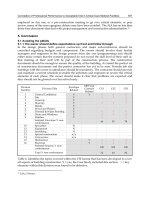
fibrogenic vascular changes (Tomasek et al., 2002) (Figure 1). The molecular interplay
regulating intercellular communication between apoptotic endothelial cells (EC) and
neointimal cells are only beginning to be unraveled.
1.2 Proteomics for studying Post Mortem Signals (PMS) exported by apoptotic EC
Apoptotic programmed cell death is classically considered a silent process. The first clues
suggesting that apoptotic endothelial cells may not "go quietly" stems from pharmacological
Proteomics – Human Diseases and Protein Functions
276
Fig. 1. Schematic diagram of the initiation of vascular remodeling characteristic of AD and
TV. Immune and non-immune factors induce endothelial apoptosis. Endothelial apoptosis
precedes neo-intima formation. The latter is accompanied by ECM degradation and
proliferation and resistance to apoptosis of neo-intimal cells (VSMC, MSC, EPC, fibroblasts
and myofibroblasts). Homing of MSC and EPC as well as myofibroblast differentiation
contribute to fibrogenic changes observed with vascular remodeling.
or genetic approaches aimed at inhibiting endothelial apoptosis in models of AD or TV.
Inhibition of endothelial apoptosis was shown to block the development of vascular
Multidimensional Proteomics for the Identification of
Endothelial Post Mortem Signals of Importance in Vascular Remodeling
277
remodeling, suggesting a paracrine role for the apoptotic endothelium in triggering
pathways of importance in neointima formation (Cailhier et al., 2006; Choy et al., 2004a;
Choy et al., 2004b; Shimizu et al., 2000a; Shimizu et al., 2000b, 2002a, b). Cell biology
approaches supported this contention and showed that medium conditioned by apoptotic
EC regulates the survival and differentiation of major cellular constituents of the vessel wall
(Cailhier et al., 2006; Laplante et al., 2005; Raymond et al., 2004; Soulez et al., 2010).
Execution of the apoptotic program relies mainly on post-translational modifications, such
as protein-protein interactions, protein translocation and proteolysis that will set in motion
the molecular pathways regulating the various phases of apoptosis (Thiede and Rudel,
2004;Wang and Chen, 2011; Mahrus et al., 2008). The caspase family of cysteine proteases is
central to the regulation of the various phases of apoptosis. Their activation in association
with mitochondrial destabilization or extracellular death receptor activation leads to
modifications in the architecture of intracellular organelles and fragmentation of the
cytoskeleton, the ER and the nucleus (Taylor et al., 2008). Apoptosis triggers changes in the
cell membrane including blebbing and extracellular exposure of PS of importance as a
phagocyte recognition signal (Leroyer et al., 2008; Martinez et al., 2005; Pober and Sessa,
2007; Verhoven et al., 1995). In addition, mounting evidence suggests that the apoptotic
program also regulates the extracellular export of a finely regulated set of signals of
importance in leukocyte trafficking, phagocytosis and coagulation (Bournazou et al., 2009;
Lauber et al., 2003; Truman et al., 2008).
The complete set of mediators released by a cell at a given time, defined as a secretome,
can be decrypted through high-throughput methods based on mass-spectrometry. Use of
technology focusing on post-transcriptional events bears special importance in dying cells
where the various levels of molecular regulation depend on protein degradation,
translocation and specific protein-protein interactions rather than gene transcription.
Proteomics was instrumental in characterizing the complex mixture of several secretomes
composed of both soluble and vesicular mediators including microparticles and exosomes
(Mathivanan and Simpson, 2009). As illustrated by the following reports, large-scale
mass-spectrometry also eased the identification of paracrine signals (lipids, proteins and
microparticles) specifically enriched within the secretome of apoptotic cells. For example,
apoptotic Burkitt lymphoma cells release lysophosphatidylcholine (LPC) through
activated caspase-3 dependent mechanisms, which in turn favors recruitment of
macrophages and clearance of apoptotic bodies (Lauber et al., 2003). Apoptotic MCF7
epithelial cells secrete lactoferrin as a means of promoting migration of mononuclear
leukocytes while inhibiting migration of polymorphonuclear leukocytes (Bournazou et al.,
2009). Apoptotic EC shed microparticles with potent immunogenic and pro-coagulant
abilities (Smalley and Ley, 2008; Smalley et al., 2007). In sum, these proteomic-based
reports suggested that a paracrine response embedded within the apoptotic program and
herein referred to as post mortem signals (PMS), controls a finely orchestrated network of
intercellular communication.
In the following sections, we will highlight the advantage of different proteomic strategies
for characterization of PMS released by apoptotic cells. The systematic analysis of the
secretome of apoptotic EC is central to gain insights into novel mechanisms of intercellular
communication of importance in TV and AD. Also, the characterization of endothelial
apoptotic secretome represents a unique opportunity to identify biomarkers of the initial
stage of vascular remodeling.
Proteomics – Human Diseases and Protein Functions
278
2. Studying the secretome of apoptotic EC: Methodological aspects
2.1 In vitro experimental systems aimed at studying endothelial apoptosis
Two major pathways, the intrinsic and extrinsic pathways, regulate the initiation of
apoptosis. The intrinsic pathway is activated by metabolic disturbances, such as nutrient
deprivation and oxidative stress, leading to mitochondrial permeabilization, release of
cytochrome C and activation of caspase-9. The extrinsic pathway is activated by death
receptors that, upon ligand-mediated activation, recruit an initiator caspase (ex. caspase-
8). The effector phase of apoptosis responsible for cleavage of key substrates that bring
about the morphological changes of apoptosis is controlled by a common phase regulated
by effector caspases (-3, -6, -7) (Taylor et al., 2008). Serum starvation (SS) is a classical
inducer of the intrinsic apoptotic pathway in EC and offers several advantages for the
characterization of an apoptotic secretome. First, four hours of SS in cultured EC induces
sequentially mitochondrial permeabilization, activation of caspases -9 and -3, PARP
cleavage and chromatin condensation characteristic of apoptotic cell death. The functional
importance of caspase activation in SS-induced apoptosis was validated with caspase
inhibitors (the pan-caspase inhibitor (ZVAD-FMK) and caspase-3 inhibitor (DEVD-FMK))
as well as small interfering RNA (siRNA) targeting caspase-3 (Sirois et al., 2011). Second,
apoptosis induced by brief SS does not induce necrotic features and cell membrane
permeabilization, as assessed by fluorescence microscopy with propidium iodide and
evaluation of lactacte dehydrogenase (LDH) activity in medium conditioned by serum-
starved EC (Laplante et al., 2010; Sirois et al., 2011). The absence of necrosis in this system
is an asset for studying secretory events in absence of uncontrolled leakage secondary to
cell membrane damage. Finally, SS circumvents contamination of the secretome by
residual components of culture medium (such as albumin) that could interfere with the
identification of less abundant proteins specifically released by apoptotic EC downstream
of caspase activation.
2.2 Identification of endothelial PMS by multidimensional proteomics
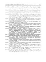
A comparative and multidimensional proteomic analysis was undertaken to characterize the
secretome of apoptotic EC (Sirois et al., 2011) (Figure 2). Proteins specifically released by
apoptotic EC were identified through comparison of the secretomes generated by equal
numbers of serum-starved apoptotic EC (SSC-apo) and serum-starved EC in which
apoptosis was blocked by the irreversible pan-caspase inhibitor ZVAD-fmk (SSC-no-apo).
Cell media were cleared of cell debris and apoptotic blebs prior to proteomic analysis
(Cailhier et al., 2008; Laplante et al., 2010; Sirois et al., 2011; Soulez et al., 2010). An
equivalent amount of proteins were fractionated either by SDS-PAGE or by HPLC anion
exchange chromatography followed by protein identification by MS/MS (Pshezhetsky et al.,
2007). The two comparative strategies were complemented by a functional approach aimed
at identifying proteins with an anti-apoptotic activity on VSMC, therefore recapitulating
induction of the neointimal anti-apoptotic phenotype (Raymond et al., 2004). Proteins
present in SSC-apo were fractionated by ultrafiltration followed by ion-exchange FPLC.
Eluted fractions were individually tested in vitro for their ability to inhibit apoptosis of
VSMC and the fraction displaying a significant anti-apoptotic activity was further
fractionated by SDS-PAGE followed by protein identification by LC-MS/MS.
Computational analysis of the peptides identified by mass-spectrometry generated three
lists built by the functional and the two semi-quantitative comparative approaches.
Multidimensional Proteomics for the Identification of
Endothelial Post Mortem Signals of Importance in Vascular Remodeling
279
Fig. 2. Schematic representation of the experimental strategy for generating serum-free
media (conditioned by equal EC numbers in equal volumes of serum-free media for 4 hours)
by apoptotic (SSC-Apo) and non-apoptotic EC (SSC-No-Apo). Secretomes were collected
Proteomics – Human Diseases and Protein Functions
280
and depleted of cell debris and apoptotic blebs prior to fractionation. Multidimensional
proteomics of the secretomes was performed using one functional and two comparative
approaches. SSC-apo was fractionated by FPLC and each eluted fraction was tested for its
anti-apoptotic activity in serum-starved VSMC. The fraction with the most significant
activity was further separated by SDS-PAGE followed by silver staining and in-gel trypsin
digestion. SSC-apo and SSC-no-apo proteins were also compared and fractionated by HPLC
or SDS-PAGE prior to protein identification by mass-spectrometry analysis. Identification of
specific components of the SSC-apo was achieved using stringent selection criteria. To be
considered a specific component of the apoptotic secretome, the protein had to meet the
following criteria: protein present in SSC-apo only; protein identified by 2 out of the 3
proteomic approaches; protein of human origin. 27 proteins were identified and classified
according to their mode of secretion and the presence of signal peptide, generating novel
hypotheses that were further validated by cell biology methods.
3. The caspase-specific endothelial secretome
A targeted screening strategy was developed to focus on the proteins with the highest
likelihood of representing caspase-specific secretome components of importance in vascular
remodeling. 1300 proteins were identified by LC- MS/MS analysis, 2385 were detected by
SDS-PAGE-MS/MS and 28 proteins were identified by the functional approach. To be
considered a specific component of the secretome of apoptotic EC, identified proteins had to
meet concomitantly the following criteria: 1) they had to be identified by at least 2 out of the
3 different MS/MS approaches, 2) they had to be found exclusively in SSC-Apo, and 3) they
had to be of human origin. According to these criteria, 27 proteins were classified as specific
components of endothelial apoptotic secretome (Table 1) (Sirois et al., 2011). In the
following section we will describe some of the observed changes and discuss the potential
function of this apoptotic secretome.
3.1 Enrichment of proteins associated with non-classical modes of secretion
Most proteins that are directed to the cell surface or the extracellular space through a
conventional secretory pathway contain a signal peptide (Nickel and Rabouille, 2009).
Recent evidence suggests alternative modes of secretion for leaderless proteins, i.e. proteins
without a signal peptide (Schotman et al., 2008) (Nickel and Rabouille, 2009). To define the
contribution of classical and non-classical secretory pathways during apoptotic cell death,
the 27 specific constituents of the endothelial apoptotic secretome were classified according
to the presence of a signal peptide in their primary amino acid sequence, their mode of
secretion, and their intracellular distribution (Table 1). This analysis showed that 25 out of
the 27 proteins appeared to be associated with non-classical modes of secretion, based on
recent literature and/or the absence of a secretion signal. 13 out of the 27 proteins were
previously identified as a component of exosomal nanovesicles. Reevaluation of the
comparative and functional proteomic results identified ten additional exosomal proteins in
SSC-apo only, whereas only two exosomal proteins were identified in SSC-no-apo (Sirois et
al., 2011). Finally, 4 proteins (Table 1 group 2) were annotated as potential components of
exosome-like nanovesicles in other cell types. In total, 31 proteins associated with exosome-
like nanovesicles were considered to be specific components of the secretome of apoptotic
EC.
Multidimensional Proteomics for the Identification of
Endothelial Post Mortem Signals of Importance in Vascular Remodeling
281
Abbreviations: Mem: Membranne; V.E.: endocytic pathway including endosomes, MVB and lysosomes;
C: cytoplasmic; N: nuclear, Ext.: Identified in the extracellular milieu; N.D.: information non available;
Mito: mitochondria; WPBs: Weibel Palade Bodies; **: shedding; Sec. Gran. : Secretory granules
Table 1. Specific components of the apoptotic secretome (SSC-apo) regrouping 27 mediators
selected according to stringent criteria (see Figure 2 and the text). Proteins were listed
according to their mode of secretion, the presence of a signal peptide and their intracellular
localization. Classical type of secretion was defined as a protein containing a signal peptide
with secretion mechanism described in the literature. Non-classical type of secretion was
defined by the absence of a signal peptide or by reports describing their non-classical secretion.
Proteomics – Human Diseases and Protein Functions
282
Initially characterized by Rose Johnstone in the 80’s, exosomes are now recognized as
important intercellular carrier devices detected in most biological liquids including plasma
and urine as well as in the media of cultured mammalian cells (Mathivanan et al., 2010; Pan
and Johnstone, 1983; Pan et al., 1985). These nanovesicles with a diameter ranging for 50-100
nm are generated from inward budding of multivesicular bodies (MVB). Exosomes contain
proteins of the MVB machinery including TSG101 and Alix, both considered classical exosome
markers (Keller et al., 2006; Thery et al., 2002). Exosomes express MHC class I and II associated
proteins and play important role in the innate immune system and in antigen presentation
(Thery et al., 2009). They also contain different cargos including proteins, lipids, microRNAs
and mRNA (Valadi et al., 2007). Their extracellular release stems from the fusion of MVB with
the cell membrane but the molecular regulation of MVB exocytosis remains ill defined. A wide
diversity of cell types have been shown to secrete exosomes but their protein composition
appears to be cell specific and/or dependent on the metabolic state of the cell.
Guided by the proteomic results, we hypothesized that apoptotic cells release nanovesicle-
associated mediators and that this process was triggered by caspase activation. This
hypothesis was further validated by several biochemical techniques, cell biology approaches
and electron microscopy (Sirois et al., 2011). Apoptotic nanovesicles were shown to express
classical constituents of exosomes. Electron microscopy with morphometry analysis
demonstrated that secreted nanovesicles are structurally and functionally distinct from
apoptotic bodies and represent a novel entity of potential significance in vascular remodeling.
3.1.1 Nanovesicular PMS as novel anti-apoptotic factors exported by apoptotic EC
Translationally Controlled Tumour Protein (TCTP) was identified by both functional and
comparative proteomics in SSC-apo (Table 1, group 1). TCTP is an evolutionarily conserved
protein of crucial importance during development (Chen et al., 2007) and for intracellular
inhibition of apoptosis (Telerman and Amson, 2009). TCTP does not contain a secretion
peptide signal and its extracellular export depends on the exosomal pathway (Amzallag et
al., 2004; Lespagnol et al., 2008). Using electron microscopy in association with immunogold
labeling we showed that TCTP was present on the outer surface of endothelial apoptotic
nanovesicles (Sirois et al., 2011). Caspase-activated apoptotic VSMC and fibroblasts also
released TCTP-positive nanovesicles in association with apoptosis, suggesting that this
pathway is active in various cellular components of the vessel wall. TCTP was found to play
a central role in the activation of an anti-apoptotic phenotype in neointimal cells. VSMC
exposed to TCTP(+) apoptotic nanovesicles mounted a robust anti-apoptotic response
whereas VSMC exposed to nanovesicles generated by TCTP-silenced EC failed to develop
an anti-apoptotic phenotype. Collectively these results suggest that TCTP released by
apoptotic nanovesicles is a novel and central inducer of resistance to apoptosis in VSMC and
a biomarker of apoptotic endothelial nanovesicles.
3.2 PMS characterized as biological mediators of vascular remodeling
We further addressed the relevance of the secretome released by apoptotic EC in vascular
remodeling. Since development of AD and TV depends initially on ECM degradation and
phenotypical changes within neointimal cells (i.e. anti-apoptotic and fibrogenic), the list of
proteins generated by the multidimensional proteomic strategy was screened for the
presence of mediators sharing these biological functions. Functional studies on EC, VSMC,
MSC and fibroblasts highlighted a multifunctional and biochemically complex paracrine
Multidimensional Proteomics for the Identification of
Endothelial Post Mortem Signals of Importance in Vascular Remodeling
283
activity of the endothelial apoptotic secretome (Cailhier et al., 2008; Laplante et al., 2006;
Raymond et al., 2004; Raymond et al., 2002; Sirois et al., 2011; Soulez et al., 2010).
3.2.1 Anti-apoptotic PMS
The importance of ECM proteolysis in association with endothelial apoptosis was
highlighted by the identification of the C-terminal perlecan fragment referred to as LG3 by
MS/MS and validated by western blot analysis (Raymond et al., 2004). This fragment
induces a significant anti-apoptotic activity on MSC through alpha-integrin-dependent
activation of the ERK1-2 pathway leading to Bcl-xl overexpression (Soulez et al., 2010). LG3
also interacts with beta-integrins on fibroblasts to induce an anti-apoptotic response but the
intermediate signaling component differs (Laplante et al., 2006). LG3–integrin interactions in
fibroblasts leads to sequential activation of Src family kinases with downstream
phosphatidylinositol 3-kinase (PI3K)-dependent induction of Bcl-xl (Laplante et al., 2006). In
support of a functionally important role for LG3 in TV, increased LG3 urinary levels were
reported in renal allograft recipients with chronic rejection (Goligorsky et al., 2007).
Comparative and functional proteomics of media conditioned by apoptotic and non-apoptotic
EC also revealed the presence of proteases, including ADAM17, ADMTS4, SPUVE, tPA and
cathepsin L of potential importance in ECM proteolysis (Cailhier et al., 2008). The extracellular
export of cathepsin L, which was validated by WB analysis and functional studies, was found
to occur through caspase-3 dependent pathways and to play a central role in cleavage of
perlecan and generation of the bioactive LG3 anti-apoptotic fragment (Cailhier et al., 2008).
Apoptotic EC export a complex array of soluble and vesicular transport-assisted mediators
sharing a common anti-apoptotic activity. Interestingly, these mediators target differentially
the cellular components of the vascular wall through non-redundant signaling mechanisms,
adding specificity to the secreted signals.
3.2.2 Fibrogenic PMS
Vascular remodeling is associated with fibrogenic changes characterized by the accumulation
of myofibroblasts within the vessel wall. Myofibroblasts represent a differentiated and
activated subset of fibroblasts characterized by de novo expression of contractile stress fibers
and alpha-smooth-muscle actin (α-SMA) and enhanced production of collagen I and II. The
accumulation of myofibroblasts plays an important role in myointimal thickening and
vascular stiffness characteristic of AD and TV. The fibrogenic mediator Connective Tissue
Growth Factor (CTGF) was identified with an abundance ratio of 2.5 in medium conditioned
by apoptotic EC as compared with medium conditioned by non-apoptotic EC (Laplante et al.,
2010). Western blotting confirmed that caspase activation significantly increased the release of
CTGF by EC during apoptosis. The central importance of CTGF in the fibrogenic response
triggered by the endothelial secretome was highlighted by injecting mice sub-cutaneously with
medium conditioned by apoptotic or non-apoptotic EC. A significant fibrogenic response with
increased skin thickness and enhanced production of collagen I developed in mice injected
with medium conditioned by apoptotic EC. Also, CTGF immunodepletion abrogated the
fibrogenic activity of medium conditioned by apoptotic EC.
3.2.3 PMS with potential biological activity on vascular repair
Besides PMS characterized and described above, other components of the secretome
released by apoptotic EC are potential regulators of vascular remodeling. PLA2G2D was
Proteomics – Human Diseases and Protein Functions
284
enriched in the secretome of apoptotic EC (Table 1, Group 1) and recent evidence suggests
that it could participate in vascular remodeling. PLA2G2D belongs to a family of secreted
phospholipases (sPLA
2
), which catalyze hydrolysis of membrane glycerophospholipids to
release fatty acids and lysophospholipids (Murakami et al., 2010). PLA2G2D secreted
through the exosomal pathway favors intercellular transfer of inflammatory molecules,
including prostaglandins (Subra et al., 2010). Tissue plasminogen activator (tPA) was also
identified in the secretome of apoptotic EC (Table 1, Group 4) (Cailhier et al., 2008). Recent
studies suggest that extracellular release of tPA fosters the development of fibrogenic
changes (Edgtton et al., 2004; Hu et al., 2008b; Zhang et al., 2007). Convincing evidence also
suggests a predominant role for tPA in atherosclerotic diseases (Gramling and Church,
2010). In fibroblasts and myofibroblasts, tPA favors myofibroblast differentiation and
induces anti-apoptotic phenotypes through phosphorylation of Bad and the inhibition of the
intrinsic apoptotic pathway (Hu et al., 2008a).
4. Conclusion
Characterizing secretomes released by apoptotic cells implies inherent experimental
challenges. Cell death is regulated by post-transcriptional events based on protein
translocation and cleavage. Failure to take into consideration the importance of proteolysis,
protein translocation and activation of non-classical secretion pathways during apoptosis
will undermine the experimental strategy. The type of initiating apoptotic signal and the
phase of apoptosis to be studied should also guide the design of the proteomic strategy.
Creative data mining based on a combination of technical and functional criteria is
necessary to gain novel insights into the modes of intercellular communication associated
with cell death. The use of a multidimensional proteomics was instrumental in
characterizing the importance of caspase activation as a novel regulator of non-classical
modes of secretion. It allowed us to demonstrate that apoptotic cells release apoptotic
nanovesicles, a novel type of membrane vesicle distinct from apoptotic bodies and
reminiscent of exosomes. Mediators of importance in vascular remodeling and of potential
use as biomarkers of endothelial injury, such as TCTP, LG3, CTGF, cathepsin L, EGF,
PLA2GD2 and tPA were also identified. Further analysis of the complex secretome of
apoptotic cells, including biochemical and functional characterization of apoptotic blebs and
nanovesicles, should provide further insights into the mechanisms of intercellular
communication between dying cells and the local microenvironment.
5. Acknowledgment
This work was supported by research grants from the Canadian Institutes of Health
Research (CIHR) (MOP-15447 and MOP-89869) and Fonds de la recherche en santé du
Québec (FRSQ) to MJH. MJH is the holder of the Shire Chair in Nephrology,
Transplantation and Renal Regeneration of Université de Montréal. We thank the J L.
Lévesque Foundation for renewed support.
6. References
Al-Lamki, R.S., Bradley, J.R., and Pober, J.S. (2008). Endothelial cells in allograft rejection.
Transplantation 86, 1340-1348.
Multidimensional Proteomics for the Identification of
Endothelial Post Mortem Signals of Importance in Vascular Remodeling
285
Amzallag, N., Passer, B.J., Allanic, D., Segura, E., Thery, C., Goud, B., Amson, R., and
Telerman, A. (2004). TSAP6 facilitates the secretion of translationally controlled
tumor protein/histamine-releasing factor via a nonclassical pathway. J Biol Chem
279, 46104-46112.
Aronov, S., Gelin-Licht, R., Zipor, G., Haim, L., Safran, E., and Gerst, J.E. (2007). mRNAs
encoding polarity and exocytosis factors are cotransported with the cortical
endoplasmic reticulum to the incipient bud in Saccharomyces cerevisiae. Mol Cell
Biol 27, 3441-3455.
Bette-Bobillo, P., and Vidal, M. (1995). Characterization of phospholipase A2 activity in
reticulocyte endocytic vesicles. Eur J Biochem 228, 199-205.
Bournazou, I., Pound, J.D., Duffin, R., Bournazos, S., Melville, L.A., Brown, S.B., Rossi, A.G.,
and Gregory, C.D. (2009). Apoptotic human cells inhibit migration of granulocytes
via release of lactoferrin. J Clin Invest 119, 20-32.
Cailhier, J.F., Laplante, P., and Hebert, M.J. (2006). Endothelial apoptosis and chronic
transplant vasculopathy: recent results, novel mechanisms. Am J Transplant 6, 247-
253.
Cailhier, J.F., Sirois, I., Laplante, P., Lepage, S., Raymond, M.A., Brassard, N., Prat, A., Iozzo,
R.V., Pshezhetsky, A.V., and Hebert, M.J. (2008). Caspase-3 activation triggers
extracellular cathepsin L release and endorepellin proteolysis. J Biol Chem 283,
27220-27229.
Chen, S.H., Wu, P.S., Chou, C.H., Yan, Y.T., Liu, H., Weng, S.Y., and Yang-Yen, H.F. (2007).
A knockout mouse approach reveals that TCTP functions as an essential factor for
cell proliferation and survival in a tissue- or cell type-specific manner. Mol Biol Cell
18, 2525-2532.
Choy, J.C., Hung, V.H., Hunter, A.L., Cheung, P.K., Motyka, B., Goping, I.S., Sawchuk, T.,
Bleackley, R.C., Podor, T.J., McManus, B.M., et al. (2004a). Granzyme B induces
smooth muscle cell apoptosis in the absence of perforin: involvement of
extracellular matrix degradation. Arterioscler Thromb Vasc Biol 24, 2245-2250.
Choy, J.C., Kerjner, A., Wong, B.W., McManus, B.M., and Granville, D.J. (2004b). Perforin
mediates endothelial cell death and resultant transplant vascular disease in cardiac
allografts. Am J Pathol 165, 127-133.
Consortium, U. (2010). The Universal Protein Resource (UniProt) in 2010. Nucleic Acids Res
38, D142-148.
Cornell, L.D., Smith, R.N., and Colvin, R.B. (2008). Kidney transplantation: mechanisms of
rejection and acceptance. Annu Rev Pathol 3, 189-220.
Dubreuil, V., Marzesco, A.M., Corbeil, D., Huttner, W.B., and Wilsch-Brauninger, M. (2007).
Midbody and primary cilium of neural progenitors release extracellular membrane
particles enriched in the stem cell marker prominin-1. J Cell Biol 176, 483-495.
Edgtton, K.L., Gow, R.M., Kelly, D.J., Carmeliet, P., and Kitching, A.R. (2004). Plasmin is not
protective in experimental renal interstitial fibrosis. Kidney Int 66, 68-76.
Gennaro, G., Menard, C., Michaud, S.E., Deblois, D., and Rivard, A. (2004). Inhibition of
vascular smooth muscle cell proliferation and neointimal formation in injured
arteries by a novel, oral mitogen-activated protein kinase/extracellular signal-
regulated kinase inhibitor. Circulation 110, 3367-3371.
Goligorsky, M.S., Addabbo, F., and O'Riordan, E. (2007). Diagnostic potential of urine
proteome: a broken mirror of renal diseases. J Am Soc Nephrol 18, 2233-2239.
Proteomics – Human Diseases and Protein Functions
286
Gonzalez-Begne, M., Lu, B., Han, X., Hagen, F.K., Hand, A.R., Melvin, J.E., and Yates, J.R.
(2009). Proteomic analysis of human parotid gland exosomes by multidimensional
protein identification technology (MudPIT). J Proteome Res 8, 1304-1314.
Gramling, M.W., and Church, F.C. (2010). Plasminogen activator inhibitor-1 is an aggregate
response factor with pleiotropic effects on cell signaling in vascular disease and the
tumor microenvironment. Thromb Res 125, 377-381.
Gutwein, P., Stoeck, A., Riedle, S., Gast, D., Runz, S., Condon, T.P., Marme, A., Phong, M.C.,
Linderkamp, O., Skorokhod, A., et al. (2005). Cleavage of L1 in exosomes and
apoptotic membrane vesicles released from ovarian carcinoma cells. Clin Cancer
Res 11, 2492-2501.
Hirata, A., Igarashi, M., Yamaguchi, H., Suwabe, A., Daimon, M., Kato, T., and Tominaga,
M. (2000). Nifedipine suppresses neointimal thickening by its inhibitory effect on
vascular smooth muscle cell growth via a MEK-ERK pathway coupling with Pyk2.
Br J Pharmacol 131, 1521-1530.
Hu, K., Lin, L., Tan, X., Yang, J., Bu, G., Mars, W.M., and Liu, Y. (2008a). tPA protects renal
interstitial fibroblasts and myofibroblasts from apoptosis. J Am Soc Nephrol 19,
503-514.
Hu, K., Mars, W.M., and Liu, Y. (2008b). Novel actions of tissue-type plasminogen activator
in chronic kidney disease. Front Biosci 13, 5174-5186.
Keller, S., Sanderson, M.P., Stoeck, A., and Altevogt, P. (2006). Exosomes: from biogenesis
and secretion to biological function. Immunol Lett 107, 102-108.
Knipe, L., Meli, A., Hewlett, L., Bierings, R., Dempster, J., Skehel, P., Hannah, M.J., and
Carter, T. (2010). A revised model for the secretion of tPA and cytokines from
cultured endothelial cells. Blood 116, 2183-2191.
Laplante, P., Raymond, M.A., Gagnon, G., Vigneault, N., Sasseville, A.M., Langelier, Y.,
Bernard, M., Raymond, Y., and Hebert, M.J. (2005). Novel fibrogenic pathways are
activated in response to endothelial apoptosis: implications in the pathophysiology
of systemic sclerosis. J Immunol 174, 5740-5749.
Laplante, P., Raymond, M.A., Labelle, A., Abe, J., Iozzo, R.V., and Hebert, M.J. (2006).
Perlecan proteolysis induces an alpha2beta1 integrin- and Src family kinase-
dependent anti-apoptotic pathway in fibroblasts in the absence of focal adhesion
kinase activation. J Biol Chem 281, 30383-30392.
Laplante, P., Sirois, I., Raymond, M.A., Kokta, V., Beliveau, A., Prat, A., Pshezhetsky, A.V.,
and Hebert, M.J. (2010). Caspase-3-mediated secretion of connective tissue growth
factor by apoptotic endothelial cells promotes fibrosis. Cell Death Differ 17, 291-
303.
Lauber, K., Bohn, E., Krober, S.M., Xiao, Y.J., Blumenthal, S.G., Lindemann, R.K., Marini, P.,
Wiedig, C., Zobywalski, A., Baksh, S., et al. (2003). Apoptotic cells induce migration
of phagocytes via caspase-3-mediated release of a lipid attraction signal. Cell 113,
717-730.
Le Gall, S.M., Auger, R., Dreux, C., and Mauduit, P. (2003). Regulated cell surface pro-EGF
ectodomain shedding is a zinc metalloprotease-dependent process. J Biol Chem
278, 45255-45268.
Leroyer, A.S., Tedgui, A., and Boulanger, C.M. (2008). Role of microparticles in
atherothrombosis. J Intern Med 263
, 528-537.
Multidimensional Proteomics for the Identification of
Endothelial Post Mortem Signals of Importance in Vascular Remodeling
287
Lespagnol, A., Duflaut, D., Beekman, C., Blanc, L., Fiucci, G., Marine, J.C., Vidal, M., Amson,
R., and Telerman, A. (2008). Exosome secretion, including the DNA damage-
induced p53-dependent secretory pathway, is severely compromised in
TSAP6/Steap3-null mice. Cell Death Differ 15, 1723-1733.
Looze, C., Yui, D., Leung, L., Ingham, M., Kaler, M., Yao, X., Wu, W.W., Shen, R.F., Daniels,
M.P., and Levine, S.J. (2009). Proteomic profiling of human plasma exosomes
identifies PPARgamma as an exosome-associated protein. Biochem Biophys Res
Commun 378, 433-438.
Lopez-Casillas, F., Payne, H.M., Andres, J.L., and Massague, J. (1994). Betaglycan can act as
a dual modulator of TGF-beta access to signaling receptors: mapping of ligand
binding and GAG attachment sites. J Cell Biol 124, 557-568.
Mahrus, S., Trinidad, J.C., Barkan, D.T., Sali, A., Burlingame, A.L., and Wells, J.A. (2008).
Global sequencing of proteolytic cleavage sites in apoptosis by specific labeling of
protein N termini. Cell 134, 866-876.
Martinez, M.C., Tesse, A., Zobairi, F., and Andriantsitohaina, R. (2005). Shed membrane
microparticles from circulating and vascular cells in regulating vascular function.
Am J Physiol Heart Circ Physiol 288, H1004-1009.
Mathivanan, S., Ji, H., and Simpson, R.J. (2010). Exosomes: extracellular organelles
important in intercellular communication. J Proteomics 73, 1907-1920.
Mathivanan, S., and Simpson, R.J. (2009). ExoCarta: A compendium of exosomal proteins
and RNA. Proteomics 9, 4997-5000.
Mechtersheimer, S., Gutwein, P., Agmon-Levin, N., Stoeck, A., Oleszewski, M., Riedle, S.,
Postina, R., Fahrenholz, F., Fogel, M., Lemmon, V., et al. (2001). Ectodomain
shedding of L1 adhesion molecule promotes cell migration by autocrine binding to
integrins. J Cell Biol 155, 661-673.
Mitchell, R.N. (2009). Graft vascular disease: immune response meets the vessel wall. Annu
Rev Pathol 4, 19-47.
Murakami, M., Taketomi, Y., Girard, C., Yamamoto, K., and Lambeau, G. (2010). Emerging
roles of secreted phospholipase A2 enzymes: Lessons from transgenic and
knockout mice. Biochimie 92, 561-582.
Nguyen, N.V., Gleeson, P.A., Courtois-Coutry, N., Caplan, M.J., and Van Driel, I.R. (2004).
Gastric parietal cell acid secretion in mice can be regulated independently of H/K
ATPase endocytosis. Gastroenterology 127, 145-154.
Nickel, W., and Rabouille, C. (2009). Mechanisms of regulated unconventional protein
secretion. Nat Rev Mol Cell Biol 10, 148-155.
Pan, B.T., and Johnstone, R.M. (1983). Fate of the transferrin receptor during maturation of
sheep reticulocytes in vitro: selective externalization of the receptor. Cell 33, 967-
978.
Pan, B.T., Teng, K., Wu, C., Adam, M., and Johnstone, R.M. (1985). Electron microscopic
evidence for externalization of the transferrin receptor in vesicular form in sheep
reticulocytes. J Cell Biol 101, 942-948.
Pisitkun, T., Shen, R.F., and Knepper, M.A. (2004). Identification and proteomic profiling of
exosomes in human urine. Proc Natl Acad Sci U S A 101, 13368-13373.
Pober, J.S., and Sessa, W.C. (2007). Evolving functions of endothelial cells in inflammation.
Nat Rev Immunol 7, 803-815.
Proteomics – Human Diseases and Protein Functions
288
Pollman, M.J., Hall, J.L., Mann, M.J., Zhang, L., and Gibbons, G.H. (1998). Inhibition of
neointimal cell bcl-x expression induces apoptosis and regression of vascular
disease. Nat Med 4, 222-227.
Pshezhetsky, A.V., Fedjaev, M., Ashmarina, L., Mazur, A., Budman, L., Sinnett, D., Labuda,
D., Beaulieu, J.F., Menard, D., Nifant'ev, I., et al. (2007). Subcellular proteomics of
cell differentiation: Quantitative analysis of the plasma membrane proteome of
Caco-2 cells. Proteomics 7, 2201-2215.
Rahmani, M., Cruz, R.P., Granville, D.J., and McManus, B.M. (2006). Allograft vasculopathy
versus atherosclerosis. Circ Res 99, 801-815.
Raymond, M.A., Desormeaux, A., Laplante, P., Vigneault, N., Filep, J.G., Landry, K.,
Pshezhetsky, A.V., and Hebert, M.J. (2004). Apoptosis of endothelial cells triggers a
caspase-dependent anti-apoptotic paracrine loop active on VSMC. Faseb J 18, 705-
707.
Raymond, M.A., Vigneault, N., Luyckx, V., and Hebert, M.J. (2002). Paracrine repercussions
of preconditioning on angiogenesis and apoptosis of endothelial cells. Biochem
Biophys Res Commun 291, 261-269.
Ross, R., Glomset, J., and Harker, L. (1977). Response to injury and atherogenesis. Am J
Pathol 86, 675-684.
Ross, R., and Glomset, J.A. (1976). The pathogenesis of atherosclerosis (first of two parts). N
Engl J Med 295, 369-377.
Rossig, L., Dimmeler, S., and Zeiher, A.M. (2001). Apoptosis in the vascular wall and
atherosclerosis. Basic Res Cardiol 96, 11-22.
Schotman, H., Karhinen, L., and Rabouille, C. (2008). dGRASP-mediated noncanonical
integrin secretion is required for Drosophila epithelial remodeling. Dev Cell 14,
171-182.
Shimizu, A., Yamada, K., Meehan, S.M., Sachs, D.H., and Colvin, R.B. (2000a). Acceptance
reaction: intragraft events associated with tolerance to renal allografts in miniature
swine. J Am Soc Nephrol 11, 2371-2380.
Shimizu, A., Yamada, K., Sachs, D.H., and Colvin, R.B. (2000b). Intragraft events preceding
chronic renal allograft rejection in a modified tolerance protocol. Kidney Int 58,
2546-2558.
Shimizu, A., Yamada, K., Sachs, D.H., and Colvin, R.B. (2002a). Mechanisms of chronic renal
allograft rejection. II. Progressive allograft glomerulopathy in miniature swine. Lab
Invest 82, 673-686.
Shimizu, A., Yamada, K., Sachs, D.H., and Colvin, R.B. (2002b). Persistent rejection of
peritubular capillaries and tubules is associated with progressive interstitial
fibrosis. Kidney Int 61, 1867-1879.
Sirois, I., Raymond, M.A., Brassard, N., Cailhier, J.F., Fedjaev, M., Hamelin, K., Londono, I.,
Bendayan, M., Pshezhetsky, A.V., and Hebert, M.J. (2011). Caspase-3-dependent
export of TCTP: a novel pathway for antiapoptotic intercellular communication.
Cell Death Differ 18, 549-562.
Smalley, D.M., and Ley, K. (2008). Plasma-derived microparticles for biomarker discovery.
Clin Lab 54, 67-79.
Smalley, D.M., Root, K.E., Cho, H., Ross, M.M., and Ley, K. (2007). Proteomic discovery of
21 proteins expressed in human plasma-derived but not platelet-derived
microparticles. Thromb Haemost 97, 67-80.