Human Musculoskeletal Biomechanics Part 12 pdf
Bạn đang xem bản rút gọn của tài liệu. Xem và tải ngay bản đầy đủ của tài liệu tại đây (1.38 MB, 20 trang )
9
Elements of Vascular Mechanics
Gyorgy L Nadasy
Clinical Experimental Research Department and Department of Human Physiology,
Semmelweis University
Budapest
1. Introduction
Between half and two thirds of human mortality in developed countries can be attributed to
vascular diseases. Financial losses, human sufferings are increasing with aging of the
population. Vascular diseases develop when some or many vessels in the body are unable to
fulfill their functions. The main function of blood vessels is essentially a mechanical one: to
conduct blood. Vessels are functioning in a unique in the body mechanical environment:
they are continuously subjected to hemodynamic forces: to shear stress of flowing blood and
to distending forces of pressure of the blood in the lumen. Vessels are so much adapted to
these hemodynamic forces that it is impossible to understand their physiology,
pharmacology and pathology without taking into consideration the unavoidable
biomechanical steps in the complicated pathways of cellular and systemic physiological
vascular feed-back control loops, to understand vascular drug action and pathomechanism
of vascular disease (Lee 2000).
Biomechanics is thus at the very core of all vascular sciences. That is reflected in the high number
of papers published in the area. 35 000 papers listed in the Ovid Medline between 1948 and
2010 included knowledge on vascular mechanics in its narrower sense (excluding papers
dealing only with physiological and pharmacological means of vascular smooth muscle
control). Deteriorating Windkessel function of the aged, of the chronic hypertensive, even
after effective treatment of mean arterial pressure, geometric, biomechanical consequences
of atheroscerotic focal remodeling of large arteries, contractile and elastic remodeling of
resistance arteries with aging, with hypertension and with diabetes, remodeling of venous
networks and the venous wall in chronic venous disease, inevitably draws the attention of
clinicians and of pathologists to biomechanical questions. Recent developments in vascular
mechanics, backed with many methodical improvements in the field (Berczi 2005, Cox 1974,
Duling 1981, Huotari 2010, Mersich 2005, Nadasy 2001, Shimazu 1986, See Fig. 1.),
integration of these results into the context of reliable older knowledge makes now a
systemic overview of the most important aspects of vascular mechanics possible. We will
see that an almost axiomatic approach to a phenomenological description of vascular
biomechanics is now in sight. Methodical advancement in the field of cellular physiology,
histochemistry and biochemistry (Discher 2009) identified many if not all extra- and
intracellular fiber types and molecules contributing to the biomechanics of the vascular wall.
Mechanical factors in intra- and extracellular fiber protein expression control are just being
identified. The emerging debate whether mechanics or biochemistry controls vascular
Human Musculoskeletal Biomechanics
212
Fig. 1. Some methodics of vascular mechanics. a. In vitro wire myography. Circumferential
vascular rings and strips are mostly studied. Frequently applied for isometric measurements
of active forces in response to different vasoactive substances. Elasticity and tensile strength
can also be studied. Geometric measurements (strip width and thickness) are needed to
compute stress and to compare the situation with in vivo pressure loads. b. In vitro pressure
arteriography. Cylindrical segments are mounted on cannulas in a glass-bottomed tissue
bath. Devices have been developed both for macroscopic and microscopic vessels.
Intraluminal pressure and flow can be altered to mimic in vivo situation, outer and inner
diameters are measured optically. Mostly pressure-diameter plots are taken at different
levels of smooth muscle tone, or diameter alterations are recorded at continuous pressures
in response to vasoactive substances. c. In vivo ultrasonographic measurement of vascular
lumen changes for biomechanical computations. Right, B-mode record of common femoral
artery and vein. Left, elastic dilation of common femoral vein diameter in response to a
controlled Valsalva attempt. M-mode record (as a function of time, courtesy of dr AÁ
Molnar and of prof V Bérczi).
Elements of Vascular Mechanics
213
protein expression, we believe, is meaningless if the question is approached from the point
of view of system physiology. Mechanical forces from hemodynamics can induce
transmitter release, which, in turn might close the physiological control loop by acting back
on hemodynamics. Or, biologically active substances inducing alterations in local tissue
function, might, at the same time induce vascular changes supporting or just speeding up
other existing vascular control loops adapting tissue circulation to altered tissue function.
Such “feed-forward” loops are very common in physiology. We believe, that in many cases
derailment of such optimized control processes in a situation that could not be
phylogenetically expected, will be the reason for the observed “pathological effect” visibly
acting against biomechanical control (Safar 2005). What is still missing now, is the
mechanics at the molecular level. The low-energy level steric deformations of force-bearing
molecules, determining the phenomenologically descriptionable mechanical behavior are not
known, maybe, with the single exception of actomyosin crossbridges of vascular smooth
muscle cells.
2. Biological background of vascular mechanics
2.1 Separation of the vascular space
The closed vascular system of vertebrates ensures fast nourishment of large neural and muscle
masses, and fast exchange of materials in gills, lungs, kidneys, liver and intestines (Schmidt-
Nielsen 1979). The term “closed” means, that blood vessels are lined internally with a fairly
continuous endothelium (some exceptions do exist). Blood cells in vertebrate tissues are not
forced to uncertain, zigzagging routes in extracellular space among neighboring cells (as e.g.
in many worms), they will move through tissues not leaving the lumen of preformed
vascular tubes (again, situations with exceptions do exist). Lesser friction makes faster blood
flows possible with the same energy expense.
2.2 Distribution of blood flow in space: the network geometry
While diffusion routes of substances from blood to cells are by confinement of blood into
vessels somewhat increased, owing to the rich network of minute capillary vessels few cells
in the body will be farther than about a hundred micrometers from a neighboring small
vessel. Such small exchange vessels, the capillaries should be very narrow and large in
number to ensure optimal diffusion, and this increases friction of blood in them. This is
minimized by the very specific molecular structure of both the luminal surface of the
endothelial cell lining and of blood cells, ensuring easy sliding along each other. Friction is
also limited by the fact that larger distances are traveled by the blood in larger vessels.
Getting closer to their target tissues such larger vessels (arteries) will divide into smaller and
smaller branches, finally forming the capillaries. Capillaries will be collected again by
repeated confluences into larger vessels, the veins. That is the basic principle how vascular
networks are built. We can also easily recognize that such a geometry ensures that blood flow
to each piece of the body can be separately controlled by adjusting the diameter of the
minute vascular tubes leading to it (Abramson 1962, Cliff 1976, Schwartz 1980).
2.3 Distribution of blood flow in time: periodic pump and elastic pressure reservoir
Convection of blood in tubes with real friction can be maintained by continuous investment
of mechanical energy. In many lower animals, a peristalsis-like movement of the blood
Human Musculoskeletal Biomechanics
214
vessel wall propagates blood in the vascular system, but in all vertebrates, motoric force is
centralized at a discrete site of the circulation, the heart. Vessels leading away from the heart
toward the tissues will be the arteries, and vessels leading and emptying the blood back into
the heart will be the veins. Motoric force of the heart is produced by the heart muscles.
Rotational pumps might be the solution for modern left ventricular assist devices, heart
chambers with muscular walls could produce pumping force only in two phases, filling and
ejection, which means that pressures and flows produced are inherently periodic. Periodic
flow in tubes is highly uneconomic. This problem is circumvented by the elasticity of the
vessels, especially of those close to the heart. These are filled with blood during the ejection
period of the heart, and they press the blood forward by their elastic contraction while the
pump is idle during its filling phase (see Windkessel function). Higher blood flow means the
possibility of a higher tissue metabolism, higher speeds of muscle contraction, higher rates
of neural, renal, splanchnic and skin functions, all advantageous for the individual. To press
viscous blood through a system of microvessels needs a pressure difference. The less is the
tissue’s hydrodynamic resistance and the higher is the difference between inlet and outlet
pressures, the higher the tissue flow will be. Diffusion will be optimal from a set of very
narrow vessels. That determines a certain resistance for the capillary segment of the
circulation. Such adaptation took place in the pulmonary circulation of mammals where
vascular resistance outside the pulmonary capillaries is negligible. An other possibility to
elevate tissue blood flow is to elevate the pressure head. In the systemic circulation of
vertebrates outlet pressure, that is venous pressure, cannot be further decreased, as blood
returning to the heart has close to atmospheric pressure. Arterial pressure, however, seems
to be increasing in more developed forms of vertebrates, mammals having higher arterial
pressures than reptilians, amphibians and fishes (Altman 1974, Schmidt-Nielsen 1979,
Schwartz 1980).
2.4 Economic and independent control of blood flow in space and in time: resistance
arteries and further elevation in blood pressure
But surprisingly, not all the energy provided by high arterial pressures will be used up to
keep tissue flows at high levels. Substantial part of this energy will be lost, seemingly
useless, in a short segment of the arterial circulation, in the resistance arteries. In healthy
humans the mean arterial pressure of approximately 95 mmHg of larger arteries (inner
diameters over 1 mm) will be halved in the small arteries and arterioles (inner diameters
from 600 m down to about 20 m), pressures in the arterial side of the capillaries being
around 40 mmHg. What might be the advantages of such a situation? For economic reasons,
tissue blood flow should be adjusted to metabolic or other physiological needs. E.g. working
muscle requires 30-50 times larger flow per unit mass than in the resting state. Large
difference between maximum and minimum blood flows will be characteristic also for the
splanchnic, renal and skin circulations. The solution is that in resting tissue small arteries
will have smaller lumina due to continuous smooth muscle contraction, which can be dilated
quickly as tissue needs increase, increasing local flow. Dilatation of a larger population of
such resistance arteries should induce the collapse of pressure in the arteries, with collapse
of blood flow to many parallelly connected tissues and organs. To ensure their blood flow
they should also dilate to a certain level, further decreasing arterial pressure, again, with
further needs for adjustments in all vessels of the body. A relative high, controlled mean
arterial pressure, however, provides a pressure reservoir, from which all capillaries are
Elements of Vascular Mechanics
215
supplied through a control segment of the resistance arteries (Abramson 1962, Cliff 1976,
Milnor 1982, Nadasy 2007a). The mean arterial pressure in the reservoir is then controlled by
feed-back mechanisms, adjusting heart pumping function and actual levels of overall
peripheral hemodynamic resistance. And now we can reach the conclusion that by this
mechanism, very high blood flows can be provided for functioning tissues, with a certain
independence from affecting the circulation of other organs and tissues.
2.5 The price: unceasing pulsatile stress on the arterial wall
We needed that flow of reasoning to touch on a central problem of mammalian biology,
which is a biomechanical one: The wall of the arteries will be subjected to continuous and
periodically changing forces arising from the pulsatile arterial pressure throughout the life of the
individual. This is a very specific problem in animal biology (Toth 1998, Nadasy 2007a,
2007b). Hearts should beat continuously, but the periods between two contractions
(diastole) guarantee some time for biochemical, metabolic and circulatory recovery. The
same can be told for periodic contractions of skeletal muscle and subsequent tendon loads
and for the compression forces in bone and cartilage. But the artery wall can never get rid of
the effect of the hard distending pressure and its periodic systolic elevations. All
components of the wall had to accommodate to the omnipresence of distending forces. One
possibility to reduce force per square millimeter section of the wall, on individual vascular
constituents is to increase the thickness of the wall. The aortic wall, with about six times
higher pressures is much thicker than that of the pulmonary trunk. Thicker wall means
larger diffusion distances to nourish the artery wall itself. Diffusion in case of large arteries
will not be sufficient, the supplying vessels (vasa vasorum) should enter the wall. Still the
innermost layers of the large arteries will be avascular, as the pressures in the wall would
compress any vasa vasorum in it. Avascular tissues are but a few in the mammalian body,
comprising geriatrically hectic areas (tooth enamel, eye cornea, lens, article hyaline
cartilage).
2.6 Force-bearing histological elements of the wall
A substantial part of the periodic stress due to the pulsatile component of the blood pressure
will be met by the elastic membranes (Apter 1966). Their amount is high in arteries close to the
heart, decreasing toward the periphery and diminishing in the smallest arteries with inner
diameters below about 120 m (true arterioles). The other connective tissue component,
collagen lends rigidity and high tensile strength to the wall. Still there is some mystery about
the omnipresence of smooth muscle in the aorta and in the large arteries. Contraction
(reduced circumference) in these vessels is not extensive, and if any, it will hardly affect
blood flows in such large vessels. It is widely accepted, that their tone sets optimal elasticity
of the artery wall. Contracting, they strengthen the cytoskeletal elements (intermediate, actin
and myosin filaments) in the wall. These cells thus are among the parallelly and serially
connected force-bearing elements of the vascular wall. The dense bodies are forming a lattice
network with intermediate filaments connecting them. Parallel bunches of thin (actin) filments
attach also to the dense bodies and to the hemidesmosomes of the smooth muscle membrane.
Thick (myosin) filaments, interconnect opposing actin filament bunches, and with the ATP-
fueled actomyosin crossbridges can pull them closer to each other. Active slide of actin and
myosin filaments upon each other ensures thus smooth muscle contraction. Vascular
smooth muscle, can characteristically form very slow cycling of cross-bridges even at
Human Musculoskeletal Biomechanics
216
actively shortened length, yielding the typical latch contraction (Rhee 2003, Somlyo 1968).
And it can be proven that at least part of vessel wall viscosity has to be attributed to passive
slide in their contractile apparatus. All smooth muscle cell is surrounded by a basement
membrane. In addition, several proteins of the mechanical transmission between intra- and
extracellular fibers and filaments forming the mechanical anchoring structures have been
identified (Clyman 1990, Gabella 1984).
In resistance arteries, however, contraction of smooth muscle will massively affect blood
flow to the affected territory. The relative thick wall of these vessels will result that a relative
slight contraction of a circumferentially positioned smooth muscle cell at the outer surface
will induce a much more effective reduction in the inner radius.
3. Mechanics of solid materials and fluids – their applicability for vascular
mechanics
Blood vessels are subjected to general laws of physics and mechanics, several of the
parameters applied to study non-living material and several of the general mechanical laws
find a broad application in the field of vascular mechanics (Bergel 1961, 1964, Fung YC 1984,
Gow 1972, Monos 1986). We must not forget, however, that vascular (living) tissue is one of the
most complicated semi-solid materials ever studied by specialists. There are some specific
characteristics rarely found in non-living material. Such is the build-up of the whole
structure under conditions of periodic and continuous distending and shear forces. The
geometry of the specimens, the amount, quality and direction of force-bearing fibers, their
mechanical interconnections with each other specially adapt to the in vivo occurring
mechanical forces. The force bearing elements in the vascular wall are mostly fibers, arranged in
direction of the forces, able to bear pulling forces only. Pushing forces are rare in the wall, maybe
they can be produced from compression of closed, deformable fluid compartments and after
pathologic calcification of the tissue. That complicates the understanding of cyclic
viscoelastic events. How then, elongation of viscous units can be restored? The ability,
never seen in non-living material, to produce active stress at the expense of chemical energy is the
solution. And in all mechanical studies, it is an ever present complicating factor. Smooth
muscle tone will massively affect not only existing geometrical appearance (lumen size and
wall thickness), but will modify elastic properties, affect tissue homogeneity and, as we will
see yield a substantial part of tissue viscosity. For this reason, biomechanical measurements
should be made either in vivo or under in vitro conditions that mimic the in vivo situation
in composition of the tissue bath in which the vascular tissue is tested. The vascular smooth
muscle tone should be set to supposed in vivo values, or, the measurements should be made
at different levels of smooth muscle tone. Unfortunately, the smooth muscle tone itself does
change in response to distending forces (myogenic response) or to endothelial shear of flow
(endothelial dilation). For many non-living material the stress-strain characteristics will be
conveniently linear at least in a certain segment of the curve. That allows the definition of a
single elastic modulus to characterize elasticity. Rigidity of vascular walls, however, always
heavily depends on actual values of wall stress, the higher is the stress, the steeper will be
the stress-strain characteristic curve, providing higher values of their locally computed ratio
(tangent), the incremental elastic modulus. Attempts to find a simple description how the
elastic modulus of the vessel wall changes with stress failed until now. Hopes that the elastic
modulus linearly changes with stress (an exponential shape for the stress-strain
relationship) did not bear the critics of more accurate measurements. According to our
Elements of Vascular Mechanics
217
experience, a double-exponential approach yields almost satisfactory results (Orosz 1999a,
1999b).
4. Network and branching geometry
We must not forget that hemodynamics will be determined at least as much by network
properties of the whole networks than by properties of individual vascular segments.
However, networks lend themselves to study and analysis with much more difficulty,
both methodical and computational, than do individual segments. For this reason
network properties are much less analyzed in the literature. For want of space we will
refrain from a more detailed analysis of the effect of mechanical factors on the
development of the network properties. Network developments seem to follow the law of
minimum energy requirement (Rossitti 1963). That can be altered in aging networks and
at chronically elevated pressure (Nadasy 2000, Lorant 2003). A well analyzed territory is
the retinal arteriolar network. Rarefaction, that is, the decreased number of parallelly
connected resistance arteries seems to be an important contributor to morphologically
elevated vascular resistance in chronic hypertension (Harper 1978). The “chaos theory”
seems to be one fruitful approach to describe general laws of geometric vascular network
development (Herman 2001).
5. Segmental geometry
5.1 Optimal cylindrical symmetry
Most vessels, especially arteries are smooth lined, long cylindrical tubes, positioned in-
between larger branchings (Schwartz 1980). This shape is optimal to ensure minimum loss
of hydrodynamic energy provided by heart contractions and homogenous distribution of
force around the circumference and along the axis, produced by intraluminal pressure. In
real situations, however, especially in pathologic ones, deviations from this optimum do
occur, in the axial, circumferential and radial directions.
5.2 Disturbances of axial symmetry
To reach their anatomical targets vessels should bend, but that axial bending is usually kept
to a minimum by adjusting the axis to an arched curve with a large radius. Anatomical
situation, however, can force the course of a vessel axis into a narrow bend. The typical
anatomical pattern of the large artery system of mammals with the aortic arch itself forms a
narrow bending for a very large mass of flowing blood. A sensitive area in human vascular
anatomy is the base of the skull, here the inner carotid artery is forced into a narrow, S
shaped bony channel, the carotid siphon. Arteries passing joints should follow the position
of the joint. In mammalian embryology, a frequent situation is that vessels originally
developing as branches deviating in an angle from mother vessel will enlarge their lumen
and taking over the role of the distal main branch, which itself then regresses. The originally
sharp angle of the axis in such cases will be later splayed to an arch as a rule. Somewhat
similar situation can be observed in adult pathology, when developing collaterals bypass the
site of slowly developing vascular stricture. Adjustment of the course of the axis is not as
effective in such cases, and a broken course of an artery will be a frequent observation on X-
ray angiography (coronary, leg). Irregular course is a frequent pathological feature in
resistance arteries, too. It can be observed in retinal arteries in hypertension and in aging
Human Musculoskeletal Biomechanics
218
and is one of the main symptoms of the venous varicosity disease. One current explanation
for pathomechanism of varicose notches is that as pressure-induced axial elongation will not
be counteracted by sufficient axial prestretch and tether, the vessel axis bends first, then
with increasing instability it irreversibly buckles into one direction.
Axial irregularities of lumen diameter and wall thickness are the very essence of vascular
pathology. In fact other irregularities of lumen shape will frequently go on unnoticed until
the events will develop toward local narrowing, disturbing flow or induce local distension,
aneurysm, compressing neighboring tissues or endangering with imminent rupture and
bleeding. However, there is a physiological disturbance of cylindrical symmetry at side
branches of arteries. An endothelial cushion just over the orifice ensures that axial blood
rich in red blood cells will be diverted into the side branch, preventing thus plasma
skimming. Focal pathologic processes typical for arteriosclerosis will typically disturb
cylindrical symmetries in all directions. On the other hand, such focal lesions in turn
typically develop where bends, angles, side branches, strictures by impressions of
surrounding tissues disturb cylindrical symmetry of vessel shape and laminar flow. Uneven
lumen and wall thickness along the axis in many resistance arteries is almost the definition
of the diabetic microangiopathy. This causes tissue flow disturbance and microaneurysms
endangering with rupture.
5.3 Circumferential deviations from cylindrical symmetry
Slight circumferential deviations from cylindrical symmetry are inherent in case of vessels
running on bony surfaces. Careful analysis shows that the thoracic and abdominal arteries
are not fully circular, but of an ovoid shape with a somewhat wider base from which the
intercostal and lumbar arteries emerge. Ellipticity of lumen cross section has been thought
to be the very essence of venous mechanics. And really, certain veins, e. g. the lumen of
human inner jugular vein forms but a narrow slit at low pressures, which is for this vessel,
in the erect body position. Other veins, however, are surprisingly circular even at fairly low
pressures. Not much deviation of the anteroposterior and mediolateral diameters of the
human brachial and axial veins could be observed by in vivo ultrasonographic
measurements in a wide pressure range (Berczi 2005). While increasing ellipticity is
characteristic for cannulated venous segments in the low pressure range in vitro, in vivo, or
even in situ, such collapse of one of the diameters is restricted by the radial tethering
provided by surrounding fat and fascial tissue down to 0 mmHg transmural pressure
(Nadasy unpublished). Disturbances of circumferential symmetry, however are occurring as
a rule in case of focal atherosclerotic lesions and in any case of mural thrombosis. Present
techniques at hand can analyze the differences of histologic composition around the vessel
circumference (and in the wall along the radius), but the biomechanical consequences,
uneven distribution of force on force bearing elements, are still poorly understood. We are
convinced, however, that it is a key issue in the pathomechanism of the progressive
development of the arteriosclerotic plaque. With destruction of the inner media in a sector of
the wall, large pulsatile forces will be transmitted to the outer layers in this segment, with
the consequence of accumulation of collagenous fibers and cessation of vasa vasorum flow.
While some remodeling of the force-bearing elements of the wall can make revascularization
possible, a necrotic nucleus, getting closer to the luminal surface and endangering with
rupture into the vascular lumen, will be the most dangerous threat caused by the focal
process. Some modern techniques raise the hope that distribution of force inside the vessel
Elements of Vascular Mechanics
219
wall could be once directly studied. Greenwald has directly demonstrated the sequential
strengthening of connective tissue elements (Greenwald 2007).
5.4 Radial asymmetry
Concerning the radial asymmetry, original views that a rigid adventitia could prevent
further distention of the elastic media (“an elastic ball in a string bag” model), still vivid in
the views of non-specialists has been opposed by direct elastic measurements on vessels
from which the adventitia has been removed. Right now it seems that the adventitia, with
its mostly loose connective tissue, is the site more for the axial tether, than for any
contribution to circumferential force-bearing. Vasa vasorum, sympathetic nerves can run
in it undisturbed by tissue pressure, the fibroblasts in it with their ability to differentiate
into vascular smooth muscle cells can ensure an “appositional” medial thickening. There
is an inherent, physiological radial inhomogeneity of the media itself in the wall of large
arteries, circumferential elastic sheets (whith holes in them) and smooth muscle cells
packed in angle with radius in a fish-bone pattern are forming alternative layers. Taking
into consideration that intraluminal pressures at the inner surface should be decreased
down to zero at the outer surface, it is really surprising to observe, still how similar are
these layers and their elastic and smooth muscle components. This supports, unproven
yet views that some equalization process in the media should exist, that distributes the
large circumferential force to the similar wall constituents in a similar manner. Some
radial inhomogeneity of force distribution, however, should exist in the wall. This can be
proven by the elegant experiments of just cutting up vessel rings in the radial directions.
The ring will be opened, the angle of which in such a state of zero stress can be measured
and analyzed (Liu 1988). We must not forget, however, that the artery wall is never at
zero at stress in vivo, fiber arrangement adapted to real pressurized wall tensions. In case
of larger vessels the contribution of the endothelium to elastic properties of the wall is
thought to be negligible. However, the basal membrane of the capillary vessels lends
sufficient rigidity and tensile strength to these vessels. In addition, intimal thickenings of
sclerotized vessels can take up a substantial part of wall stress, relieving thus the outer
layer of the affected segments.
5.5 Vascular diameter
Vascular specialists with biomechanical backgrounds are rarely satisfied when “the”
diameter of a vessel is mentioned. All vessels alter their diameter as a result of acute smooth
muscle contraction, and the same measured intraluminal diameter could mean very
different vessels at different levels of vascular smooth muscle tone (a larger vessel but with a
larger tone), and different measured intraluminal diameters could mean a morphologically
identical vessel segment but with a somewhat altered tone. The diapason between
maximum and minimum contractions is routinely measured now in wire and pressure
angiography, and such practice is more and more frequently applied in in vivo
measurements and to some degree, even in clinical practice.
For the exact biomechanical analysis, we have to discriminate between the morphological
diameter of the segment, best characterized by its fully relaxed state, its diameter in full
contraction, which in healthy arteries below about 1 mm of inner diameter will be the fully
closed segment, and the actual diameter measured in a discrete state at a given level of
Human Musculoskeletal Biomechanics
220
muscle tone. Things will be even more complicated when we realize that vascular lumen
will also be dependent on transmural pressure, elasticity of the wall and also even on axial
distension. Fortunately, relaxed vessels pressurized close to or somewhat over physiological
pressures, turn to fairly rigid structures and do not further change much their lumina as a
function of pressure. This stable diameter will well characterize the morphological lumen. In
the scientific practice it is even more accurate to characterize morphological lumen with the
whole course of the relaxed pressure-diameter curve.
5.6 Physiological control of the morphological lumen
The “passive” (morphological) lumen will be differently controlled and with more delay
than the actual lumen is determined by the actual level of smooth muscle tone. The
morphological control process needs a reorganization of the histological components of the
wall. The terms “remodeling” (segmental remodeling, geometrical remodeling, wall
remodeling) or “long term control” are used to describe it (Fig.2.and 3.).
It had been known for ages that vessels with larger flows have larger lumina. The problem
can be reduced to the question, that branching of a larger mother vessel how will effect the
lumens of the smaller and smaller daughter branches? Early analysis of pressure and flow
in vascular networks have shown that while mean linear velocities are decreasing toward
smaller branches (30 cm/sec in the aorta, a few hundred micrometers in the capillaries)
there is an elevation of mean pressure drop per unit length. There is hardly any drop in
mean arterial pressure in large arteries, substantial pressure drop occurs along a few cm
length of small arteries with a few hundred m of diameter, finally, a sharp drop of pressure
happens in arterioles, a few mm of lengths, but with diameters between 30-150 m.
(Abramson 1962, Cliff 1976, Milnor 1982, Schwartz 1980). In the simplest case of symmetric
branching, to maintain the mean linear velocity in daughter branches would need a ratio of
radii of daughter (r
d
) to mother (r
m
) branches of r
d
2
+ r
d
2
=r
m
2
; from whence 2 r
d
2
=r
m
2
and r
d
/
r
m
=1/2 = 0.707. To maintain the unit pressure drop per unit length (with unaltered
viscosity, following the Hagen-Poisseuille law) would need daughter to mother radius
ratios of Q=/8* r
m
4
/*p/l=2*/8* r
d
4
/*p/l; from whence 2 r
d
4
=r
m
4
and
r
d
/
r
m
=1/
4
2 = 0.841. Hemodynamic analysis of existing arterial networks thus leads us to the
conclusion, that in case of symmetrical branching daughter to mother branch ratios should
be in-between these two values, m 0.707 < r
d
/
r
m
< 0.841. Measuring many arterial
diameters Murray has suggested, that in case of any types of branchings, the equation of
r
m
3
= r
d1
3
+ r
d2
3
+ r
d3
3
+ r
d4
3
+…. will be valid. This seems a fairly good approach even in our
days. How the vessel wall should “know” how much is the flow in its lumen? The answer
was given in two classic works by the great American cardiologist, Rodbard, who supposed
that endothelial shear is somehow sensed by the endothelial cells and is kept constant by
chronic morphogenetic processes adjusting vascular lumen to flow. The value of endothelial
shear rate (dv/dr) computed based on the Hagen-Poiseuille law is dv/dr= 4Q/r
3
(where
Q is the volume flow and r is the inner radius) being in accordance with Murrays law. For
our symmetric bifurcation, dv
d
/dr
d
= dv
m
/dr
m
and 4(Q/2)/r
d
3
=4Q/r
m
3
from whence
2 r
d
3
=r
m
3
(the form corresponding to Murray’s law) and finally, r
d
/
r
m
=1/
3
2 = 0.794.
This latter number is just in-between 0.707 and 0.841 as required by common hemodynamic
experience. Validity of such computations has been proven in analysis of several types of
vascular branchings (Lorant 2003, Nadasy 1981, Pries 2005, Rodbard 1970, 1975, Zamir
1977).
Elements of Vascular Mechanics
221
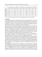
Fig. 2. Morphological (long-term) control of vascular lumen. a. Shear rate and shear stress in
a cylindrical vessel with continuous flow. b. Geometry of a symmetric branching. c.
Relevance of the Murray-Rodbard law: Pressure diameter plots of normal double and single
(morphologically malformed) human umbilical artery segments. The ratio of the cubes of
inner radii at physiological pressures is around, 2 which can be expected in case of doubled
flow in single arteries. (From Nadasy 1981, with permission of Karger) c. Relevance of the
Murray-Rodbard law: In vivo microprepared popliteal confluence of the rat saphenous vein.
Video-microscopic records at two magnifications with normal pressure and flow in the
lumen of anesthetized animals. The ratio of the cube of diameter of the mother branch to the
sum of cubes of diameters of daughter branches is close to the expected 1 (from Lorant 2003,
with permission of Physiol Res, Czech Academy of Sciences).
Human Musculoskeletal Biomechanics
222
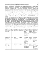
Fig. 3. Long-term control of vascular wall thickness. a. Vessels with thicker walls can more
effectively control their inner diameter, a feature characteristic for resistance arteries. b.
Series of intramural coronary resistance arteries from the rat. Contours of cross sections are
shown. From left to right, morphologically different segments with diameters around 4-500,
300, 200 and 100 micrometers. From up to down, at different transmural pressures. Passive
and contracted segments are shown. Note typical increasing thickness-to-diameter ratio
toward smaller arteries, increasing effectivity of lumen control. (From Szekeres 1998, with
permission of Karger) c. Parameters determining tangential wall stress. d and e. Wall stress
and wall thickness of a resistance sized small artery from normotensive and hypertensive
animals. Note that elevation of wall thickness (and reduction of lumen) just ensured
unaltered wall stresses at elevated in vivo pressures as could be expected by the Folkow-
Rodbard-Mulvany law (from Nadasy 2010, with permission of Karger).
At a more scrutinizing analysis, however, the situation will be more complicated than that.
In fact, not shear rates but shear forces will be sensed. This latter will be the function of
blood viscosity, a fairly elusive factor supposing its dependence on shear stress (the blood
being a non-Newtonian fluid) and on the level of accumulation of red blood cells in the axial
Elements of Vascular Mechanics
223
flow (according to the Fahreus-Lindquist law). At a fairly good flow, and especially in
smaller vessels, almost clear plasma, devoid of cellular elements will slide along the luminal
surfaces of endothelial cells, yielding a fairly continuous viscosity of 2-3 cP. The other
problem is the changing levels of flow (depending on pressure and on more distal
resistance) and changing levels of arterial tone. Based on experience on skeletal muscle
circulation, we have to suppose that relative short periods with high flows will be sufficient
stimuli to increase the morphological lumen. On the contrary, when maximum flows do not
come for a while, shortened circumferences could get morphologically stabilized reducing
the range of luminal vascular smooth muscle control (Nadasy 2010a). An other problem will
be prominent when we compare arteries and veins. Mean velocity of blood flow is about
half as fast as in corresponding arteries. We have to suppose that either other factors than
blood flow contribute substantially to morphological lumen control in veins or, the venous
endothelium is differently tuned to flow sensation than is the arterial. To solve these
questions would be essential to reveal the pathomechanism of chronic venous diseases. An
other complication is rising from wall thickness adjustment to elevated pressure, which, can
reduce lumen below levels required by flow inducing thus resistance elevation and having a
stabilizing effect on the high blood pressure. Which is the cause and which is the effect?
Fine network analyses in different states of circulation and in different stages of the
hypertension disease will be needed fully to describe the intermingling feedback networks
of flow, resistance and pressure. We believe that a disturbed endothelium will not be
sufficient to restore morphological lumen to rare flow maxima, or to counteract the lumen-
narrowing effects of adaptive wall thickening. Extensive studies revealed a set of
cytophysiologic and even molecular mechanisms how shear is sensed at endothelial luminal
surfaces.
Summing up this chapter, we have to conclude that despite many remaining questions, the
law, describing the long term morphological control of vascular lumen formation, originally
found by Murray and by Rodbard (1970, 1975) has proven its validity for a substantial
period of time (Kamiya 1980, Lorant 2003) and can be accepted as one basal law of normal
vascular functioning. It can now stated with a high level of certainty that long term control
processes in the vascular wall do exist that adjust morphological lumen to flow so that they
tend to stabilize endothelial shear.
5.7 Physiological control of vessel wall thickness
Vessels with higher pressures have thicker walls. The pulmonary arteries have thicker walls
than the caval veins and the aorta than the pulmonary trunk, despite similar flows. Higher
pressure means higher tangential force per unit length (F) on vessel circumference, F= p*r
i
,
where p is the transmural pressure and r
i
is the inner radius (Fig.3.). That can be distributed
on a vessel wall thickness of h, = p*r
i
/ h, where is the tangential stress. Folkow
observed that arterial wall thickness increases in a compensatory manner in hypertension
(1971, 1990, 1995). An other assumption published by Rodbard (1975) stated that
morphological thickness of the vascular wall is controlled to stabilize the value of tangential
stress. But there are more problems with this observation. As there is hardly any drop in
mean arterial pressure along Windkessel and distributing arteries (to about 600 m of inner
diameter) inner radius to wall thickness ratios should have remained unaltered along the
whole arterial tree to make unaltered, too. In fact radius to wall thickness ratios decrease
toward smaller arteries. What is even more contradicting, in more distal resistance arteries
Human Musculoskeletal Biomechanics
224
following a substantial pressure drop, radius to wall thickness ratios should increase to
compensate for lower pressures to ensure stable values of tangential stress. Just the opposite
is the case: small resistance arteries have relative thicker walls, and computed in vivo values
for tangential stress decrease along the arterial tree reaching very low values in smallest
resistance arteries. First we have to see what is the advantage of such a difference (Fig. 3a).
Smooth muscle in large vessels will not contract to induce substantial reduction in diameter.
That would be useless, as no substantial pressure drop could be reached taking into
consideration real flow and viscosity values. But there is an opposite situation at the level of
the resistance arteries: fast and effective acute changes in lumen are the very essence of their
physiological functioning. Contraction of a helical smooth muscle cell ring at the outer
surface of a resistance artery wall will be much more effective than the contraction at the
inner site of the wall, the difference will be the higher the thicker the resistance artery wall
is. A 20 % contraction at the inner circumference will induce 20% reduction of the lumen
(2.4fold increase in segmental resistance). A similar 20% contraction at the outer
circumference will induce 32.9, 47.0 and 100% reduction in lumen in vessels with 10:1, 8:1
and 6:1 radius to wall thickness ratios, respectively as they will push the inner vascular wall
layers into the lumen. This will result respective segmental resistance elevations of 4.9 and
12.7 times in the first and second cases, while the lumen will be fully closed and flow will
cease in the third situation. The question, however, can be raised, that as tangential stresses
are so much less in smaller arteries, are the smooth muscle cells themselves in this vessels so
much different? Several differences between smooth muscles of these vessels could be listed,
but one outstanding difference is the presence and amount of elastic tissue which is
diminishing toward the peripheral arteries practically synchronously with the reduction of
the r
i
/h ratio. A simple solution can be that while smooth muscle is similarly stressed in
large and small arteries, the parallelly connected elastic membranes will bear a substantial
part of the tangential strain. But later we will see that amount of elastic tissue will develop
in response to pulsatile not steady stress. And we will also see that elastic tissue has also its
impact in the lower part (below diastolic pressures) of the arterial pressure-diameter
characteristics. And with that restriction the Folkow- Rodbard-Mulvany’s law on long term
control of the thickness of the vascular wall can now be valid (Rodbard 1970, 1975): Vessel
wall thickness develops to stabilize tangential wall stress – valid, when tissue composition is
unaltered.
Thickened wall of affected vessels is a main alteration in case of chronic arterial
hypertension (Folkow 1971, 1990, 1995), chronic pulmonary hypertension and venous
pressure elevation in chronic venous disease. In several clinical and experimental studies,
hypertensive remodeling of arteries seemed just to stabilize in vivo tangential wall stress.
Such control mechanisms should exist at least in a certain phase of the hypertension disease
(Albinsson 2004, Dickhout 2000, Frisbee 1999, Hayashi 2009, Nadasy 2010a, Pries 2005).
6. Vascular elasticity
6.1 Significance of vascular elasticity
As we could see above, elasticity is a very important, inherent property of blood vessels, it
ensures the fairly continuous pressure and flow in small vessels despite periodic functioning
of the heart pump (Bergel 1961, 1964, Fung 1984, Gow 1972). Actual geometric properties of
a vessel, determining their hydrodynamic resistance are in turn determined by their
Elements of Vascular Mechanics
225
morphological geometry, the actual level of smooth muscle tone, transmural pressure,
luminal flow inducing endothelial dilation, their axial stretch and the elasticity of the wall.
The role of elasticity in forming the actual geometry will be more important in vessels with
high distensibility, where small changes in transmural pressure will induce large changes in
lumen volume in such vessels. As we will see later, most vessels are relative rigid at and
especially above physiological pressures. That means that elasticity will be a central
parameter in determining lumen size when pressures in the lumen decrease below
physiological pressures. We have a good reason to think that elasticity of force-bearing
fibers in the vascular wall helps to maintain a fairly even distribution of distending forces
and stretches among parallelly and serially stressed wall components: elongation of a
stressed fiber transfers part of the force onto the parallelly connected poorly stressed fibers.
More elongation in the less stressed serially connected fibers will help an even distribution
of stretch among the serially connected circumferential elements of the wall. One central
problem in vascular pathology, we believe, is the elastic force-bearing capacity of the inner
layers of larger vessels: by their force-bearing they relieve the outer layers. Decreasing
hydrostatic pressures in the outer layers of the vessel wall make possible the proper
functioning of the vasa vasorum and proper nourishment of the whole wall.
6.2 Parameters measuring elasticity
We can determine the elasticity of a vascular segment in vivo directly, by measuring
geometrical parameters (inner, outer diameter, wall thickness) at different levels of pressure
(Fung 1984). Modern ultrasonography (from the surface of the body and also intravascular)
provides sufficient means for larger vessels (Berczi 2005, Molnar 2006, Mersich 2005,
Shimazu 1986). A certain level of manipulation of pressure and smooth muscle tone is
possible even in human volunteers (See Fig. 1c.). In vivo animal experiments of course give
a wider and more accurate potential for that. Delicate measurements can be made on
isolated, axially isometric, cylindrical vascular segments mounted in the tissue bath of a
pressure angiometer (Cox 1974, Duling 1981, Nadasy 2001, see 1b.). The pressure-diameter
characteristics can be recorded at different levels of smooth muscle contraction (with
vasoconstrictors added) and in the so called passive state (fully relaxed smooth muscle e.g.
with calcium-free incubation medium). We can cut rings, circular or helical strips and study
them in a wire myograph (Fig. 1a.). To characterize circumferential (tangential) elasticity we
can compute compliance, C=V/p, that is volume alteration in response to unit alteration in
(transmural) pressure. The question, answered by the compliance value is, that how much
the pressure will change if we press a certain amount of blood into the vessel. This
parameter is frequently used to characterize venous elasticity and even for large arteries.
One problem with it is, that a rigid, large vessel will have larger compliance than a small
elastic one. To circumvent this problem the term distensibility has been applied, simply
normalizing volume change to the initial volume (V
o
), D=V/ V
o
*(1/p). Distensibility
almost fully describes the elastic properties of the wall, the way it is taking part in
hydrodynamic processes, and is used frequently in hydrodynamic models for this reason.
However, it will not properly characterize the elasticity of the wall material as more elastic,
but thicker walls can have the same distensibility. The Young’s elastic modulus will be
computed, which is the tangent of the stress-strain relationship. For cylindrical segments
with inner and outer radii of r
i
and r
o
, respectively, with not negligible, but relative thin
walls (valid for most vessels) the equation given by RH Cox (1974, 1975a, 1975b), the
Human Musculoskeletal Biomechanics
226
inventor of pressure angiometer (Fig. 1b.) is mostly accepted: E= 2r
o
r
i
2
/ (r
o
2
-r
i
2
)*(p/r
o
),
where p is the pressure change inducing an alteration of the outer radius, r
o
. As we
described it earlier, elasticity of the vascular wall will be heavily dependent on the
conditions under which we have measured it. For this reason, we have to repeat the
measurements at different levels of wall stress (intraluminal pressure) and at different levels
of smooth muscle tone. Typically, pressure-diameter characteristics will be recorded at
different levels of smooth muscle tone. If pressure alterations are slow enough, we can
suppose that the wall is transiting a series of equilibrium states and each further
infinitesimal elevation in pressure (stress) will induce an infinitesimal rise in circumference
(strain) and the tangent of the normalized pressure-volume curve (incremental
distensibility) and tangent of the stress-strain curve (incremental elastic modulus) can be
computed and plotted as a function of pressure (“isobaric” parameters). Or, in case of the
incremental elastic modulus, it will frequently be given as a function of computed wall
stress. Such elastic modulus-tangential stress characteristics will characterize best the
elasticity of the wall material itself. The vertical axis is usually logarithmic, but will not be
linear even in this form. The question here can be raised how reproducible and how
characteristic for the in vivo situation the elastic parameters measured this way will be?
One problem is the different levels of smooth muscle tone. That has to be somehow
stabilized, which is not so easy as it itself changes with changing wall stress. In case of a well
developed myogenic response, as it is the case with many resistance arteries, elevated
pressures will not produce elastic dilation but myogenic contraction (Kuo 1988, Osol 1985,
Szekeres 2004). The term plasticity is used for a typical behavior pattern of vascular smooth
muscle: the material of the wall somehow adapts to lasting pressure loads or pressure
patterns. The mechanical past of the vessel (in the last few minutes) determines to some
degree its present mechanical behavior.
For this reason, reproducibility should be ensured by preliminary incubation of the segment
under controlled contractile and mechanical conditions (continuous or cyclic stress or
pressure load). Stress or pressure changes during elastic measurements should be applied
following a reproducible pattern: rises at continuous rates, stepwise rises, cyclic sinusoid or
triangle patterns are widely used. Because of viscosity, characteristics taken with increasing
and decreasing loads might differ. Elastic moduli in the range of 10
4
Pa can be considered as
very low, found only at very low wall stresses, at a few mmHg intraluminal pressure in
arteries, and close to 0 mmHg pressure in veins. Values of 10
6
Pa (10
5
-10
7
) are typical for
many vessels in their physiological pressure range. Further elevating the pressure values in
the lumen 10
7
Pa can be reached with any type of vessels (veins included!), but a damage to
the wall (mostly reflected by reduced smooth muscle contractility) is in such cases
imminent. The tensile strength of healthy vessels is surprisingly high, veins can be sutured
as bypasses into the arterial system and arteries will endure for a shorter period 1
atmospheric transmural pressure. Tangential stresses in vivo range from the very low values
of a few 10
4
Pa (e.g. 30 mmHg = 4 kPA pressure, r
i
/h values around 3, for a thick walled,
contracted arteriole, = 1.2*10
4
Pa) to highest measured values (e.g. 150 mmHg=20 kPa
elevated systolic pressure for a thin walled large artery with r
i
/h ratios around 10, = 2*10
5
Pa). Stresses in the range of 10
6
Pa will damage the wall as maximum forces produced by the
smooth muscle are in the range of 3-5 atm (3-5*10
5
Pa, 200-400 mmHg for a large vessel
with a radius to thickness ratio of 10).
In in vivo experiments and in the clinical practice, too, vascular elasticity is frequently
measured indirectly. The shape of the aortic pressure curve can be analyzed. The so called
Elements of Vascular Mechanics
227
augmentation index gives an indirect information about the rigidity of the large arteries.
Even more popular is the determination of the pulse wave velocity. These will be discussed
in more detail in the chapter on the Windkessel arteries.
6.3 Effect of histological composition on vascular elasticity
After several decades of systemic investigations now we have a fairly good if not final
picture how the histological composition of the vascular wall affects its elasticity (Apter
1966, Bergel 1961, 1964, Cox 1975a, 1978, Dobrin 1978, Fung 1984, 1995, Gow 1972,
Greenwald 2007, Koens 2010, Oxlund 1986, Roach 1957, VanDijk 1984, Vidik 1982). The
endothelium will not much contribute to the mechanical properties with the exception of
capillaries and maybe, of the smallest other vessels. But we must not forget, that it is the
endothelium that senses the shear at the inner surface of vessels and adjusts the diameter to
it (see endothelial dilation, and vascular lumen control). Also, the endothelium, with the
basement membrane underneath it, is a fairly mechanically stable structure forming the
capillary walls. Capillaries in the renal glomeruli seem to be enforced by the leg processes of
the podocytes there. Contrary to earlier expectations, the loose connective tissue of the
adventitia will not restrict pressure-induced dilation of an elastic media (“elastic balloon in a
string bag” model, Burton 1954). The collagenous fibers running in it will ensure some axial
tether when, with decreasing pressures the axial extension of the arteries decreases. In case
of veins at very low pressures, and in arteries at extremely large levels of medial smooth
muscle contraction, we can suppose even some adventitial radial tether. Vascular smooth
muscle if relaxed does not resist distending force until the intracellular fiber structure is not
stretched. Vascular structures containing abundant smooth muscle (umbilical artery,
resistance arteries) have very low elastic moduli at low stresses and high moduli at high
stresses. At least part of the elasticity of the stretched vascular smooth muscle will be
determined by elasticity in the actomyosin crossbridges (“series elasticity”, Mulvany 1981,
Siegman 1976), and should reflect the mechanical deformation of the myosin head or neck
in response to distending forces. In fact, as each smooth muscle cell is surrounded by a
socket of basal lamina, composed of collagenous fibers, and there is an intracellular scaffold
in them formed of intermediate filaments and dense bodies, and to go further, bundles of
actin filaments are more abundant in them than thick myosin filaments, contribution of
these latter structures to “Series elasticity” cannot be excluded. We have found that elasticity
of vascular tissue, rich in smooth muscle can be explained as originating from a “unit
elasticity”, possibly the elasticity of the actin filaments. Upon cyclic loading, the number of
serially and parallely connected elastic units could adapt by breaking up and passive slide
of overstressed latching actomyosin crossbridges as well as by spontaneous shortening in
understressed ones. That ensured uniform stretch and load in affected filamentous units
(Nadasy 1987, 2007b, Szekeres 1998).
Collagen lends rigidity and high tensile strength to the vascular wall, again, at high
distending forces, while very moderate forces are sufficient to strengthen the coiled up
collagenous bundles in the vessel wall. More collagen in the wall usually means higher
rigidity (Apter 1966, Bergel 1961, 1964, Cox 1978, Dobrin 1978, Gow 1972, Greenwald 2007,
Hegedus 1984, Oxlund 1986, Vidik 1982), but usually only at higher stresses. Collagen
accumulation is typical in many types of diseased vessels (“sick vessel syndrome”, Heistad
1995). Contrary to the other two wall constituents, elastin will resist distension even at very
low stretches but will not be fully stretched even at high distending forces. The result is that
Human Musculoskeletal Biomechanics
228
its presence elevates elastic modulus (increases rigidity!) at low pressures, but decreases it at
high tangential forces (Fig. 4b.). Frequent contradictions about connective tissue
composition and measured elastic modulus can be prevented by exact analysis of the
pressure and tangential stress levels where the measurements have been made. Plots of
tangential elastic modulus against tangential stress usually are in good accordance with
histological composition. Such plots are accepted as reflecting the elastic properties of the
wall material itself. Still it is poorly understood how the elastic laminae are mechanically
connected to smooth muscle. In rabbit aortic strips we have found that series and parallel
elastic components of this elastic tissue changed parallelly upon passive stretch (Nadasy
2007b). But similar observations were made in the aneurysmic tissue fully devoid of muscle
and elastic components (Toth 1998).
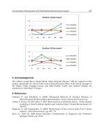
Fig. 4. Long and short term control of vascular elasticity. Comparisons of pressure-diameter
characteristics of different vessels. Relative values of diameter are shown. Black lines
represent the characteristics of a typical distributing artery in the relaxed state (same in each
diagram). Shaded areas mark the range of in vivo pressures of vessels being compared
(drawn in color) a. Red, the same artery in the contracted state. Smooth muscle contraction
reduces elastic modulus as a function of pressure but increases it as a function of strain.
(Cox-Dobrin law) b. Comparison with a more elastic (Windkessel) artery (red). Note
decreased distensibility at low pressures and increased distensibility at high pressures c.
Comparison with a hypertensively remodeled artery (red). Note upward dislocation
(toward elevated in vivo pressures) of the transition between more and less distensible parts
of the characteristic curve. d. Comparison with a resistance artery in the relaxed and
contracted state (red). Note transition between more and less distensible parts of the
characteristic curve toward lower pressures existing in vivo in these more distal resistance
vessels. e. Comparison with elasticity of upper body (left, blue) and leg (right, blue) veins.
Transition between more and less distensible parts of the pressure diameter characteristic
curves also corresponds to in vivo occurring pressures in these vessels (Burton-Roach-Kadar
law).
Elements of Vascular Mechanics
229
6.4 Typical shape of the vascular elasticity curve
As a result of the elastic properties of their components, all vessels, without exception show,
elastic characteristics with relative similar shapes (Burton 1954, Dobrin 1978, Roach 1957,
Wolinsky 1967, Fig. 4.). Their elastic modulus increases with stress in a such a manner that
they are distensible at lower than commonly occurring physiological pressures and turn rigid at
higher pressures (Burton-Roach-Kadar law). (Berczi 2005, Busse 1981, Cox 1975b, Gow 1972,
Molnar A 2006. Molnar G 2010, Roach 1957, Stooker 2003, Szentivanyi 1998) The
physiological working point of the vessel is somewhere at the turn of the pressure radius
characteristic curve. The work-points, of course will be different for aortas, small resistance
vessels, veins, embryonic vessels and hypertensive vessels (Fig. 4.). But that will hardly
affect the validity of the above statement. Such an organization of vascular elasticity can be
considered biologically logical: potential rises in pressure will not induce unlimited
distension, while unexpected volume reductions will be “followed” by the elastic shrinkage
of the wall, stabilizing to some degree against fast pressure drop. Larger smooth muscle
tone can cause some complications (see next chapter).
6.5 Control of vascular elasticity
In addition to reduce inner radius (and to increase wall thickness) vascular smooth muscle
contraction substantially alters the elastic properties of vessels (Apter 1966, Busse 1981,Cox
1975a, Dobrin 1969, 1978, Gow 1972, Greenwald 1982, Hudetz 1980, Monos 1979, Nadasy
1987, vanDijk 1984, Fig 4a.). In case of large arteries, where the lumen reducing effect
practically will not affect flow-resistance, the setting of the elastic modulus can be
considered one of the main functions of the smooth muscle in the wall. Effects of smooth
muscle contraction on vascular elasticity have been described in some classical publications
on vascular mechanics. We can set the rule that smooth muscle contraction reduces the elastic
modulus if plotted against pressure and increases it if plotted against radius (Cox-Dobrin law)
(Busse 1981, Cox 1975a, Dobrin 1969, 1978, Hudetz 1980, Monos 1979). One reason for the
reduction of isobaric elastic modulus is the decreased stress (reduced inner radius-elevated
wall thickness). The contracted pressure radius curves themselves can be less steep (lower
pressures) and more steep (high pressures) than the relaxed ones. Right now we do not have
a clear picture how the contractile apparatus of the smooth muscle cells is mechanically
connected to the elastic lamina and to the more rigid collagenous components. Contracted
pressure radius curves at high level of contraction can be very complicated resisting
attempts to describe them as elasticity curves. Segments contracted at low pressures will
give a very large hysteresis, that is the difference between the upward and downward
routes of the pressure-radius curves will form a large loop. Under in vivo conditions, in
arteries, periodic pulsatile pressure gives a “conditioning” effect, that reduces but does not
diminish hysteresis. This effect can be mimicked in vitro by repeated pressure-radius cycles.
In vessels with a pronounced myogenic effect (resistance arteries) elevating pressure will be
responded by active myogenic contraction (Bayliss 1902, Jackson 1989, Kuo 1988, Szekeres
2004 ).
There is also a long-term control of vascular elasticity. Connective tissue components of the
wall can be degraded and rebuilt, their amount can change with altering hemodynamic
conditions and pathology (Arribas 1999, Briones 2003, Cox 1988, Greenwald 2007, Vidik
1982). Such alterations are very typical for segmental vascular remodeling processes, and of
Human Musculoskeletal Biomechanics
230
course, they will also affect the elastic properties of the vessels. As a rule we can state that
such mechanically driven remodeling processes will ensure, with the exceptions of extreme
pathologies, just that the shape of the pressure-radius curves will be adjusted to in vivo
pressures as described above. The sole cellular component of the media is the vascular
smooth muscle cell. In answer to different biomechanical and biochemical stimuli such cells
will be transferred to the secretory state and secrete the components of the extracellular
matrix. Elastin production by them will be stimulated by pulsatile pressure changes with
consequent alterations in the pressure-radius characteristic curve (Kadar 1969). Several
pathological processes will stimulate the superfluous production of collagen, increasing the
vessel wall’s rigidity. We have a good reason to think that this is the lesser harm: the
high tensile strength of collagen helps prevent fatal rupture of the vascular wall (Toth
1998).
7. Vascular contractility
With the exception of the capillaries and of such extreme pathological states as late
fibrosclerotic plaqes and advanced aneurysms, all vascular walls contain of smooth muscle
cells in their media, able to contract. Smooth muscle contraction follows the biophysical laws
characteristic for isometric and isotonic contraction of other smooth muscle (Herlihy 1973,
Lundholm1966). Smooth muscle cells can contract to about 40% of their fully relaxed length.
They are able to produce active shortening up to about 5 kp/cm
2
stress, a value similar to
skeletal muscle, which latter is much richer in contractile proteins. The rate of vascular
smooth muscle contraction is relative slow, full contractions are reached in a few tens of
seconds. Typical are the tonic contractions, but certain vascular muscles also do produce
periodic contractions (lymph vessels, portal vein, umbilical vessels?). A typical feature is the
spontaneous contraction, which is more pronounced in microvessels than in large vessels. The
smooth muscle slowly contracts in a medium to which no specific vasoconstrictor agent has
been added. This spontaneous contraction is, at least partly responsible for the above
mentioned “memory” effect. Vascular specimens should be subjected to a standardized
equilibration process, lasting about 20-30 minutes to yield reproductive contractile and
biomechanical responses. The explanation is the specific control of vascular smooth muscle
contractility ensuring certain level of IC calcium without further stimulation. The other
feature is the remaining tone. After contraction, in an in vitro contractility study, when the
agonist has been washed off, the original resting level of strain or force will not return. This
will be typical at low stresses. There is a temptation to apply larger than physiological
stresses to prevent this to occur. The other frequent technical solution is the continuous
readjustment of resting tone during the measurement. Because of the spontaneous and
resting tones, it is a common requirement nowadays to test the passive length or force after
incubation in calcium-free medium with fully to relaxed vascular smooth muscle. Vascular
smooth muscles are prone to form “latch” contractions. That means that after some
shortening their lengths against moderate stresses, the muscle will be able to resist very high
passive stresses not distending at all, or distending only at a very slow rate. A slow
cyclization of the actomyosin crossbridges is thought to be in the background of such a
behavior (Somlyo 1968). Some new observations raised the possibility of a rearrangement of
the cytoskeleton, ensuring such “plastic” behavior. We can also mention here that slow
yielding to large stresses after latch-type contraction gives base for the stress-relaxation and
creep. That is, latch contraction is responsible for at least part of viscotic behavior of the