Proteomics Human Diseases and Protein Functions Part 13 docx
Bạn đang xem bản rút gọn của tài liệu. Xem và tải ngay bản đầy đủ của tài liệu tại đây (1.2 MB, 25 trang )
Multidimensional Proteomics for the Identification of
Endothelial Post Mortem Signals of Importance in Vascular Remodeling
289
Soulez, M., Sirois, I., Brassard, N., Raymond, M.A., Nicodeme, F., Noiseux, N., Durocher, Y.,
Pshezhetsky, A.V., and Hebert, M.J. (2010). Epidermal growth factor and perlecan
fragments produced by apoptotic endothelial cells co-ordinately activate ERK1/2-
dependent antiapoptotic pathways in mesenchymal stem cells. Stem Cells 28, 810-
820.
Subra, C., Grand, D., Laulagnier, K., Stella, A., Lambeau, G., Paillasse, M., De Medina, P.,
Monsarrat, B., Perret, B., Silvente-Poirot, S., et al. (2010). Exosomes account for
vesicle-mediated transcellular transport of activatable phospholipases and
prostaglandins. J Lipid Res 51, 2105-2120.
Taylor, R.C., Cullen, S.P., and Martin, S.J. (2008). Apoptosis: controlled demolition at the
cellular level. Nat Rev Mol Cell Biol 9, 231-241.
Telerman, A., and Amson, R. (2009). The molecular programme of tumour reversion: the
steps beyond malignant transformation. Nat Rev Cancer 9, 206-216.
Thery, C., Boussac, M., Veron, P., Ricciardi-Castagnoli, P., Raposo, G., Garin, J., and
Amigorena, S. (2001). Proteomic analysis of dendritic cell-derived exosomes: a
secreted subcellular compartment distinct from apoptotic vesicles. J Immunol 166,
7309-7318.
Thery, C., Ostrowski, M., and Segura, E. (2009). Membrane vesicles as conveyors of immune
responses. Nat Rev Immunol 9, 581-593.
Thery, C., Zitvogel, L., and Amigorena, S. (2002). Exosomes: composition, biogenesis and
function. Nat Rev Immunol 2, 569-579.
Thiede, B., and Rudel, T. (2004). Proteome analysis of apoptotic cells. Mass Spectrom Rev 23,
333-349.
Tomasek, J.J., Gabbiani, G., Hinz, B., Chaponnier, C., and Brown, R.A. (2002).
Myofibroblasts and mechano-regulation of connective tissue remodelling. Nat Rev
Mol Cell Biol 3, 349-363.
Truman, L.A., Ford, C.A., Pasikowska, M., Pound, J.D., Wilkinson, S.J., Dumitriu, I.E.,
Melville, L., Melrose, L.A., Ogden, C.A., Nibbs, R., et al. (2008). CX3CL1/fractalkine
is released from apoptotic lymphocytes to stimulate macrophage chemotaxis. Blood
112, 5026-5036.
Valadi, H., Ekstrom, K., Bossios, A., Sjostrand, M., Lee, J.J., and Lotvall, J.O. (2007).
Exosome-mediated transfer of mRNAs and microRNAs is a novel mechanism of
genetic exchange between cells. Nat Cell Biol 9, 654-659.
Valantine, H.A. (2003). Cardiac allograft vasculopathy: central role of endothelial injury
leading to transplant "atheroma". Transplantation 76, 891-899.
Verhoven, B., Schlegel, R.A., and Williamson, P. (1995). Mechanisms of phosphatidylserine
exposure, a phagocyte recognition signal, on apoptotic T lymphocytes. J Exp Med
182, 1597-1601.
Wang, L., and Chen, G. (2011). Current advances in the application of proteomics in
apoptosis research. Sci China Life Sci 54, 209-219.
Wang, P., Tortorella, M., England, K., Malfait, A.M., Thomas, G., Arner, E.C., and Pei, D.
(2004). Proprotein convertase furin interacts with and cleaves pro-ADAMTS4
(Aggrecanase-1) in the trans-Golgi network. J Biol Chem 279, 15434-15440.
Wubbolts, R., Leckie, R.S., Veenhuizen, P.T., Schwarzmann, G., Mobius, W.,
Hoernschemeyer, J., Slot, J.W., Geuze, H.J., and Stoorvogel, W. (2003). Proteomic
Proteomics – Human Diseases and Protein Functions
290
and biochemical analyses of human B cell-derived exosomes. Potential implications
for their function and multivesicular body formation. J Biol Chem 278, 10963-10972.
Yu, X., Harris, S.L., and Levine, A.J. (2006). The regulation of exosome secretion: a novel
function of the p53 protein. Cancer Res 66, 4795-4801.
Zhang, G., Kernan, K.A., Collins, S.J., Cai, X., Lopez-Guisa, J.M., Degen, J.L., Shvil, Y., and
Eddy, A.A. (2007). Plasmin(ogen) promotes renal interstitial fibrosis by promoting
epithelial-to-mesenchymal transition: role of plasmin-activated signals. J Am Soc
Nephrol 18, 846-859.
14
The Microtubule-Dissociating
Tau in Neurological Disorders
Francisco José Fernández-Gómez,
Susanna Schraen-Maschke and Luc Buée
Inserm UMR837 - Alzheimer & Tauopathies -
Jean-Pierre Aubert Reserch Center,
Université de Lille Droit & Santé, Lille
France
1. Introduction
Around 24 million of people worldwide have some kind of dementia, and most of them are
diagnosed to suffer from Alzheimer's disease (AD). In fact, every seven second a new case of
dementia is identified, arriving to the rate of 4,6 new million cases per year. It is expected
that by 2040 over 80 million of people will be affected. Neurological diseases are therefore a
major public health problem due to the rise in the aging population, only in Europe these
disorders cover approximately 35% of the burden of all diseases. In economical terms, brain
diseases in Europe cost a total of 386 billion of euros per year, with an average of 829€ per
inhabitant. AD and other dementias represent the second-leading cause of brain disorders
after affective ones and equal with addiction diseases (Wittchen and Jacobi, 2005).
Altogether dementias, and in particular AD represent a huge socio-economical impact, not
only regarding the cost from the pharmacological point of view but also familiar cares
which increase in an alarming rate in the last stages of the disease. Worthy to mention is the
role of the family during the progression of this kind of diseases, relatives have to watch the
patient every moment above all during the first lapses of memory and some of them need
psychological help to assume the situation and the change in their lifestyle.
The incidence and prevalence of this group of diseases explain the need to understand
mechanisms underlying dementia to uncover early and discriminative diagnostic markers
as well as new therapeutic targets in order to improve the quality of life of these patients
and the efficacy of the treatements. For these reasons research in AD is currently considered
as a priority. At this time, the pharmacological treatments available aim to enhance the
cognitive impairments once the disease is diagnosed, only cholinesterase inhibitors and one
NMDA receptor antagonist are commercialized. Despite these products can alleviate the
symptomatology, they are far away to constitute an effective remedy to cure or prevent the
deleterious effect of the disease. In line of these observations, methods for improving
diagnosis are needed, the search of biomarkers and neuroimaging techniques might help to
support clinical diagnosis and detect the disease in the earliest stages. The identification of
potential genetic and environmental risk factors as well as protective ones may provide a
new window of action even if interventions at this level are more complex and controversial
(Ballard C et al., 2011).
Proteomics – Human Diseases and Protein Functions
292
Despite AD covers between 60 to 80% of the causes of dementia, there are many other
causes: vascular dementia, mixed dementia, dementia with Lewy bodies, Parkinson's
disease, frontotemporal dementia, Creutzfeldt-Jakob disease, Huntington's disease and
Wernicke-Korsakoff syndrome are some of them (). Current available
diagnosis of AD is based mainly on the severity of cognitive impairments. However, even
with the help of several neuroimaging techniques it is not simple to discriminate among AD
and other age-related cognitive impairments. Unfortunately only an accurate diagnosis of
AD can be reached after autopsy examination. Nonetheless, it is necessary and desirable to
incorporate new biomarkers that are more sentitive, specific and may facilitate the diagnosis
not only among the different disorders but also to discern the clinical progression (Seshadri
S et al., 2011).
As it is described along this chapter the field of proteomics provides a powerful tool, which
might enable to identify new proteins for early diagnostic and potentially therapeutic
targets in AD. It is also remarkable the mandatory use of animal models in order to
elucidate new pathways involved in the pathogenesis. Transgenic mouse models provide
biochemical modulable approches where in a dependent or independent way several
parameters can be studied (Sowell RA et al., 2009).
2. Historical input of proteomics to Alzheimer´s disease and other
neurological disorders
AD is a progressive neurodegenerative disorder that leads to dementia. This pathology is
characterized by two histopathological features: senile plaques and neurofibrillary
degeneration (NFD) (Alzheimer A et al., 1907). Senile plaques are an extracellular
accumulation of amyloid deposits formed by Aβ peptide. Aβ is a small 39 to 43 amino acid
peptide produced by the complex catabolism of a type I transmembrane glycoprotein
precursor named amyloid precursor protein (APP). Despite in AD only 1% of the cases have
a familial history or inherited, most of the mutations described are related to APP, presenilin
1 (PSEN1), PSEN2 and SORL1 genes. Indeed the amyloid hypothesis of AD is considered
almost like a dogma regarding the number of therapeutical research focused on this event
(Hardy J and Selkoe DJ 2002). NFD has been consistently found in many neurodegenerative
diseases among which the most prevalent is AD. Others include corticobasal degeneration
(CBD), dementia pugilistica, fronto-temporal dementia with parkinsonism linked to
chromosome 17 (FTDP-17), head trauma, Down syndrome, postencephalic parkinsonism,
progressive supranuclear palsy (PSP), myotonic dystrophy (DM) and in Pick´s disease (Buee
L et al, 2000). Nonetheless, the vast majority of studies have been performed in AD.
At the molecular level NFD corresponds to the aggregation of hyper- and abnormally
phophorylated Tau proteins into filaments referred to paired helical filaments (PHFs) (Brion
JP et al., 1985; Ihara Y et al., 1986). The spatiotemporal distribution of NFD in the diseased
human nervous system is well correlated with the clinical expression of cognitive deficits
(Delacourte A et al., 1999). However, there is a long and clinically silent period during
which the lesions slowly developed and progress in several brain areas and are yet clinically
silent. Neuropathological studies show that NFD is already detected in locus coeruleus of
some people under 30. Moreover, the entorhinal cortex of non-demented individuals aged
over 50 years, and the hippocampus are also often affected. During the earliest stages of AD
with cognitive functions impairment, NFD is quite specific, spreading from the
hippocampal formation to the anterior, inferior, and mid temporal cortex. NFD follows a
The Microtubule-Dissociating Tau in Neurological Disorders
293
stereotyped, sequential and hierarchical pathway. The progression is categorized into ten
stages according to the brain regions affected: transentorhinal cortex (S1), entorhinal (S2),
hippocampus (S3), anterior temporal cortex (S4), inferior temporal cortex (S5), medium
temporal cortex (S6), polymodal-association areas (prefrontal, parietal inferior and temporal
superior) (S7), unimodal areas (S8), primary motor (S9a) or sensory (S9b, S9c) areas and all
neocortical areas (S10). Up to stage 6, the disease can be asymptomatic (Figure 1).
Fig. 1. NFD evolution in AD and cognitive decline. Watches represent the perception of the
objects depending on the stage of the disease.
Despite tau proteins are heat stable, acid stable and very soluble in its native unfolded form
(Cleveland DW et al., 1997), numerous methods have been used in order to dissect tau
aggregates. First, PHFs in AD were initially observed by electron microscopy in 1963 (Kidd
M. 1963). Then, in chronological order, Selkoe and collaborators described in 1982 a partial
purification of PHFs from human brain tissue. PHFs showed a small solubility in urea,
guanidine and detergents as sodium dodecyl sulphate (SDS), representing an example in
neurons of a rigid intracellular polymer maybe as a consequence of covalent bonds that
avoid a molecular separation by gel electrophoresis (Selkoe DJ et al., 1982). The first
commonly used PHF preparation is that described by Nukina N and Ihara Y in 1985 and
consists to have PHF in Sarkosyl insoluble fractions. Further purification of Sarkosyl pellets
was described by Hasegawa and collaborators in 1992. Pellets were suspended in a small
volume of 50 mM Tris-HCI (pH 7.6), and dissolved with 6 M guanidine HCI for further
purification. The guanidine HCI suspension was centrifuged at 500,000 X g for 30 min on a
TL100.3 microcentrifuge (Heckman). The supernatants were treated with iodoacetate after
Proteomics – Human Diseases and Protein Functions
294
reduction and fractionated on a TSK gel G-3000 SW column (7.8 X 600 mm, Tosoh)
equilibrated with 6 M guanidine HCI in 10 mM phosphate buffer (pH 6.0), at a flow rate of
1.0 ml/min. The TSK fractions contain full-length tau with unusually slow mobilities in
SDS-PAGE. The second commonly used preparation is that of Greenberg and Davies: about
50% of PHF immunoreactivity can be obtained in 27,200 x g supernatants following
homogenization in buffers containing 0.8 M NaCl. Further enrichment was made by taking
advantage of PHF insolubility in the presence of zwitterionic detergents and 2-
mercaptoethanol, then removal of aggregates by filtration through 0.45-microns filters, and
sucrose density centrifugation. PHF-enriched fractions contained proteins of 57-68 kDa that
displayed the same antigenic properties as PHFs. The next stept was to develop an amino
acid sequencing technique for PHFs combining a purification and solubilization procedure.
After electrophoresis the insoluble fraction presented identical amino acid composition
despite successive electrophoresis. Electron microscopy confirmed no changes in PHFs
structures for the insoluble fraction even after electrophoresis. Moreover, this insoluble
fraction displayed immunoreactivity against purified PHFs antibodies. Almost totally
solubilization for the insoluble part was achieved by increasing the time of electrophoresis
till almost 35 h showing one predominant band at 66 kDa and three additional bands
between 50 and 70 kDa (Vogelsang GD et al., 1990).
Further studies based on the soluble and insoluble fractions after sucrose density gradient
showed tau amino-terminal epitopes were more abundant in the soluble part and almost
nonexistent in the insoluble one, in the other way around carboxy-terminal epitopes were
observed in both fractions. These last observations pointed out the proteolytic degradation
involved tau amino-terminal region and not in the carboxy-terminal part in the formation of
PHFs in NFD (Ksiezak-Reding H et al., 1994).
Apart from characterization of PHFs from the solubility point of view, the development of
additional approaches as electronic microscopy has definitely contributed to elucidate their
ultrastructure. For instance, scanning transmission electron microscopy (STEM) provides
accurate measurements of samples purified from human tissue and allows quantitave
comparison between aggregated and dispersed population (Ksiezak-Reding H et al., 2005).
Information regarding the filamentous conformation contributes to uncover the
phosphorylation role in their formation. PHFs display ultrastructural different
characteristics in AD and other neurological disorders. One possible classification is
according to the straight or twisted filaments, based on the width of them along the length.
Particularly twisted filaments are more abundant in AD and straight ones in PSP and both
can be easily differentiated in CBD.
Along this section it has been described the main attempts to solubilize PHFs in order to
clarify their composition, structure and their role in the aetiology in neurodegenerative
disorders, mainly focused on AD. It can be considered that these were the first proteomics
contribution to uncover the NFD progress involved in the cognitive impairments and loss of
memory. In the next section we will discuss about the more modern and current proteomics
methods and their application in the field of neurodegeneration.
3. Proteomic methods
Proteomics is the study of proteome, which are the whole set of proteins expressed by a genome
of a cell, tissue or organism. So the analysis of a proteome is any study directed to level
expression, degradation or post-translational modifications of proteins. Proteomics methods
enable the identification and composition of these proteins from diverse biological samples.
The Microtubule-Dissociating Tau in Neurological Disorders
295
Proteomics field may be divided into two main areas: protein profiling and functional
proteomics. Profiling proteomics provides all the proteins of a sample, level of expression
and global profile. At a functional level proteomics afford a lot of new and challenge
pathways that may be related to disease aetiology and development of the symptoms.
Identification of theses pathways and protein changes in expression or post-translational
modifications might lead to a novel window of therapeutical targets. A better knowledge of
the evolution in these proteins during the pathological process may also increase the
accuracy for an early clinical diagnosis. In that sense, the most challenging discovery would
be to find characteristic biomarkers of each disease and their modifications concerning the
worsening of the symptoms during the progress of the illness. The study of the human brain
proteome is one of the most challenging aspects in science during the last decades. Brain
functions and their involvement in process like memory, behavior, and emotions in
physiological as well as in pathological orchestration remain far from understood.
Independently where samples come from tissue, cells or body fluids as cerebrospinal fluid
(CSF), the extraction of proteins is the caput anguli in all experiments. It is mandatory to
establish the brain area, neuronal population or affected region, which is object of study.
Moreover, thanks to the enormous protocols available for protein isolation, it is possible to
achieve material enough from subcellular regions such as mitochondria or lipid rafts.
Nowadays it is very useful and worldwide use the microdissection that enables to select a
homogenous tissue or neuronal population, using a laser-dissecting microscope.
Noteworthy that proteome analysis is not always reliable, not only because of changes in the
expression profile as a consequence of genomic modifications, but also due to variability in
extraction protocols and the quality of the sample after autopsy.
Proteomics analyses include two key steps, on one hand the separation and isolation of the
protein to study and on the other hand the identification of proteins by mass spectrometry.
In addition to separation and identification methods, there are also many well characterized
technology to quantify protein as 2D differential gel electrophoresis (2D-DIGE), iTRAQ-
Isobaric Tags for Relative and Absolute Quantification or SILAC-Stable Isotope Labeling by
Amino Acids. Proteomics and bioinformatic developing technologies run in pararell since it
is not possible to achieve hight standards in protein quantification and reliable identification
if softwares do not allow discriminating among the possible variants and erasing the
background that all the experimental conditions generate. Filters and integrators constitutes
a general paradigm for signal detection in biology (Ideker T et al., 2011). In any case the
researcher owns the most powerful weapon that is the capacity to assume the feasibility of a
biological data, it means how the system is constructed and the functions carried out.
Software enables to have update database easily accessible on internet including genome,
transcriptome, metabolome, interactome and of course proteome (Brewis IA and Brennan P,
2010). There are several databases available for the research community dedicated to the
analysis of protein sequences and structures, some of them are NCBI Peptidome, Expert
Protein Analysis System (ExPASy), PeptideAtlas, the PRoteomics IDEntifications database
(PRIDE) and Global Proteome Machine Database (GPMDB) (Vizcaíno JA et al., 2010).
3.1 Identification methods
Mass spectrometry (MS) is one of the most widespread developed analytical technique in
biological sciences. Analysis of the amino acid sequence, tridimensional structure and
characterization of post-translational modifications has allowed elucidating protein
functions. Despite it is not the aim of this chapter it is useful to say that MS is also used in
Proteomics – Human Diseases and Protein Functions
296
DNA studies (Murray KK, 1996). MS is nowadays used in a large number of fields including
from biochemistry to genome studies (Pandey A and Mann M, 2000). In combination with
separation techniques, MS due to its sensitivity and speed may have an important role in
identifying and monitoring biomarkers in physiological fluids as well as in drug discovery.
This approach enables to identify therapeutic targets present at low concentrations in
complex biological samples.
From the theorical point of view MS is not a measure of the mass, indeed it is a mass-to-
charge (m/z) ratio of gas-phase ion. The values should be represented in terms of Daltons
(Da) per unit of charge and the unit in the International System are Kilograms per Columb.
In spite of the information obtained with this analysis is directly associated with the
molecular weight and amount of protein, the results offered the possibility to acquire
additional information as structural disposition (Zellner M et al., 2009).
MS are composed by three different parts: an ionization source, a mass analyser and a
detector. The development of this technique is strengthly linked to the introduction of new
and more sensitive components in these equipments.
Ionization source
Ionization can be defined as any process by which electrically neutral compounds are
converted into ions (electrically charged atoms or molecules). Samples must be ionised and
transferred to the gas phase, as a consequence of this step sample is destroyed. Classically
ionization takes places in two separate steps, one in which the sample is volatilized and
another one where it is ionized. The improvement in ionization methods permits to ionise
large, non-volatile and thermally labile biomolecules and convert them into a gas phase
without dissociation (Chait BT and Kent SB, 1992). The importance of these improvements
was awarded in 2002 by the Nobel Prize in Chemistry "for the development of methods for
identification and structure analyses of biological macromolecules" with one half jointly to
John B. Fenn and Koichi Tanaka "for their development of soft desorption ionisation
methods for mass spectrometric analyses of biological macromolecules" and the other half to
Kurt Wüthrich "for his development of nuclear magnetic resonance spectroscopy for
determining the three-dimensional structure of biological macromolecules in solution".
Electrospray ionization (ESI) and matrix-assisted laser desorption/ionization (MALDI) are
the most worldwide ionization sources used nowadays.
In ESI the ion transfer from the solution to the gas phase ocurrs at atmospheric pressure
(Zellner M et al., 2009). It is a process by which an aerosol is generated between two
electrodes through a capillary held at a high potential (classically 3–4 kV), ions are separated
of the solvent and get into the mass analyser. This method does not present a limit of size of
the molecule to ionize and it can be easily coupled to MS and liquid separation techniques.
Another variation of ESI is nanospray that owns a higher ionisation efficacy and it is less
sensible to salt contamination. ESI might be the technique of choice for the design and
development of quenchers against α,β-unsaturated aldehydes that are strongly associated
with the oxidative stress (Beretta G et al., 2008), it has been used for instance to identify a
human T-cell activation RhoGTPase-activating protein in a high frequency electromagnetic
field irradiation model to induce AD features (Chang IF and Hsiao HY, 2005), to identify
phosphorylation sites on tau (Reynolds CH et al., 2008) and analysis of phospholipids in
CSF of AD patients (Kosicek M et al., 2010).
MALDI is maybe the most common ionization source used at the present time in proteomics
era. Above all because it can be easily coupled to time-of-flight (TOF) mass analysers.
The Microtubule-Dissociating Tau in Neurological Disorders
297
MALDI was introduced by Hillenkamp and Karas and currently is like ESI a suitable
technique to the study of complex biological samples (Hillenkamp F and Karas M, 1990).
MALDI produces mostly singly charged ions by a pulsed-laser irradiation. Moreover,
MALDI owns a really high sensibility with almost no sample wasting and no desalting
process is necessary since it works at physiological concentration of salts. In addition,
MALDI requires relatively cheap equipment and quite easy to handle. MALDI TOF mass
spectrometry is the technique of choice for protein identification separated by two-
dimensional gel electrophoresis. MALDI TOF is widely used in the study of AD in different
cellular compartment as synaptosomes proteins (Yang H et al., 2011), Aβ isoforms and their
effect on tau phosphorylation in transgenic mouse model overexpressing Aβ1-40 and Aβ1-
42 (Mustafiz T et al., 2011), evalutation of a vaccine specifically targeting the pathological
amino-truncated species of Aβ42 that induces the production of specific antibodies against
pathological Aβ products (Sergeant N et al., 2003), the possible role of heavy metal as copper
(II) in the formation of PHFs (Zhou LX et al., 2007), identification of lipids containing in the
PHFs from human brain as phosphatidylcholine, cholesterol, galactocerebrosides and
sphingomyelin (Gellermann GP, et al 2006), identification of post-translational changes of
proteins involved in AD as JNK-interacting protein 1 that is hyperphosphorylated following
activation of stress-activated and MAP kinases (D'Ambrosio C et al., 2006), enrichment of
more truncated glycans in PHFs (Sato Y et al., 2001) and decrease in the expression of M2
acetylcholine receptor (Zuchner T et al., 2005) are some examples.
Mass analyser
Once ions have been originated they are transported to the mass analyser region and
separated according to their m/z. The election of one o the type of analyser will depend on
their resolution, when more resolutive high capacity to defferentiate two close signals. Mass
analysers available in the market are electric- and magnetic-field, depending on the way to
separate the ions. The choise among them will depend on the application needed and the
budget since each analyzer type has its strengths and weaknesses. Mass analysers systems
are Quadrupoles, Sectors, Fourier transform cyclotrons and TOF. Quadrupole analysers are
normally coupled to ESI ion sources and TOF analysers are often used with MALDI ion
sources. Anyway, hybrid systems are also employed as ESI–TOF and MALDI–QTOF.
TOF spectrometer separates ions based on their velocity with a theorical mass gap
unlimitated. TOF consists basically of a flight tube in high vacuum where ions are
accelerated with equal energies and fly along the tube with different velocities. The flight
time is related to the m/z values of the ions. The combination of high m/z range and
compatibility with pulsed-ionization methods has made TOF the most commonly used
analyser for MALDI experiments.
In Peptide Mass Fingerprinting approach gel-separated proteins are digested in the gel with
a site-specific proteinase as trypsin (Hellman U et al., 1995). Then MS measurement of the
cleavaged proteins is performed generally by MALDI TOF equipment. Finally Fingerprint
peptides are compared to databases in which protein sequences have been already digested
with the same proteinase. This is the method of choise for highthroughput identification of
numerous samples. Moreover, robotic systems launched onto the market make possible the
automation from detection spot in the gel till MS identification (Henzel WJ et al., 1993).
Tandem Mass Spectrometry (MS/MS) is another identification method predominantly
suitable for analysing complex samples and a routine method used in research. This
technique permits the identification of unkown proteins by sequencing their peptides.
Proteomics – Human Diseases and Protein Functions
298
MS/MS involved two steps of MS. In the first analyser ions with a desired m/z are
separated (product ions) from the rest of the ions coming from the ionization source, and in
the second type of analyser the mass spectrum is measured. Furthermore, MS/MS
experiments improve the ratio signal/noise facilitating the resolution.
The product ions can be used to find out the primary structure of the peptide but nowadays
most efforts are directed towards identification of post-translational modifications. In the
case of tau protein is particulary special, since phosphorylation provides an additional
negative charge to the sample. This fact complicates the analysis by MS because of detection
of phosphopeptides is highly dependant on the equipment used as well as the software
applied to analyze the spectra. Moreover, the existence of several adjacent serine or
threonine residues allows MS/MS not to attribute the exact position of a phophate group as
a result of the fragmentation of the peptide data.
The team of Hasegawa performed the earliest application for identification of Tau into PHFs.
They used different fractions: purified PHF-tau, AD-soluble tau, or normal tau treated or not
with alkaline phosphatase. The digests were applied to a Superspher Select B column (2.1 X
125 mm, Merck) and eluted with a linear gradient of 4-48% acetonitrile in 0.1% trifluoroacetic
acid in 20 min at a flow rate of 0.2 ml/min. Amino Acid Sequence and Mass Spectrometric
Analyses of the API Peptides-Fractionated peptides were sequenced on an Applied
Biosystems 477A Protein Sequencer equipped with an on-line 120A PTH Analyzer or on an
Applied Biosystems 473A Protein Sequencer. Mass spectral analysis was performed on a PE-
SCIEX API 111 Hiomolecular Mass Analyzer (triple-stage quadrupole mass spectrometer)
equipped with a standard atmospheric pressure ion source. Detailed comparison of peptide
maps of PHF-tau and normal tau before and after dephosphorylation pointed to three
anomalously eluted peaks which contained abnormally phosphorylated peptides, residues
191-225,226-240,260-267, and 386-438, according to the numbering of the longest tau isoform.
Protein sequence and mass spectrometric analyses localized Thr-231 and ser-235 as the
abnormal phosphorylation sites and further indicated that each tau 1 site (residues 191-225)
and the most carboxyl-terminal portion of the protein (residues 386-438) carries more than two
abnormal phosphates. Ser-262 was also phosphorylated in a fraction of PHF-tau. Modifications
other than phosphorylation, removal of the initiator methionine, and Nu-acetylation at the
amino terminus and deamidation at 2 asparaginyl residues were found in PHF-tau, but these
modifications were also present in normal tau (Hasegawa et al., 1992).
NMR spectroscopy is an alternative to MS and it has been used to uncover physiological
and pathological roles of tau protein. However, this is challenging since tau protein has 441
amino acids and an unfavorable amino acid composition. Quantification of phosphorylated
tau samples is complex and studies are being performed in vitro using recombinant kisases
(Landrieu I et al., 2010).
3.2 Separation methods
Analysis of a sample is always a challenge, it depends on the origin and of the aim of the
experiment. Separation of the components of a sample offers the possiblility to establish a
pre-selection and to perform a study concerning parameters as molecular weight (MW) and
isoelectric point (pI). The separation methods available today have the enormous advantage
that they can be coupled to other quantification techniques, including in this way not only
the identification of the protein of interest, but also it relative amount compared to the
control conditions. During this section we will converse abouth two separation approaches
such as bidimensional electrophoresis and liquid chromatography.
The Microtubule-Dissociating Tau in Neurological Disorders
299
3.2.1 Two-dimensional gel electrophoresis in AD brain
Two-dimensional gel electrophoresis (2D) is one of the most often-used separation methods
in proteomics since first description by O´Farrell PH in 1975. This approach combines two
electrophoretic methods: in the first dimension proteins are separated on an immobilized
pH gradient strip with isoelectric focusing and migrate to the point on the strip at which
their net charge is zero or pI, and in the second dimension or SDS-PAGE, proteins are
separated according to their MW and thus isolating isoforms and isovariants of a certain
protein.
This approache provides two kinds of information depending on the aim of the study. On one
hand it can offer the global proteome profile with a high resolution containing nearly one
thousand protein spots. However, the main limitation of the 2D is that several replicates of the
same gel should be performed in order to reach statistically differences. The lack of a loading
control makes complicated to rule out between differences in protein expression and loading
variability among gels (Molloy MP et al., 2003). In addition, absence of an internal control for
loading makes this approche very hand variable. On the other hand, this method is quite
indicated if qualitative analysis is pointed out, ie if post-translational modifications are
searched, the performance of a 2D western blot for two different conditions may supply
changes in pI and /or MW. More specifically in the case of the tau protein, this method might
give interesting data about the acidification or alkalinization as a consequence of
phosphorylation process, which is the most common post-translational modification. For
instance in figure 2 is shown 2D western blots for human total tau and phospho dependent
AD2 antibodies in AD brain sample. Remarkably in the acidic part of the membrane it can be
observed the characteristic triplet of phosphorylated tau (2A) in AD (60,64,69 kDa), while in
the basic region all the tau isovariants dephosphorylated with postmorten delay are revealed
(2B). Interestingly, in a recent study of our group it has been shown that the use of 2D may
provide evidence that tau mutations dysregulate tau phosphorylation status. This event could
be one of the first steps in the NFD cascade (Bretteville A et al., 2009).
Fig. 2. 2D profiles of phospho-tau (A) and total tau (B) antibodies. Number 1 represents the
hyperphosphorylated isovariants of tau while number 2 shows the low phosphorylated
ones. Number 3 displays the native form of tau (Fernandez-Gomez FJ et al., personal
unpublished data).
3.2.2 Quantitative proteomics by Two-Dimensional Differential Gel Electrophoresis
(2D-DIGE)
2D-DIGE method is based on the same principle as “classical” 2D. The main differences rely
on the fact that proteins are labeled with fluorescent dyes and all the samples are separated at
Proteomics – Human Diseases and Protein Functions
300
the same time in the same gel reducing spot pattern variability and the number of gels in an
experiment. The reduction in number of gels during the manipulation increases the cost
effectiveness and accurate spot matching. 2D-DIGE presents also the advantage that it is a
quantitative approche since each protein spot has its own internal standard (IS), which ensure
that the differences found are real and not due to a gel-to gel variation. Moreover, 2D-DIGE is
a very sensitive technique with a detection threshold of around 1 femtomole of protein (Gong
L et al., 2004). In the minimal labeling proteins are stained by cyanines, these dyes has a N-
hydroxysuccinimidyl ester reactive group which forms a covalent bond with the epsilon
amino group of the lysine in proteins via an amide connection. The single positive charge of
the cyanine replaces the single positive charge of the lysine and the pI of the protein is not
altered. This labeling reaction is minimal since only affects between 1-3% of the lysine
residues. Using different cyanines dyes as Cy2, Cy3 and Cy5 covalently coupled to one protein
sample each, then they can be mixed and loaded in the same gel (Viswanathan S et al., 2006) as
it is shown in figure 3. A pool of all the samples is labeled with Cy2 and in this way the
loading variability among gel is reduced to about 7% (Tannu NS et al., 2006). Differences will
be observed after measurement of the intensity of the fluorescence for each cyanine. The 2D
analysis software using the IS achieves a fast detection of less than 10% of differences between
samples with more than 95% of statistical confidence (Gharbi S et al., 2002).
Fig. 3. Cy2, Cy3 and Cy5 merged (A) Cy2 labels IS (B) Cy3 pool of control (C) and Cy5 pool
of AD samples (D). The software overlaps Cy2, Cy3 and Cy5 in order to establish the
statistical differences among the replicates of the gels for each spot (Fernandez-Gomez FJ et
al., personal unpublished data).
Despite the fact that it is far less used, there is in the market another 2D-DIGE method called
saturation labeling where only two cyanines are used. Cy3 is the pool of samples and it
The Microtubule-Dissociating Tau in Neurological Disorders
301
constitute the IS and Cy5 is the sample object of study. In this technique saturation dyes
have a maleimide reactive group, which is designed to form a covalent bond with the thiol
group of cysteine residues on proteins via a thioether linkage, and a high dye-to-protein
labeling ratio is required. This type of labeling approach tries to label all available cysteines
on every protein. This method has the main inconvenient that only one sample can be
loaded in a gel apart from the IS and not two sample like in the minimal labelling. The big
adventage is that cyanines offer great sensitivity with detection over 5 orders of magnitude
(Shaw J et al., 2003).
The main limitation inherent to 2D method is that the gap of separation is among pH 3-10.
As a consequence of this, poor solubilisation of highly acidic and basic proteins is reached.
Proteins strongly attached to the biological membranes and samples with high
concentration of salt own difficulty to be separated by isoelectric focusing, for this reason it
is strong recommended to perform a purification step previous to the first dimension.
2D-DIGE application accomplishes one of the new perpectives in the medical research. This
approach is been widely used for many studys in neurodegenerative disorders including
AD. 2D-DIGE has been utilized in the search for biomarkers in CSF in amyotrophic lateral
sclerosis (Brettschneider J et al., 2008), in Creutzfeldt-Jakob disease (Brechlin P et al., 2008) in
AD patients (Maarouf CL et al., 2009), in frontal cortex brain samples of AD (Müller T et al.,
2008) and in animal models.
3.2.3 Quantitative proteomics by Liquid Chromatography linked to Mass Spectrometry
(LC-MS)
Liquid chromatography (LC) consist in separating proteins eluted from a LC column after
the peptides are enzimatically digested, then they can be measured by MS. LC separation
takes place when the sample components interact to a different extent with a mobile or
stationary phase and elute at different times from this system. Normally several
chromatographic systems are used in order to achieve a high resolution separation since
only one system may not separate the complex mixture of peptides successfully. Then
eluted fractions are undertaken to MS. The biggest adventage of LC coupled with MS is that
this system presents a high-speed identification of the sample in an automatically way
avoiding interindividual variability (Zellner et al., 2009). LC-MS is not a quantitative
method per se, the peptide products coming from the proteolytic cleavage may alter the
intensity of the signal in MS analysis due to their physicochemical characteristics. In order to
discard this problem the use of stable isotopes has had a wide acceptance in the science
community to achive accurancy in the quantification. The approach is based on the idea that
a stable isotope-labeled peptide is chemically identical to its native counterpart and behaves
identically during fractionation, digestion, chromatographic and MS analysis, but is
distinguishable in a MS due to the mass diference. The ratio of signal intensities for the
labeled and unlabeled peptide pairs provides an accurate measure of relative abundance of
peptides from different samples. Stable isotopic tags can be introduced onto selective sites
on peptides via metabolically, chemically, enzymatically, or provided by adding synthetic
peptide standards to the sample. Strategies for isotope-based quantitative proteomics can be
divided into two groups, depending on whether the isotopic tag is incorporated in vitro
during sample preparation (iTRAQ, ICAT) or in vivo (SILAC) (Colucci-D´Amato et al., 2011).
Isotope Coded Affinity Tagging (ICAT) reagents consist of an affinity biotin tag for selective
purification, a linker that incorporates stable isotopes and an iodoacetamide group that
specifically reacts with free thiol of cysteines. Proteins from two different samples are
Proteomics – Human Diseases and Protein Functions
302
labeled with either light or heavy ICAT reagents obtaining a distinctive mass (eight or nine
Da). To minimise the error, the labeled mixture of protein samples are combined, digested
with protease to peptides and fractionated by multidimensional chromatography and
analysed by LC-MS. The ratios of signal intensities of differentially mass-tagged peptide
pairs are quantified to determine the relative levels of proteins in the two samples. An
interesting application of this technique is for the redox proteomic since ICAT labels
cysteine residues (Sethuraman M et al., 2004). However, this method is not suitable for
quantifying proteins that do not contain enough residues of cysteine and it presents the
limitation that only two samples can be done at once (Shiio Y and Aebersold R, R 2006). For
this reason this approach is limited for studying of post-translational modifications and
splice isoforms.
Another amino group-based isotope labeling approach is isobaric tagging for relative and
absolute protein quantification (iTRAQ). Unlike ICAT this method allows identification and
quantification as well as comparison of up to eight conditions at the same time. This strategy
has been developed in order to overcome the limitations of the previous one, so this method
targets the peptide N-terminus of the residues (Ross PL et al., 2004). The iTRAQ reagent
consists of a reporter group that is a tag with a specific mass in each individual reagent and
a balance group to ensure that the reporter and balanced groups remain invariant without
changing the mass. After collision-induced dissociation reporter ions spectra is correlating
with the protein-sequence database and relative quantification of proteins with high
accuracy is reached (Gevaert K et al., 2008).
Stable Isotope Labeling by Amino Acids (SILAC) is a metabolic stable isotope labeling
during cell growth and division in bacteria and afterwards was adapted to amino acids in
cell cultures (Ong SE et al., 2002). SILAC is a simple procedure in which natural variants of
essential amino acids are replaced by deuterated, carbon-13 or more currently by nitrogen-
15. Using nitrogen-15 the number of incorporate labels is defined and not dependent of the
number of carbons that constitute the peptide sequence, this facilitates the analysis of the
results. The advantage of this method relies on it accurate quantification since stable
isotopes are incorporated very early in the sample. The main inconvenient of this technique
is that isotopes can only be incorporated during protein synthesis. This is a huge limitation
for the study of CSF and human brain tissue taking into account that neurons are post-
mitotic cells (Bantscheff M et al., 2007). Despite this handicap SILAC is a powerful tool to
study cellular pathways as polyubiquitin involment in the aetiology of AD (Dammer EB et
al., 2011), neuroinflamation (McGeer EG and McGeer PL, 2010), reactive microglia (Klegeris
A et al., 2008), neurotrophin signaling (Zhang G et al., 2011), oxidative stress (Akude E et al.,
2011), TDP-43 proteinopathy in frontotemporal lobar degeneration and amyotrophic lateral
sclerosis (Seyfried NT et al., 2010) mitochondrial alterations in dopaminergic cells (Jin J et
al., 2007) and modulation of ion channels by phosphorylation (Park KS et al., 2006).
Other methods for protein quantification are multiple reaction monitoring (MRM) that has
been successfully used for low abundant proteins in plasma (Anderson L and Hunter CL,
2006) and phosphopeptides quantification (Lange V et al., 2008). The absolute quantification
of proteins (AQUA) technology uses a known quantity of heavy isotope labeled peptides as
IS added as soon as possible in the analytical process (Kettenbach AN et al., 2011).
3.2.4 Surface-enhanced laser desorption/ionization mass spectrometry
Surface-enhanced laser desorption/ionization mass spectrometry (SELDI) method combines
retention chromatography with MS detection, and it can be used in biological samples such
The Microtubule-Dissociating Tau in Neurological Disorders
303
as cancer cells, CSF and tissue lysates. A few microliters of a sample of interest are deposited
on the chromatographic surface. The protein chip arrays are incubated and then washed
with a suitable buffer. SELDI protein chip surfaces are uniquely designed to retain proteins
from complex mixtures according to their specific properties using chromatographic-based
selectivity. The proteins of interest are captured on the chromatographic surface by
adsorption, partition, electrostatic interaction or affinity chromatography depending on
their properties, and analyzed by MS. SELDI is frequently coupled to MALDI-TOF and
possess the significative advantage that minimal amount of sample is consuming and
consequently not destroyed.
The main application of this technique is in the search of biomarker in cancer as well as in
neurodegenerative disorders. In the field of AD, SELDI has been used to find significantly
higher levels of amyloid-beta peptides monomer and dimer in the blood of AD subjects
compare to controls (Villemagne VL et al., 2010) and in CSF the enrichment in Aβ10-40
paralleled by depletion of the fragment Aβ1-42 seems to be a common event in familial AD
(Ghidoni R et al., 2009).
4. Contribution of proteomics to Tauopathies classification
Classification and characterization of neurodegenerative disorders have been one of the
biggest achievements in proteomic field. Proteomics enable to separate, identify and study
protein-protein interactions within the different pathologies. Nowadays the term
tauopathies includes more than twenty well-characterized diseases. The high resolution
separations of tau proteins in electrophoretic profiles as well as the immunoreactivity with a
wide range of antibodies provide substantial information to discriminate among the
different diseases. Major post-translational modification in tau proteins is phophorylation.
For this reason vast of studies are focused on the role of this modification in the structure,
function, pI and signalling pathways of tau proteins during the progression of the diseases.
4.1 Tau proteins
Tau (tubulin associated unit) is the major component of PHFs. Weingarten MD et al. described
this protein for the first time in 1975 as an essential factor for the organization, stabilization,
and dynamics of microtubules (Weingarten MD et al., 1975). Tau is essentially a neuronal
phosphoprotein located within the axonal compartment (Butler M and Shelanski ML, 1986).
Tau is prone to modulate the axonal transport and neuronal plasticity (Sergeant N et al., 2005).
Recently, it has been established that tau regulates the motility of dynein and kinesin motors
proteins by an isoform-dependent mechanism. Indeed, the shortest tau isoform lacking exon 2,
3 and 10 impedes the motility of both kinesin and dynein whereas the longest tau isoforms
with all exons less affects motor protein motility (Dixit R et al., 2008). Therefore, a modified
pattern of tau isoform expression/ratio, due to tau aggregation for instance, may profoundly
affect the axonal transport and could possibly lead to neurodegeneration (Crosby AH, 2003).
Besides its known role as a microtubule-stabilizer and organizer, tau may exert several other
functions as signalling pathway in neurons (Ittner LM et al., 2010 and Leugers CJ, 2010) and
DNA protection under stress stimuli (Sultan A et al., 2011).
A unique human tau (MAPT) gene is located on chromosome 17 at the band position 17q21.
The restriction analysis and sequencing of the gene shows that it contains two CpG islands,
one associated with the promoter region and the other with the exon 9 (Andriadis A et al.,
1992). The human tau primary transcript contains 16 exons and in the adult human brain,
Proteomics – Human Diseases and Protein Functions
304
alternative splicing of exons 2, 3 and 10 gives rise to six tau isoforms where exon 3 never
appears independently of exon 2. Alternative splicing is regulated during development and
differentially between tissues. A single isoform lacking the 3 alternative exons 2, 3 and 10 is
expressed in the foetal brain. Exon 10 encodes an additional microtubule-binding motif
numbered R1 to R4. Half of tau proteins contain three microtubule-binding motifs and the
other halves have four microtubule-binding motifs (figure 4A). Constitutive exons are 1, 4, 5,
7, 9, 11, 12 and 13 and the start codon is located in exon 1. There are two alternate stop
codons located either following exon 13 or inside exon 14 (Andreadis A, 2005 and Sergeant
N et al., 2008). Human brain tau isoforms have a range from 352 to 441 amino acids and a
molecular weigth between 45 to 65 kDa in polyacrylamide gel electrophoresis (figure 4B).
Primary sequence analysis of tau protein shows that it can be subdivided in four structural
regions. The amino-terminal region is acidic and variable, depending on the presence or
absence of exons 2/3 and a proline-rich domain follows it. The latter is followed by 3 or 4
imperfect repeat motifs (R1 to R4; see figure 4A) - depending on the presence or absence of
exon 10 - and corresponding to the microtubule-binding domain of tau. Finally, a short
carboxy-terminal region is found and it is the basic region of the protein (figure 4C).
Fig. 4. Six tau isoforms are presented in human brain. These isoforms differ by the absence
or presence of one or two 29 amino acids inserts encoded by exon 2 (green box) and 3 (violet
box) in the amino-terminal part. Exon 3 is always incorporated with exon 2. R2 corresponds
to the presence of exon 10 (orange box) that encodes an additional microtubule-binding
motif numbered R1 to R4 in the carboxy-terminal part and they are represented as black
boxes. (A). Molecular weight in mono-dimensional electrophoresis for the six isoforms of
tau (B) and tau protein regions corresponding to the full-length isoform (C).
The Microtubule-Dissociating Tau in Neurological Disorders
305
The amino-terminal region together with the proline-rich domain is referred to as the
“projection domain”. This unstructured and negatively charged region detaches from the
surface microtubules (Hirokawa N et al., 1988) and can interact with the plasma membrane
or cytoskeletal proteins (Brandt R et al., 1995). Tau may therefore contribute to spacing in
between microtubule lattice and to the parallel ordered organization of microtubules in
axons (Chen J et al., 1992). Amino-terminal region of tau also interacts with a growing panel
of polypeptides including motor proteins such as kinesin-1 (Utton MA et al., 2005) and
dynactin/dynein complex (Magnani E et al., 2007). All interacting polypeptides constitute
the interactome of tau and indicate the functions in which tau may be implicated. The
application of 2D gel electrophoresis method has been used to study tau (Janke C et al.,
1996). The six main isoforms of tau are separated as several isovariants with isoelectric
points comprised between 9.5 and 6.5 due to the alternative splicing and to post-
translational modifications. The amino-terminal region has a pI of 3.8, proline domain has a
pI of 11.4 and carboxy-terminal has a pI of 10.8. Regarding to the primary structure, the
polypeptide sequences encoded by exons 2/3 add to tau acidity, whereas exon 10 encodes a
positively charged sequence that adds to the basic character of tau. Thus tau is rather a
dipole with two domains with opposite charge modulated either by post-translational
modifications or tau proteolysis (Wischik CM et al., 1988).
Tau stabilizes oligomers of tubulins, it is partially folded while interacting with
microtubules and it was shown to link laterally protofilaments made of tubulin (Santarella
RA et al., 2004). NMR investigations showed that residues between Val226 to Glu372 are
binding to microtubule surface involving the all four repeat binding motifs showing that
amino- and carboxy-terminal domains do not participate in the binding properties of tau
to microtubules (Sillen A et al., 2007). Tau mutations like in FTDP may impair the binding
of tau to microtubules (Delobel P et al., 2002). Regarding the physic-chemical properties of
tau protein it has been addressed that tau protein owns pro-aggregative motifs called
PHF6 and PHF6* in its carboxi-terminal region at the level of R2 and R3. The amino acids
sequence of these motifs (306)VQIVYK(311) and (275)VQIINK(280) are prone to promote
aggregation by the formation of beta-structure (von Bergen M et al., 2001). This
aggregation and accumulation of misfolded proteins might have a common cause and
pathological pathway in several neurodegenerative disorders resulting in neuronal loss
(Tyedmers J et al., 2010). Several studies have revealed that truncated tau drive NFD in
vivo (Zilka N et al., 2006) and caspase activation lead to tangles formation (de Calignon et
al., 2010).
4.2 Post-translational changes of Tau proteins
Phosphorylation of tau is instrumental to NFD and it is the main post-translational
modification in tau isovariants as it was shown by 2D immunoblots (Butler M and Shelanski
ML, 1986). These data shed light to the impact of tau protein for tau biology. There are 85
potential phosphorylation sites on the longest brain tau isoform. Phosphorylation sites were
identified with proteomic approaches as MS, NMR, phospho-peptide mapping and the use
of site-specific phosphorylation dependent tau antibodies (Hanger et al., 2007). Among them
around 71 correspond to putative phosphorylation sites in physiological and pathological
conditions. It is worthy to remark that most of the phosphorylation sites surround the
microtubule-binding domains in the proline-rich region and carboxi-terminal region of tau.
Phosphorylation regulates several functions of tau such as its binding to microtubules, the
axonal transport of tau as well as its interactions with amino-terminal partners’ particularly
Proteomics – Human Diseases and Protein Functions
306
SH3-containing proteins (Rosenberg KJ et al., 2008). For instance, tau transport along the
axon is negatively regulated by its phosphorylation by GSK3β leading to a reduced binding
to kinesin-1 (Cuchillo-Ibanez I et al., 2008). By phosphorylating amino-terminal serines 212
and 217, GSK3β also reduces the binding of SH3-containing proteins, such as Fyn, PLC-γ1,
p85α (Reynolds CH et al., 2008). Once tau proteins are phosphorylated they cannot
polymerize tubulin into microtubules and do not stabilize the latter.
Tau phosphorylation is mainly regulated through kinases and phosphatases, but other
enzymes are also involved, such as Pin1 isomerase (Buee L et al., 2000). A total of more than
20 protein kinases can phosphorylate tau proteins (Sergeant N et al., 2008). This includes
four groups of protein kinases. (a) Proline-directed protein kinases (PDPKs), which
phosphorylate tau on serines or threonines that are followed by a proline residue. This
group includes CDK1 and 5 (Hamdane M et al., 2003), MAPK and several SAPKs (Ferrer I et
al., 2005). (b) The non-PDPK group includes tau-tubulin kinases 1 and 2, casein kinases 1
and 2, DYRK1A (dual-specificity tyrosine-phosphorylated and –regulated kinase 1A),
phosphorylase kinase, Rho kinase, PKA, PKB/Akt, PKC and PKN (Sergeant N 2005). (c) The
third group includes protein kinases that phosphorylate tau on serine or threonine residues
followed or not by a proline. GSK (glycogen synthase kinase) 3α and GSK3β and AGC
kinases (such as MSK1 (mitogen- and stressactivated protein kinase) belong to this group
and have recognition motifs SXXXS or SXXXD/E and RXRXXS/T respectively (Buée L et al.,
2010). (d) The fourth group corresponds to tyrosine protein kinases such as Src kinases, c-
Abl and c-Met ( The principal role of tau
phosphorylation is related to microtubule binding. However, phosphorylation or
dephosphorylation of tau may also contribute to the cell localization of tau. For instance,
phosphorylation of tau by GSK3β regulates its axonal transport by reducing its interaction
with kinesin. In sharp contrast, dephosphorylated tau is located to the cell nucleus and is
suggested to contribute to nucleolar organization and/or contribute to chromosome
stability. Mutations in TAU gene lead to a change in the affinity of kinases that
phosphorylate tau near the site of the mutation. Some mutations like R406W may reduce the
phosphorylation of tau at Ser404, which is necessary for GSK3-β to phosphorylate tau at
Ser396 afterwards (Tatebayashi Y et al., 2006). However, this priming putative
phosphorylation site is not a prerequisite for JNK3 to phosphorylate tau at Ser396. These
data provide evidence that tau mutations may potentially modify the global phophorylation
state of tau.
Abnormal phospho sites on PHF-tau were identified on constitutive exons, such as Ser212–
214 together and Ser422. These three new sites were identified on the alternative sequence
encoded by exon 2. As tau isoforms expression may be different in subneuronal
populations, these phospho epitopes would be of interest in identifying such subneuronal
populations or the laminar distribution of NDF in AD (Delacourte A et al., 1996).
In normal brains the phospho-epitopes are rapidly dephosphorylate during postmorten
delay, this effect may be due to the drop in ATP and inactivation of phophatases. However,
in AD brains this dephosphorylatyon does not occur. Some of the hypotheses are that
aggregation of tau proteins into filaments render them inaccessible to phosphatases,
phosphatases are not activated any more or their activity is suddenly decreased.
Other post-translational modification of tau proteins is O-glycosylation. O-glycosylation
results from the attachment of a sugar on the hydroxyl radical of serine or threonine residue
in the vicinity of the proline-rich domain. Glycosylation decreases tau phosphorylation by
CDK5, PKA and and GSKβ, probably due to a competition between phosphorylation and
The Microtubule-Dissociating Tau in Neurological Disorders
307
glycosylation for the same sites. In fact, tau proteins from AD brains present abnormally
glycosylation in comparison with controls. Using a recombinant O-GlcNAc modified tau,
MS has mapped O-GlcNAc on tau at Thr-123, Ser-400 sites and a third one on either Ser-409,
Ser-412, or Ser-413 (Yuzwa SA et al., 2011). The identification of these sites may provide
evidence to elucidate the role of glycosylation in tau function.
The microtubule-associated protein tau is known to be post-translationally modified also by
acetylayion. Recent studies reported that tau is acetylated and this acetylation avoids its
degradation. Tau acetylation impares tau-microtubules interactions and facilitates tau
aggregation. In fact, specific antibodies for acetylated tau showed an increase in acetylation in
several Braak stages with the involment of histone acetyltransferase p300 and the deacetylase
SIRT1 (Min SW et al., 2010). MS provides specific lysines within the microtubule-binding
domain including lysine 280 (K280) that are main sites of tau acetylation. One model shows
that K280 is exclusively acetylated in pathological conditions (Cohen TJ et al., 2011).
4.3 Tau as a bar code for neurodegenerative diseases
The most obvious pathological event in tauopathies is the presence of aggregates of tau
isoforms into intraneuronal filamentous inclusions. The evolution in the proteomics era
allows to establish different physiological and pathological electrophoretical patterns to
distinguish among the diversity of tauopathies. Comparative biochemistry of tau aggregates
differs in both isoform phosphorylation and content, which enables a molecular
classification of tauopathies. In postmorten brain tissue tau proteins are resolved as six
bands (figure 4B) whereas more acidic hyperphosphorylated isoforms present four bands
between 60 and 74 kDa depending on the disorder (figure 5). The classification presented
here is composed by five classes of tauopathies, depending on the type of tau aggregates
that constitute the bar code for neurodegenerative diseases (Sergeant et al., 2005).
Class 0: frontal lobe degeneration non-Alzheimer non-Pick
Frontal lobe degeneration is the second more common presinile disorder that leads to
dementia after AD. This class is genetically linked to mutations in the progranulin gene
(Baker M et al., 2006 and Cruts M et al., 2006). Frontal lobe degeneration presents no specific
neuropathological hallmarks, no tau aggregation and a loss of expression in tau proteins.
The transactive response (TAR)-DNA-binding protein with a molecular weight of 43 kDa
(TDP-43), encoded by the TARDBP gene, has been recently identified as a major
pathological protein of frontotemporal lobar degeneration with ubiquitin-positive and tau-
negative inclusions. It is the most common underlying pathology in frontotemporal
dementias with and without motor neuron disease. In fact TDP-43 pathology is identified
till the 50% of AD cases and it is the main component in the amyotrophic lateral sclerosis
(Wilson AC et al., 2011). This pathology from the clinical point of view is quite similar to
Pick´s disease. It is characterized by a frontal distribution of morphologic changes involves
neuronal cell loss, spongliosis and gliosis mainly in the superficial cortical layers of the
frontal and temporal cortex (Delacourte A et al., 1977).
Class I: all brain Tau isoforms are aggregated
Class I is characterized by a pathological tau quartet at 60, 64 and 69 kDa, and a minor
pathological tau at 72/74 kDa (figure 5). This pathological tau quartet corresponds to the
aggregation of the six tau isoforms (Sergeant N et al., 1997b and Goedert M et al., 1992). The
pathological tau 60 is composed of the shortest tau isoform (2–3-10-). The pathological tau 64
Proteomics – Human Diseases and Protein Functions
308
and 69 are each composed of two tau isoforms: tau isoforms with either the exon 2 or exon
10 alone compose the pathological tau 64, while the pathological tau 69 is made of tau
isoforms with either exon 2 + 10 or 2 + 3. The longest tau isoform containing exons 2, 3 and
10 (2 + 3 + 10) constitutes the 72/74-kDa pathological component, as determined by 2D gel
electrophoresis coupled to western blotting using exon-specific tau antibodies (Sergeant N
et al., 1997a). This typical tau profile was first characterized in AD, but now includes nine
additional neurological disorders AD as cerebral aging (over 75 years), ALS/parkinsonism–
dementia complex of Guam, Parkinson with dementia of Guadeloupe, Niemann–Pick
disease type C, Postencephalitic parkinsonism, Familial British dementia, Dementia
pugilisticia, Down’s syndrome and FTDP-17. Using histochemistry, aggregates of this class
can be observed with AD2 and antibodies against exon 2 and exon 10 (Buee L et al., 2000
and Sergeant N et al., 2008).
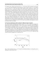
Fig. 5. Bar Code for neurodegenerative diseases. Schematic representation of the
modifications leading to tau proteins aggregation in Tauopathies. Native tau proteins are
detected as a triplet of bands ranging between 60 and 74 kDa by numerous
phosphorylation-dependent antibodies. Tau proteins are shown by western blotting as three
major bands between 60 and 69 kDa, and a minor band at 74 kDa. AD pattern is also found
in Down’s syndrome, post-encephalitic parkinsonism, ALS/parkinsonism–dementia
complex of Guam among others (class I). The doublet tau 64, 69 represent the aggregation of
hyperphosphorylated tau isoforms with exon 10 (orange box) typical for CBP and CBD
(class II), the exclusion of exon 10 (only black boxes) in hyperphosphorylated tau
aggregation lead to tau 60, 64 doublet characteristic for Pick’s disease (class III). The
aggregation of Tau isoforms lacking exons 2 (green box) and 3 (violet box) is found in
myotonic dystrophy (class IV).
The Microtubule-Dissociating Tau in Neurological Disorders
309
Class II: Tau isoforms containing the exon 10 encoding sequence aggregate
Aggregation of tau proteins with four microtubule-binding domains is the characteristic of
class II (figure 5). This pathological tau profile is observed in CBD, argyrophilic grain
dementia, PSP and FTDP-17 due to tau gene mutations (Sergeant N et al., 1999 and Tolnay
M et al., 2002). PSP, CBD and argyrophilic grain dementia are rare atypical parkinsonism
disorders.
Class III: Tau isoforms lacking the exon 10 encoding sequence aggregate
This class of tauopathies includes Pick’s disease and autosomal dominant inherited FDTP-17
(figure 5). Pick’s disease is a rare form of neurodegenerative disorder characterized by a
progressive dementing process. Early in the clinical course, patients show signs of frontal
disinhibition. Neuropathologically, Pick’s disease is characterized by the presence of typical
spheroid inclusions in the soma of neurons called Pick bodies. Pick bodies are labeled by tau
antibodies, with a higher density in neurons of the dentate gyrus of the hippocampal
formation than in the temporal and frontal cortices. The pathological tau profile of Pick’s
disease contrasts with that of class II tauopathies, with the pathological tau isoforms
consisting essentially of the 3R tau isoforms.
Immunohistologic staining of these aggregates is positive for AD2 and exon 2 antibodies but
negative for exon 10 antibodies. In addition, aggregated tau proteins in Pick’s disease are
not detected by the monoclonal antibody 12E8 raised against the phosphorylated residue
Ser262/Ser356, whereas this phosphorylation site is detected in other neurodegenerative
disorders. The lack of phosphorylation at Ser262 and Ser356 sites is likely to be related to
either a kinase is not active in neurons that degenerate in Pick’s disease or those neurons do
not constitutively express these kinases within degenerating neurons (Mailliot C et al., 1998).
Class IV: Tau isoform lacking exon 2, 3 and 10 principally aggregate
This group is represented by a single neurological disorder: myotonic dystrophy (DM) of
types I and II (figure 5). DM is the commonest form of adult-onset muscular dystrophy.
Genetically it is an inherited autosomal dominant disorder caused by a single gene mutation
consisting of expansion of a CTG trinucleotide motif in the 3V untranslated of the myotonic
dystrophy protein kinase gene (DMPK), located on chromosome 19q. It is a multisystemic
disease affecting many systems as the central nervous system (cognitive and
neuropsychiatric impairments), the heart, the genital tract, the eyes, the ears, gastrointestinal
tract, endocrine system, thus leading to a wide and variable complex panel of symptoms
(Meola G, 2000). Cognitive impairments, as memory, visuo-spatial recall and verbal scale,
cortical atrophy essentially of the frontal and the temporal lobe and white matter lesions are
often described in both DM1 and DM2 (Sansone V et al., 2007).
Neuropathological lesions, as neurofibrillary tangles (NFTs), have been observed in adult
DM1 individuals aged over 50 years. The pathological tau profile of DM1 is characterized by
a strong pathological tau band at 60 kDa and, to a lesser extent, a pathological tau
component at 64 and 69 kDa. This typical pathological tau profile is reflected by a reduced
number of tau isoforms expressed in the brain of individuals with DM1, both at the protein
and mRNA levels (Sergeant N et al., 2001). In addition, tau protein expression is also
demonstrated to be altered in transgenic mice with human DM1 locus (Gomes-Pereira M et
al., 2007). Using specific immunological probes against exon 2 and exon 3 corresponding
amino acid sequences, the neurofibrillary lesions were shown to be devoid of tau isoforms
with amino-terminal inserts (Maurage CA et al., 2005). An altered splicing of tau
Proteomics – Human Diseases and Protein Functions
310
characterized by a reduced expression of tau isoforms containing the amino-terminal inserts
characterizes both DM1 and DM2. Overall, it demonstrates that the central nervous system
is affected and that DMs are real tauopathies (Dhaenens CM et al., 2011). The direct
relationship between the altered splicing of tau and NFD in DM remains to be established.
Indeed, such an altered splicing of tau is commonly observed in FTDP- 17 and considered as
reminiscent to NFD and tauopathies.
5. Use of proteomics to investigate the mechanisms leading to Tauopathies
Induction of tau fibrillization in cells remain unsatisfactory, this is a limiting factor since
NFD cannot be totally reproduced in vitro (Sibille N et al., 2006). The development of in vivo
models has provided an important tool to precise sequence of molecular events leading to
tau aggregation. The use of proteomics in these transgenic animals has permitted to go
further in the uncovering of the cellular and molecular pathways involved in NFD
spreading within the brain and its relationship with the clinical expression of neurological
disorders. In this section we will focus on the overexpresion either several isoforms of tau
protein or mutated forms in animal models.
5.1 Tau models
Several animal models have been created to recapitulate the two main hallmarks of AD,
refearing as amyloid plaques and PHFs. Despite the numerous models existing to mimic the
features of this disease, none of them cover all the neuropathological, biochemical and
behaviour alterations so far. There are models focus on overexpression of APP and/or
presenilin containing one or more mutations linked to familial AD but they do not present
NFD. Inspite tau mutations have not been described in AD patients, mutations in tau result
in NFTs in an inherited form of FTDP and this dysfunction can lead to neurodegeneration
and dementia. Taking into account that AD is a complex disorder and the perfect model
does not exist, the large number of tau transgenic models with their strengths and
weaknesses may allow for both understanding tau pathology and developing innovative
therapeutic strategies. Nowadays there are several transgenic models which own
combination of mutant APP, presenilin and tau (Chin J 2011). However, this triple model
presents the “limitation” that tau pathology cannot be studied independently of the amyloid
effects (Sergeant N and Buée L 2011).
5.1.1 Caenorhabditis elegans
The nematode Caenorhabditis elegans is widely being used to study neurodegenerative
disorders despite the evolutionary difference. C. elegans has a short lifespan and it is easy to
manipulate genetically. Modelling tauopathies is achieved through pan-neuronal
overexpression either wild-type or mutated tau leading to a progressive uncoordinated
locomotion which is directly correlated with the nervous system alterations in worms. This
model is very useful to identify new genetic targets (Wolozin B et al., 2011). Recent data
point out that tau pathology may lead to specific interference with intracellular mechanisms
of axonal outgrowth and pathfinding (Brandt R et al., 2009).
5.1.2 Drosophila melanogaster
Another model used is the fruitfly Drosophila melanogaster. Regarding tauopathies, many
groups developed fruitfly models by overexpressing wild-type and mutant forms of human
The Microtubule-Dissociating Tau in Neurological Disorders
311
tau. Transgenic fruitflies showed key features of tauopathies as tissue- and temporal-specific
effects as adult onset, progressive neurodegeneration, early death, enhanced toxicity of
mutant tau, accumulation of abnormal tau and relative anatomic selectivity coupled with
differential effects of distinct tau isoforms (Papanikolopoulou K and Skoulakis EM, 2011).
5.1.3 Zebrafish
The novel use of the vertebrate zebrafish as a model system for AD research offers a
powerful platform for genetic and chemical screens as well as developmental studies
(Tomasiewicz HG et al., 2002). The transgenic expression of the human tau mutation P301L
in zebrafish neurons by Gal4/UAS–based vector system recapitulates most pathological
features of tauopathies as abnormally phosphorylated reactivity with the epitopes AT180,
AT270, 12E8, PHF1, 422, and AT8 in spinal cord neurons, aggregation and behavioral
impairments (Paquet D et al., 2010). Application of inhibitors of human GSK3β reduced tau
phosphorylation showing that zebrafish kinases are sufficiently conserved with respect to
their human orthologues. Current evidence point out that zebrafish tau models recapitulate
pathological and biochemical events that occur in tauopathies and therefore may be useful
tools for further studies in the aetiology of dementia (Bai Q and Burton EA, 2011).
5.1.4 Tau knock out mice and transgenic mice with wild-type human Tau
Tau mouse models where tau expression is suppressed by MAPT deletion or invalidation
present no major changes and animals are physiologically normal (Harada A et al., 1994). It
seems other microtubule-associated proteins such as MAP1A probably compensate tau
deficiency. Among the mice models available with wild-type human tau it is remarkable to
note that overexpression of 3R tau isoforms lead to an accumulation of
hyperphosphorylated tau proteins in spinal cord neurons and axonal degeneration as well
as a reduction in axonal transport (Brion JP et al., 1999). Similar data were observed in
transgenic mice expressing the longest human brain tau isoform under the control of the
human Thy-1 promoter. Hyperphosphorylated human tau protein was present in nerve cell
bodies, axons and dendrites (Gotz J et al., 1995). Furthermore, recent studies in transgenic
mouse models that express the entire human MAPT gene in the presence and absence of the
mouse Mapt gene show differences between mouse and human tau in the regulation of exon
10 inclusion during development and in the young adult. In addition, it was observed
species-specific variations in the expression of 3R- and 4R-tau within the frontal cortex and
hippocampus during the development as well as in cell distribution of the isoforms
(McMillan P et al., 2008).
5.1.5 Transgenic mice with mutated human Tau
Mutated tau transgenes have been used under various promoters (2’,3’-cyclic nucleotide 3’-
phosphodiesterase, CaMKII, PDGF, Prion, or Thy1.2) with or without inducible systems.
The most common phenotype of transgene tau animal is the motor alterations. Tau
transgenic mice rTg4510 present P301L mutation in an inductible way and develop NFTs,
neuronal loss and behavioural impairments (Santacruz K et al., 2005). Nonetheless the
suppression of the expression of this mutated tau revers behavioural impairements despite
the NFTs formation keeps on, indeed it seems soluble tau rather than NFTs may be
deleterious. These observations are in agreement with a recent report in which brain extract
injection from mutant P301S tau expressing mice into brain of transgenic wild-type tau-
Proteomics – Human Diseases and Protein Functions
312
expressing animals induces assembly of wild-type human tau into filaments and spreading
of pathology from the site of injection to neighbouring brain regions (Clavaguera et al.,
2009).
Another transgenic mice model is TauRD/ΔK280 that expresses only the 4R tau domains
and carry the ΔK280 mutation with a deletion of the amino- and carboxy terminal regions of
tau protein. This mutation leads to tau aggregation followed by astrogliosis and neuronal
loss. When the transgene is switched off the aggregation of the exogenous tau disappears
within around one month and a haf and only aggregated murine tau proteins remain acting
as a nucleation factor for tau aggregation (Mocanu MM et al., 2008). Other study suggest a
“prion –like” propagation since aggregation continues even if the original tau species have
disappeared (Sydow A and Mandelkov EM, 2010).
The K3 transgenic mouse strain expresses human tau carrying the K369I mutation under the
Thy1 promoter (Ittner LM et al., 2008). This tau mutation was found in a family of patients
presenting with Pick’s disease without parkinsonism and amyotrophy (Neumann M et al.,
2001). The transgenic mice present early-onset memory impairment and amyotrophy in the
absence of overt neurodegeneration. Tau transgene is mainly expressed in the substantia
nigra and such expression leads to an early-onset parkinsonism phenotype. Interestingly,
motor performance of young, but not old K3 mice improves upon L-dopa treatment.
Amyotrophy is probable to be related to tau expression in the sciatic nerve in the same way
as in Tg30tau model where pathogenic mutations (P301S and G272V) are expressed in the
forebrain and the spinal cord showing progressive motor impairment with neurogenic
muscle atrophy besides the hippocampal atrophy (Leroy K et al., 2007). Moreover,
transgenic mouse model overexpressing human 1N4R double-mutant tau (P301S and
G272V) and invalidated endogenous TAU gene show an accelerated human mutant tau
aggregation (Ando K et al., 2011) suggesting that murine tau proteins may act as inhibitors
of tau aggregation.
Thy-Tau22 mouse transgenic line exhibits progressive neuron-specific AD-like tau
pathology devoid of any motor deficits (Schindowski K et al., 2006). In addition to
neurofibrillary tangle-like inclusions and mild astrogliosis, this model shows hyper- and
abnormally phosphorylated tau on several Alzheimer’s disease-relevant tau epitopes that
accumulates within the somato-dendritic area in the hippocampus (Schindowski K et al.,
2008). A progressive development of NFTs is observed in the hippocampus and amygdala,
which parallels behavioural impairments as well as electrophysiological alterations (Van der
Jeugd et al., 2011). These latter changes are observed despite any striking loss of
neuronal/synaptic markers until 12 months of age in the hippocampus. Interestingly, at that
time point, THY-Tau22 mice exhibit septo-hippocampal tau pathology accompanied by
altered retrograde transport from hippocampus to medial septum (Belarbi K et al., 2009)
with an accumulation of the nerve growth factor (NGF) levels in the hippocampus
consistent with a decrease of its uptake or retrograde transport by cholinergic terminals
(Belarbi K et al., in press). Recent data indicate that voluntary exercise prevented memory
alterations in these transgenic mice and increased mRNA levels of genes involved in
cholesterol trafficking such as NPC1 and NPC2 (Belarbi K et al., 2011).
6. Tau proteins as biomarkers of Tauopathies
Searching for biomarkers is one of the most challenges in current medicine. Biomarkers
must be not only specific for a single pathology but also indicative of its progression
The Microtubule-Dissociating Tau in Neurological Disorders
313
(Mayeux R et al., 2011). This is extremely complex in diseases concerning elderly since many
symptoms are common and indistinguishable among them as the dementia sign. It is
compulsory to find proteins and their post-translational modifications that may provide
accuracy on the early diagnosis of the disease and eventually could serve as a therapeutic
target. Successfully the development in neuroimaging techniques enables to facilitate and
establish a preliminary diagnosis of different neurodegenerative disorders.
Focusing on tauopathies, the presence of tau in CSF was first described in 1993. In AD, tau
inclusions in the brain associated with neuronal damages lead to the leakage of abnormal
forms of tau in the CSF resulting in quantitative and qualitative changes in CSF-tau
composition. Numerous studies demonstrated increased CSF total tau and phosphorylated
tau levels in AD, with mean levels 2-3 times higher compared to healthy controls. Tau is
now a validated biomarker for AD, it improves the clinical diagnostic accuracy and its
assessment for AD diagnosis is now proposed (Dubois B et al., 2010). As the brain lesions
develop very early during the disease course even before the first clinical symptoms appear,
CSF tau is not only a useful diagnostic marker in the advanced stages of the disease but also
a usefull predictive marker in the earliest stages when clinical expression is weak (Hertze J
et al., 2010). However, for differential diagnosis of dementia, the actually available tests
measuring tau and phosphorylated tau levels in CSF are not sufficient and the identification
of more specific postranslational modifications of tau in AD by proteomic approaches is
needed. In the future, for the use of tau as biomarker in large clinical trials or in clinical
practice, one important goal will be to develop sensitive methods to detect the very low
concentration of tau in the blood (<1 pMol). Therefore, sample pre-treatment and handling
will be crucial in developing a reliable tau assay in blood/plasma.
7. Conclusion
Tau is a neuronal protein that promotes neuronal survival, it is essentially located within the
axonal and indispensable for the organization, stabilization, and dynamics of microtubules.
The interaction between tau and microtubules is regulated by phosphorylation. It is widely
reported that abnormally and hyperphosphorylated tau proteins lead to insoluble
aggregates. The presence of these aggregates is clinically correlated with cognitive decline in
a process called NFD; this event common to more than twenty diseases is referred as
tauopathies.
The development of the proteomics era has achieved to go further in the characterization of
tauopathies and shed light to the mechanism involved in their aetiology. Proteomics
approaches as chromatography, mono- and bi-dimensional gel electrophoresis have reached
to separate proteins with a quite high resolution after fractioning precedures, selecting a
concrete population of cells or organelle isolation. The use of additional reagents to the
extraction buffer such as detergents and the evolution of concomitant technologies as
microscopy have provided a broad spectrum to characterize the structure and size of a large
number of biological complex samples. The combination of protein separation methods with
fluorescence dyes and radioactive isotopes (ICAT, iTRAQ, SILAC) makes possible not only
more sensitive and reproducible results but also provides a quantitative analysis among
samples (2D-DIGE, LC-MS, SELDI).
The previous hallmark is extremely linked to the identification of the separated or isolated
proteins. MS has provided the composition of the molecules and also their post-translational
modifications since changes in amino acid residues may be identified and characterized by