NOMENCLATURE AND STANDARD REPORTING TERMINOLOGY OF INTERVERTEBRAL DISK HERNIATION
Bạn đang xem bản rút gọn của tài liệu. Xem và tải ngay bản đầy đủ của tài liệu tại đây (796.6 KB, 24 trang )
<span class="text_page_counter">Trang 1</span><div class="page_container" data-page="1">
<b>Nomenclature and Standard Reporting Terminology of Intervertebral Disk Herniation </b>
<i><b>Author: Richard F. Costello, Douglas P. Beall </b></i>
<i>Magnetic Resonance Imaging Clinics of North America. Volume 15, Issue 2, May 2007, Pages 167-174 - doi:10.1016/j.mric.2006.12.001 </i>
<b>Danh pháp và thuật ngữ viết báo cáo chuẩn của thoát vị đĩa đệm </b>
<i><b>Người dịch: BS Cao Thiên Tượng - radiocr.vn </b></i>
<i><b>Biên tập song ngữ: HÌNH ẢNH Y KHOA - hinhanhykhoa.com </b></i>
<i><b>Abstract </b></i>
<i>Spine pathology is ubiquitous and is encountered by nearly all medical specialties. The anatomy of the spine is complex, but the language used to describe pathology may be even more complex. Many of the common references differ in their nomenclature used to report intervertebral disk herniation. This article summarizes and relates the standard recommendations for reporting terminology in regard to herniation of the intervertebral disk. This standard reporting terminology may be used with CT or MR imaging and is useful to report the location and size of the disk herniation. The diagnostician must also be aware of the various pitfalls associated with disk herniation to avoid the scenario of surgical intervention at the incorrect spinal level. </i>
<i><b>Tóm tắt </b></i>
<i>Bệnh lý cột sống rất phổ biến và gặp ở gần như ở tất cả các chuyên khoa. Giải phẫu cột sống phức tạp, nhưng ngôn ngữ sử dụng để mô tả bệnh lý có thể cịn phức tạp hơn. Nhiều tham khảo thông thường khác nhau về danh pháp dùng để viết báo cáo thốt vị đĩa đệm. Bài này tóm tắt và đưa ra các khuyến cáo chuẩn để viết báo cáo các thuật ngữ liên quan đến thoát vị đĩa đệm. Thuật ngữ viết báo cáo chuẩn này có thể sử dụng cho CT và MRI và giúp báo cáo vị trí và kích thước thốt vị đĩa đệm. Các nhà chẩn đoán cần phải nhận thấy nhiều cạm bẫy liên quan đến thoát vị đĩa đệm để tránh trường hợp can thiệp phẫu thuật ở tầng cột sống không đúng. </i>
</div><span class="text_page_counter">Trang 2</span><div class="page_container" data-page="2"><b>Introduction </b>
Anatomic derangement of the spine is extremely common and may be seen in symptomatic and asymptomatic individuals. The terminology used to describe intervertebral disk herniations is complex and many of the common references differ in their nomenclature used to report disk pathology. The nomenclature contained in this article is based on recommendations from The North American Spine Society, The American Society of Neuroradiology, and The American Society of Spine Radiology [1].
The intent of the recommendations are to standardize the reporting of the size and location of intervertebral disk lesions and to simplify the anatomic descriptions of findings on CT and MR imaging. Recommendations are to report abnormalities in zones on axial images and in levels on the sagittal and coronal images. The size of the herniation may be described in words or numbers. The terminology may be used in the cervical and thoracic spine, but was primarily developed for use in the lumbar spine.
<b>Mở đầu </b>
Xáo trộn giải phẫu cột sống rất thường gặp và có thể thấy ở người có triệu chứng cũng như khơng có triệu chứng. Thuật ngữ dùng để mơ tả thoát vị đĩa đệm phức tạp và nhiều tham khảo thông thường khác nhau về danh pháp dùng để báo cáo bệnh lý thoát vị đĩa đệm. Danh pháp trong bài này dựa trên khuyến cáo của hội cột sống Bắc Mỹ, hội X quang thần kinh Mỹ và hội X quang cột sống Mỹ.
Mục đính của khuyến cáo là chuẩn hóa viết báo cáo về vị trí và kích thước của tổn thương đĩa đệm và làm đơn giản hóa việc mơ tả giải phẫu các dấu hiệu trên CT và MRI. Các khuyến cáo là để báo cáo các bất thường về các vùng trên hình ảnh cắt ngang và các tầng trên hình sagittal và coronal. Kích thước của thốt vị có thể mơ tả theo từ ngữ hoặc số lượng. Thuật ngữ này có thể dùng trong cột sống cổ và ngực, nhưng chủ
<i>yếu đưa ra để dùng trong cột sống thắt lưng. </i>
</div><span class="text_page_counter">Trang 3</span><div class="page_container" data-page="3"><b>Discussion </b>
There are 23 intervertebral disks between adjacent vertebral bodies from C2 to S1. The disks are thicker anteriorly in the cervical and lumbar spine, which contributes to the lordosis normally seen in these regions of the spine. In the thoracic spine the intervertebral disks are uniform in thickness. The intervertebral disks contribute approximately one fourth of the length to the vertebral column.
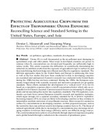
The normal intervertebral disk has an inner nucleus pulposus and an outer annulus fibrosus and is bordered by the cartilage end plates (Fig. 1). The nucleus pulposus contains hydrophilic glycosaminoglycans with a lattice of collagen fibers. Over time the gelatinous glycosaminoglycans is replaced by fibrocartilage [2]. The annulus fibrosus contains 15 to 20 collagenous laminae that are organized obliquely to one another and are weakest posterolaterally where the collagen bundles are less organized. The cartilage end plates are composed of hyaline cartilage and are the anatomic limit of the disk with the posterior portion of disk forming part of the anterior wall of the neural foramen. The intervertebral disc is avascular and aneural and obtains its nutrition by diffusion from the adjacent vertebral body and end plates.
<b>Bàn luận </b>
Có 23 đĩa đệm giữa các thân đốt sống từ C2 đến S1. Đĩa đệm dày hơn về phía trước ở cột sống cổ và cột sống thắt lưng. Ở cột sống ngực, đĩa đệm có độ dày như nhau. Các đĩa đệm góp một phần tư chiều dài cột sống.
Đĩa đệm bình thường có nhân đĩa bên trong và vịng xơ bên ngồi, và được bao bọc bởi sụn bề mặt thân sống (hình 1). Nhân đĩa chứa các glycosaminoglycans ái nước với một mạng lưới sợi collagen. Theo thời gian, các lycosaminoglycans gelatin bị thay thế bởi sụn xơ. Vòng xơ gồm 15-20 lá collagen được sắp xếp chéo nhau và yếu nhất ở phía sau ngồi, ở đó các bó collagen được sắp xếp ít hơn. Sụn bề mặt thân sống bao gồm sụn trong, vô mạch và khơng có thần kinh, được ni dưỡng bằng sự khuếch tán từ thân sống và bề mặt thân sống kế cận.
</div><span class="text_page_counter">Trang 4</span><div class="page_container" data-page="4"><b>Fig. 1. Axial (A) and sagittal (B) T2-weighted MR images demonstrate </b>
the increased signal of the normal nucleus pulposus (N) and decreased signal of the normal annulus fibrosis (A)
<b> Hình 1. Axial (A) và Sagittal (B) T2W thấy tăng tín hiệu của nhân đĩa </b>
bình thường (N) và giảm tín hiệu của vịng xơ bình thường (A).
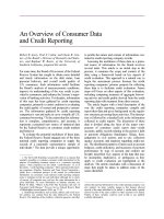
</div><span class="text_page_counter">Trang 5</span><div class="page_container" data-page="5">An annular tear is a concentric, radial, or transverse separation of the annular fibers. Annular tears ( Fig. 2) can be found in a minority of individuals over 40 years of age and are considered pathologic and a precursor to intervertebral disk herniation. Most annular tears are not visible on MR imaging, but those tears that are visible demonstrate a high intensity zone that is often crescent shaped and is commonly seen in the posterior portion of the disk at L4-5 and L5-S1. Radial tears demonstrate disruption perpendicular to the axis of the collagen fibers. The disruption extends from the gelatinous nucleus pulposus through the annulus fibrosis to the periphery of the disk and may be painful, especially if the tear is located immediately adjacent to the dorsal nerve root ganglion. The dorsal nerve root ganglion gives rise to the sinuvertebral nerve that innervates the posterolateral portion of the intervertebral disk. Tears of the annulus fibrosis to the periphery of the disk also allow nerve and granulation tissue ingrowth into the disk. Concentric tears are disruption parallel to the axis of the collagen fibers. This forms a high intensity zone between adjacent collagen fibers. The significance of an annular tear is controversial and the presence may not correlate with the need for treatment or with clinical symptoms [3].
Rách vòng xơ là sự tách đồng tâm, tỏa vòng hoặc ngang của vòng xơ. Rách vịng xơ (hình 2) có thể gặp ở một số ít ngưới trên 40 tuổi và được xem như là bệnh lý và là yếu tố dự báo thốt vị đĩa đệm. Hấu hết rách vịng xơ khơng nhìn thấy trên MRI, nhưng rách nhìn thầy được là vùng tín hiệu cao thường có hình liềm và thường gặp ở phần sau đĩa đệm L4-L5 và L4-L5-S1. Rách tỏa vòng là sự phá vỡ vng góc với trục các sợi collagen. Sự phá vỡ này lan từ nhân collagen đến vòng xơ ở phía ngoại vi đĩa đệm và có thể gây đau nếu rách nằm ngay cạnh hạch rễ thần kinh phía lưng. Hạch rễ thần kinh lưng cho ra dây thần kinh xoang cột sống, dẫn truyền thần kinh phần sau bên của đĩa đệm. Rách đồng tâm là sự phá vỡ song song với trục các sợi collagen. Điều này hình thành nên vùng tín hiệu cao giữa các sợi collagen. Ý nghĩa của rách vòng xơ còn tranh luận và khi có có thể khơng liên quan với nhu cầu điều trị hoặc với các triệu chứng lâm sàng.
</div><span class="text_page_counter">Trang 6</span><div class="page_container" data-page="6"><b>Fig. 2. Axial (A) and sagittal (B) T2-weighted MR images demonstrate </b>
a crescentic area (arrows) of increased signal (high intensity zone) in the posterior portion of the disk at L4-5. This is characteristic appearance of an annular tear.
<b> Hình 2. Axial (A) và sagittal (B) T2W thấy một vùng hình liềm (mũi </b>
tên) tăng tín hiệu (vùng tăng tín hiệu) ở phần sau củ đĩa đệm L4-L5. Đây là hình ảnh đặc trưng của rách vòng xơ.
</div><span class="text_page_counter">Trang 7</span><div class="page_container" data-page="7">Herniation is the term used most commonly to describe the displacement of disk material. This can involve displacement of the nucleus pulposus, end plate cartilage, fragmented apophyseal bone, or annular tissue beyond the normal confines of the disk space. Intervertebral disk herniations may occur centrifugally out from the periphery of the disk or in a superior or inferior direction. A disk herniation is a localized (involving less than 50% of the circumference of the disk or less than 180<sup>o</sup> of the periphery of the disk) displacement of disk material beyond the normal confines of the disk. This is in contrast to a disk bulge that involves more than half of the circumference (more than 180<sup>o</sup> of the disk) or the entire disk. Herniation is a general term that can be further characterized as a protrusion or extrusion based on the shape and amount of the displaced disk material [4].
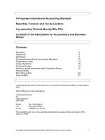
A herniated disk is a protrusion if the greatest distance of the edges of the protruded material is less than the edges of the normal disk material as determined on the same plane. For example, if the greatest superoinferior distance of the edges of the herniated disk material is less than or equal to the edges of the normal disk in the same plane (as measured on the sagittal or coronal plane to assess superoinferior extension), then this is classified as a disk protrusion (Fig. 3). The superoinferior measurement is nearly always best determined on the sagittal plane. A protrusion, as determined on the axial plane, is a herniated portion of the disk that involves less than half of the disk (<180<sup>o</sup> of the disk circumference). Protrusions may be further classified as focal or broad, based on their degree of disk involvement. A focal protrusion involves less than 25% (or 90<small>o</small>) of disk circumference, whereas broad-based protrusions involve between 25% and 50% (90 – 180<small>o</small>) of the disk circumference (Fig. 4).
Thoát vị là thuật ngữ thường dùng nhiều nhất để mô tả sự di lệch của chất đĩa đệm. Điều này có thể liên quan với sự di lệch nhân đĩa sụn bề mặt thân sống, mấu xương bị phân mảnh hoặc mơ vịng xơ vượt khỏi giới hạn bình thường của khoang đĩa đệm. Thốt vị đĩa đệm có thể gặp dạng ly tâm ra phía ngoại vi đĩa ệm, hoặc hướng lên hoặc hướng xuống. Thoát vị đĩa đệm là một sự di lệch khu trú của đĩa đệm ra xa khỏi giới hạn đĩa đệm bình thường (ít hơn 50% chu vi đĩa đệm hoặc ít hơn 180 độ phía ngoại vi đĩa đệm). Điều này ngược với lồi đĩa đệm là liên quan với hơn một nửa chu vi (hơn 180 độ của đĩa đệm) hoặc tồn bộ đĩa đệm. Thốt vị là thuật ngữ chung có thể xác định đặc điểm thêm là protrusion hoặc extrusion dựa vào hình dáng và lượng chất đĩa đệm bị di lệch.
Thoát vị là protrusion nếu khoảng cách lớn nhất của các bờ chất bị lồi nhỏ hơn các bờ của chất đĩa đệm bình thường khi xác định trên cùng một mặt phẳng. Ví dụ, nếu khoảng cách trên dưới của các bờ chất đĩa đệm bị thoát vị nhỏ hơn hoặc bằng các bờ của chất đĩa đệm bình thường trên cùng một mặt phẳng (khi đo ở mặt cắt sagittal hoặc coronalđể đánh giá độ lan trên dưới), thì nó được phân loại là protrusion (hình 3). Đo chiều trên dưới gần như luôn tốt nhất ở mặt phẳng sagittal. Lồi khu trú khi xác định trên mặt phẳng axial là phần thốt vị liên quan với ít hơn một nửa đĩa đệm (< 180 độ chu vi đĩa đệm). Protrusion có thể được phân loại thêm thành khu trú hoặc đáy rộng dựa vào mức độ đĩa đệm bị tổn thương. Protrusion khu trú liên quan với ít hơn 25% (hoặc 90 độ ) của chu vi đĩa đệm,trong khi protrusion đáy rộng nằm giữa 25% đến 50% (90-180 độ) của chu vi đĩa đệm (hình 4).
</div><span class="text_page_counter">Trang 8</span><div class="page_container" data-page="8"><b>Fig. 3. Sagittal T2-weighted MR image of the lumbar spine </b>
demonstrates a disk protrusion at the L5-S1 level ( white arrow). Note that the protruded disk material does not extend beyond the confines of the superior and inferior disk borders (ie, beyond the disk/end plate junction). As defined on the sagittal plane, this disk herniation may be described as a protrusion.
<b> Hình 3. Hình sagittal T2W mơ tả protrusion đĩa đệm L5-S1 (mũi tên </b>
trắng). Ghi nhận chất đĩa đệm thốt vị khơng vượt qua giới hạn của bờ trên và bờ dưới đĩa đệm (tức là vượt qua chỗ nối đĩa đệm/bề mặt thân sống). Khi xác định trên mặt cắt sagittal, thoát vị đĩa đệm này là protrusion.
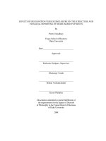
</div><span class="text_page_counter">Trang 9</span><div class="page_container" data-page="9"><b>Fig. 4. Axial T2-weighted MR images taken through the L4-5 </b>
intervertebral disk show disk protrusions. Focal (A) and broad-based disk (B) protrusions are outlined by the dotted white line. A focal protrusion involves less than 25% (or 90<sup>o</sup>) of disk circumference, whereas broadbased protrusions involve between 25% and 50% (90 – 180<sup>o</sup>) of the disk circumference.
<b> Hình 4. axial T2w qua đĩa đệm L4-5 cho thấy protrusion đĩa đệm. </b>
Protrusion khu trú (A) và đáy rộng (B) được vẽ bởi đường gạch chấm trắng. Protrusion khu trú liên quan ít hơn 25% (hoặc 90 độ) của chu vi đĩa đệm, trong khi protrusion đáy rộng nằm trong khoảng 25% đến 50% (90-180 độ) của chu vi đĩa đệm.
</div><span class="text_page_counter">Trang 10</span><div class="page_container" data-page="10">In contrast to a protrusion, a herniated disk is an extrusion if the greatest distance of the edges of the herniated material is greater than the distance between the edges of the base of the herniation as measured on the same plane. For example, if disk material is displaced beyond the confines of the superior or inferior border of the disk or end plates as seen on the sagittal plane, this would constitute a disk extrusion (Fig. 5). If the extruded disk material has a narrow neck and a wider extruded portion as seen on the axial plane, this would also be best described as a disk extrusion (Fig. 6). In contrast, a protrusion would not extend beyond the superior or inferior border of the intervertebral disk on the sagittal images and would have a broad-based neck with a protruded portion of the disk that was not as wide as the neck of the protrusion (see Figs. 5 and 6).
Ngược với protrusion, thoát vị đĩa đệm là extrusion nếu khoảng cách lớn nhất giữa các bờ của chất bị thoát vị lớn hơn các bờ của đáy thoát vị khi đo trên cùng một mặt phẳng. Ví dụ, nếu chất bị thoát vị vượt qua giới hạn của bờ trên hoặc bờ dưới đĩa đệm hay bề mặt thân sống thấy trên mặt phẳng sagittal thì đây là extrusion đĩa đệm (hình 5). Nếu chất đĩa đệm bị extrusion có cổ hẹp và phần thốt vị rộng hơn khi thấy trên mặt phẳng axial thì đó cũng là một minh hoạ rõa nhất cho extrusion (hình 6). Ngược lại, protrusion thì khơng thì vuợt qua bờ trên và bờ dưới đĩa đệm trên hình sagittal và có thể có cổ đáy rộng với phần protrusion của đĩa đệm không rộng như cổ của protrusion (xem hình 5, hình 6).
</div><span class="text_page_counter">Trang 11</span><div class="page_container" data-page="11"><b>Fig. 5. Sagittal MR images of the lumbar spine demonstrate normal </b>
intervertebral discs (A), a disk protrusion at L5-S1 (arrow) (B), disk extrusion with inferior displacement at L5-S1 ( arrow) (C). The superior and inferior borders of the L5-S1 intervertebral disks are indicated by the solid white lines in B and C. Protrusions do not extend beyond the superoinferior dimension of the disk in the sagittal plane (B), whereas extrusions extend beyond this border (C).
<b> Hình 5. Hình MRI sagittal cột sống thắt lưng cho thấy đĩa đệm bình </b>
thường (A), protrusion L5-S1 (B), extrusion kèm di lệch xuống dưới ngang mức L5-S1 (mũi tên) (C). Các đường trắng chỉ ra bờ trên và bờ dưới đĩa đệm L5-S1 ở hình B và C. Protrusion khơng vượt qua chiều trên – dưới đĩa đệm trên hình sagittal (B), trong khi extrusion vượt qua bờ này (C).
</div><span class="text_page_counter">Trang 12</span><div class="page_container" data-page="12"><b>Fig. 6. Axial T1-weighted MR images of the lumbar spine at the L4-5 </b>
level show posterior disk herniations. (A) The herniation (white dotted line) shows a wider neck (white arrows); (B) herniation (white dotted line) shows a narrow neck (white arrows).
<b> Hình 6. Hình axial T1W của cột sống thắt lưng ngang mức L4-5 cho </b>
thấy thoát vị đĩa đệm ra sau. (A) thoát vị (đường chấm trắng) thấy cổ rộng hơn (các mũi tên trắng); (B) thoát vị (đường chấm trắng) thấy cổ hẹp (các mũi tên trắng)
</div>