Atomic Force Microscopy in Cell Biology_Methods in Cell Biology Volume 68 pptx
Bạn đang xem bản rút gọn của tài liệu. Xem và tải ngay bản đầy đủ của tài liệu tại đây (10.29 MB, 430 trang )
Methods in Cell Biology
VOLUME 68
Atomic Force Microscopy in Cell Biology
Series Editors
Leslie Wilson
Department of Biological Sciences
University of California, Santa Barbara
Santa Barbara, California
Paul Matsudaira
Whitehead Institute for Biomedical Research and
Department of Biology
Massachusetts Institute of Technology
Cambridge, Massachusetts
Methods in Cell Biology
Prepared under the Auspices of the American Society for Cell Biology
VOLUME 68
Atomic Force Microscopy in Cell Biology
Edited by
Bhanu P. Jena
Department of Physiology and Pharmacology
Wayne State University School of Medicine
Detroit, Michigan
J. K. Heinrich H ¨orber
Cell Biology and Biophysics Program
European Molecular Biology Laboratory
Heidelberg, Germany
Amsterdam Boston London New York Oxford Paris
San Diego San Francisco Singapore Sydney Tokyo
Paperback edition cover photo credit: The image is the surface topology
of the apical plasma membrane in live pancreatic acinar cell, depicting
fusion pores (dark circles). Courtesy of Dr. Bhanu P. Jena, Departments
of Physiology & Pharmacology, Wayne State University School of
Medicine, Detroit, MI, USA.
This book is printed on acid-free paper.
∞
Copyright
C
2002, Elsevier Science (USA).
All Rights Reserved.
No part of this publication may be reproduced or transmitted in any form or by any
means, electronic or mechanical, including photocopy, recording, or any information
storage and retrieval system, without permission in writing from the Publisher.
The appearance of the code at the bottom of the first page of a chapter in this book
indicates the Publisher’s consent that copies of the chapter may be made for
personal or internal use of specific clients. This consent is given on the condition,
however, that the copier pay the stated per copy fee through the Copyright Clearance
Center, Inc. (222 Rosewood Drive, Danvers, Massachusetts 01923), for copying
beyond that permitted by Sections 107 or 108 of the U.S. Copyright Law. This consent
does not extend to other kinds of copying, such as copying for general distribution, for
advertising or promotional purposes, for creating new collective works, or for resale.
Copy fees for pre-2002 chapters are as shown on the title pages. If no fee code
appears on the title page, the copy fee is the same as for current chapters.
0091-679X/2002 $35.00
Explicit permission from Academic Press is not required to reproduce a maximum of
two figures or tables from an Academic Press chapter in another scientific or research
publication provided that the material has not been credited to another source and that
full credit to the Academic Press chapter is given.
Academic Press
An imprint of Elsevier Science.
525 B Street, Suite 1900, San Diego, California 92101-4495, USA
Academic Press
84 Theobalds Road, London WC1X 8RR, UK
International Standard Book Number: 0-12-544171-1 (hb)
International Standard Book Number: 0-12-383851-7 (pb)
PRINTED IN THE UNITED STATES OF AMERICA
020304050607MM987654321
CONTENTS
Contributors xi
Preface xiii
1. Local Probe Techniques
J. K. Heinrich H
¨
orber
I. Introduction 1
II. Scanning Tunneling Microscopy 4
III. Atomic Force Microscopy 7
IV. Force Spectroscopy 13
V. Photonic Force Microscopy 21
References 30
2. The Atomic Force Microscope in the Study of Membrane Fusion
and Exocytosis
Bhanu P. Jena and Sang-Joon Cho
I. Introduction 33
II. Methods 35
III. AFM Studies on Live Cells 37
IV. Identification of New Plasma Membrane Structures Involved in Exocytosis 39
V. Future of AFM in the Study of Live Cells 47
References 48
3. Atomic Force Microscope Imaging of Cells and Membranes
Eric Lesniewska, Pierre Emmanuel Milhiet, Mar ie-C
´
ecile Giocondi,
and Christian Le Grimellec
I. Introduction 52
II. AFM Equipment 52
III. AFM Operating Modes 53
IV. Requirements for the Imaging of Intact Cells 53
V. Imaging of Cells 56
VI. Imaging of Isolated Membranes 63
VII. Conclusion and Perspectives 63
References 64
v
vi Contents
4. Measuring the Elastic Properties of Living Cells by the Atomic
Force Microscope
Manfred Radmacher
I. Introduction 67
II. Principles of Measurement 70
III. Application to Cells 74
IV. Mechanics of Cellular Dynamics 84
V. Summary 86
References 87
5. Cell Adhesion Measured by Force Spectroscopy on Living Cells
Martin Benoit
I. Introduction 91
II. Instrumentation 92
III. Preparations of the Force Sensor for Measurements with Living Cells 94
IV. Cell Culture 109
V. Final Remarks 110
References 111
6. Molecular Recognition Studies Using the Atomic Force Microscope
Peter Hinterdorfer
I. Introduction 115
II. Experimental Approach 117
III. Dynamic Force Spectroscopy 124
IV. Recognition Imaging 133
References 137
7. The Biophysics of Sensory Cells of the Inner Ear Examined by Atomic
Force Microscopy and Patch Clamp
Matthias G. Langer and Assen Koitschev
I. Introduction 142
II. Morphology and Function of Cochlear Hair Cells 143
III. AFM Technology 147
IV. Applications 155
V. Discussion 165
VI. Outlook 166
References 167
8. Biotechnological Applications of Atomic Force Microscopy
Guillaume Charras, Petri Lehenkari, and Mike Horton
I. Introduction 172
II. Methods 174
Contents vii
III. Analysis 178
IV. Application Examples 182
V. Future Directions and Improvements 187
References 190
9. Cellular Membranes Studied by Photonic Force Microscopy
Arnd Pralle and Ernst-Ludwig Florin
I. Introduction 193
II. Photonic Force Microscopy 194
III. Experimental Considerations 199
References 211
10. Methods for Biological Probe Microscopy in Aqueous Fluids
Johannes H. Kindt, John C. Sitko, Lia I. Pietrasanta, Emin Oroudjev,
Nathan Becker, Mario B. Viani, and Helen G. Hansma
I. Introduction 214
II. Substrates/Surfaces 215
III. Basic Methods for Atomic Force Microscopy in Aqueous Fluids 215
IV. Molecular Force Probing 223
V. Advanced Fluid Handling 225
VI. Conclusion 228
References 228
11. Supported Lipid Bilayers as Effective Substrates for Atomic
Force Microscopy
Daniel M. Czajkowsky and Zhifeng Shao
I. Introduction 231
II. Preparation of the Supported Bilayer Substrates 232
III. Examples of Applications 236
IV. Summary 240
References 240
12. Cryo-Atomic Force Microscopy
Sitong Sheng and Zhifeng Shao
I. Introduction 243
II. Designs and Instrumentation 244
III. Applications in Structural Biology 248
IV. Deep Etching as the Preferred Sample Preparation Method 252
V. New Directions 253
References 254
viii Contents
13. Conformations, Flexibility, and Interactions Observed on Individual
Membrane Proteins by Atomic Force Microscopy
Daniel J. M
¨
uller and Andreas Engel
I. Introduction 258
II. High-Resolution AFM Imaging 260
III. Identification of Membrane Proteins 264
IV. Observing the Oligomerization of Membrane Proteins 270
V. Unraveling the Conformational Variability of Membrane Proteins 272
VI. Comparing AFM Topographs to Atomic Models 275
VII. Conformational Changes of Native Membrane Proteins 278
VIII. Observing the Assembly of Membrane Proteins 285
IX. Detecting Intra- and Intermolecular Forces of Proteins 287
X. Conclusions and Perspectives 289
References 292
14. Single-Molecule Force Measurements
Aileen Chen and Vincent T. Moy
I. Introduction 301
II. Experimental Design 302
III. Applications 306
References 308
15. Forced Unfolding of Single Proteins
S. M. Altmann and P F. Lenne
I. Introduction 312
II. The Biological System 313
III. Forced Unfolding 317
IV. Analysis 321
V. Models 324
VI. Conclusions and Prospects 328
VII. Appendices 328
References 334
16. Developments in Dynamic Force Microscopy and Spectroscopy
A. D. L. Humphris and M. J. Miles
I. Introduction 337
II. Active Q Control 340
III. Application of Active Q-Control AFM 344
IV. Transverse Dynamic Force Techniques 351
V. Conclusions 354
References 354
Contents ix
17. Scanning Force Microscopy Studies on the Structure and Dynamics
of Single DNA Molecules
Giampaolo Zuccheri and Bruno Samor
`
ı
I. Introduction 358
II. The Control of Adsorption of DNA on Surfaces 359
III. Air Imaging of DNA: Which Present, Which Future? 366
IV. Imaging DNA in Fluid 370
V. DNA Manipulation with the SFM: The Controlled Dissection
of DNA 377
VI. The Study of DNA Conformations and Mechanics:
Curvature and Flexibility 379
VII. An Interesting Issue: The Shape of Supercoiled DNA 385
VIII. Conclusions and Perspectives 388
References 389
Index 397
Volumes in Series 409
This Page Intentionally Left Blank
CONTRIBUTORS
Numbers in parentheses indicate the pages on which authors’ contributions begin.
S. M. Altmann (311), Cell Biology and Biophysics Program, European Molecular
Biology Laboratory, D-69117 Heidelberg, Germany
Nathan Becker (213), Department of Physics, University of California, Santa Barbara,
Santa Barbara, California 93106
Martin Benoit (91), Center for Nanoscience, Ludwig-Maximilians-Universität
München, D-80799 Munchen, Germany
Guillaume Charras (171), Bone and Mineral Center, Department of Medicine, The
Rayne Institute, University College London, London WC1E 6JJ, United Kingdom
Aileen Chen (301), Department of Physiology and Biophysics, University of Miami
School of Medicine, Miami, Florida 33136
Sang-Joon Cho (33), Department of Physiology and Pharmacology, Wayne State Uni-
versity School of Medicine, Detroit, Michigan 48201
Daniel M. Czajkowsky (231), Department of Molecular Physiology and Biological
Physics, University of Virginia School of Medicine, Charlottesville, Virginia 22908
Andreas Engel (257), M. E. Müller Institute, Biocenter, University of Basel, CH-4056
Basel, Switzerland
Ernst-Ludwig Florin (193), Cell Biology and Biophysics Program, European Molecular
Biology Laboratory, D-69117 Heidelberg, Germany
Marie-C´ecile Giocondi (51), Center of Structural Biochemistry, French National
Institute for Health and Medical Research U414, 34090 Montpellier Cedex, France
Christian Le Grimellec (51), Center of Structural Biochemistry, French National
Institute for Health and Medical Research U414, 34090 Montpellier Cedex, France
Helen G. Hansma (213), Department of Physics, University of California, Santa
Barbara, Santa Barbara, California 93106
Peter Hinterdorfer (115), Institute for Biophysics, University of Linz, A-4040 Linz,
Austria
J. K. Heinrich H ¨orber (1), Cell Biology and Biophysics Program, European Molecular
Biology Laboratory, D-69117 Heidelberg, Germany
Mike Horton (171), Bone and Mineral Center, Department of Medicine, The Rayne
Institute, University College London, London WC1E 6JJ, United Kingdom
A. D. L. Humphris (337), H. H. Wills Physics Laboratory, University of Bristol, Bristol
BS8 1TL, United Kingdom
Bhanu P. Jena (33), Department of Physiology and Pharmacology, Wayne State Uni-
versity School of Medicine, Detroit, Michigan 48201
Johannes H. Kindt (213), Department of Physics, University of California, Santa
Barbara, Santa Barbara, California 93106
xi
xii Contributors
Assen Koitschev (141), Department of Otorhinolaryngology, Universität Tübingen,
D-72076 Tübingen, Germany
Matthias G. Langer (141), Division of Sensory Biophysics, Universität Tübingen,
D-72076 Tübingen, Germany
*
Petri Lehenkari (171), Departments of Surgery and Anatomy, Univer sity of Oulu, FIN-
90014 Oulu, Finland
P F. Lenne (311), Cell Biology and Biophysics Program, European Molecular Biology
Laboratory, D-69117 Heidelberg, Germany
Eric Lesniewska (51), Laboratory of Physics, National Center for Scientific Research
URA 5027, UFR Sciences et Techniques, 21078 Dijon Cedex, France
M. J. Miles (337), H. H. Wills Physics Laboratory, University of Bristol, Bristol BS8
1TL, United Kingdom
Pierre Emmanuel Milhiet (51), Center of Structural Biochemistry, French National
Institute for Health and Medical Research U414, 34090 Montpellier Cedex, France
Vincent T. Moy (301), Department of Physiology and Biophysics, University of Miami
School of Medicine, Miami, Florida 33136
Daniel J. M ¨uller (257), Max-Planck-Institute of Molecular Cell Biology and Genetics,
D-01097 Dresden, Germany
Emin Oroudjev (213), Department of Physics, University of California, Santa Barbara,
Santa Barbara, California 93106
Lia I. Pietrasanta (213), Department of Physics, University of California, Santa Barbara,
Santa Barbara, California 93106
Arnd Pralle (193), Cell Biology and Biophysics Program, European Molecular Biology
Laboratory, D-69117 Heidelberg, Germany
†
Manfred Radmacher (67), Drittes Physics Institute, Georg-August Universität, 37073
Göttingen, Germany
‡
Bruno Samor
`
ı (357), Department of Biochemistry, University of Bologna, 40126
Bologna, Italy
Zhifeng Shao (231, 243), Department of Molecular Physiology and Biological Physics,
University of Virginia School of Medicine, Charlottesville, Virg inia 22908
Sitong Sheng (243), Department of Molecular Physiology and Biological Physics, Uni-
versity of Virginia School of Medicine, Charlottesville, Virg inia 22908
John C. Sitko (213), Department of Physics, University of California, Santa Barbara,
Santa Barbara, California 93106
Mario B. Viani (213), Depar tment of Physics, University of California, Santa Barbara,
Santa Barbara, California 93106
Giampaolo Zuccheri (357), Department of Biochemistry, University of Bologna, 40126
Bologna, Italy
*
Present address: HNO-Klinik, D-72076 T¨ubingen, Germany
†
Present address: Department of Molecular Cell Biology, University of California, Berkeley, Berkeley,
California 94720
‡
Present address: Department 1, Universit t Bremen, D-28359 Bremen, Germany
PREFACE
In the last decade, the atomic force microscope (AFM) has emerged as a powerful tool
for cell biology research giving ultrahigh resolution in real time under near physiolog-
ical conditions. Studies revealing nanometer-scale details of the living cell, subcellular
organelles, and biomolecules, previously impossible due to the resolution limits of light
microscopes, are now accessible using the AFM. Pioneering work and instrumental de-
velopment were carried out by the groups of Paul Hansma, University of California,
Santa Barbara; and Gerd Binnig, IBM Physics, Munich. For the first time, in 1989,
Binnig visualized the process of pox virus release on living cells. Meanwhile, many
other groups contributed exciting new insights at the cellular and molecular levels using
the AFM. This book contains examples of more recent studies done with instruments that
have reached a stage of development in which the biological question and the preparation
procedures become the major objectives. The first section focuses on the application on
cells and their membrane structures. The contribution by Benoit deals with cell adhesion,
whereas Radmacher demonstrates how the elastic properties of cells can be determined
using the AFM. The elastic properties of single stereocilia of haircells are studied by
Langer and Koitschev, who with a combined AFM/patch-clamp setup simultaneously
measure membrane potentials. The last two contributions of this section deal with mem-
brane structures. Lesniewska et al. investigate special lipid structures, and Jena and Cho
identify new cellular structures involved in exocytosis combining, for the first time,
biochemical and AFM techniques. The second section focuses on extracted molecular
structures. The contribution by M¨uller and Engel demonstrates the resolution possibili-
ties of the instrument on two-dimensional protein crystals. Czajkowsky and Shao explain
how supported lipid bilayers can be used as substrates for AFM investigations of various
molecular structures. The contribution by Sheng and Shao introduces cryo preparation
for the AFM, a procedure developed for electron microscopy. Zuccheri and Samor`ı de-
scribe DNA studies with the AFM, and in the last contribution of this second section,
Kindt et al. provide a more general overview on AFM studies on molecular structures
and introduce a force-measuring technique, which is the main theme of the third section.
The last section focuses on actual instrumental developments and new methods. The con-
tribution by Hinterdorfer describes how, at the AFM tip, ligands can be used to measure
specific interactions even on cell surfaces. Humphris and Miles developed a new type of
AFM which is able to measure forces in a dynamic way, whereas Chen and Moy describe
static force measurements with a conventional AFM. Altmann and Lenne invented a new
type of active stabilization for the AFM making force-clamp measurements, used for
protein unfolding studies, more accessible.
Recently, the photonic force microscope (PFM) was developed (see first chapter by
H¨orber) by combining the principles of AFM, confocal microscopes, and optical twee-
zers into a new nanotechnological tool. The advantage of the PFM is its capability
xiii
xiv Preface
of entering the force range from 50 pN down 1/10 pN. This allows imaging of very
soft membrane structures. Furthermore, the instrument provided new methods to study
molecular structures with the observation of the thermal movement of the small particles
used, e.g., the tip in an AFM. This became possible by using a new optical technique
to detect the three-dimensional position of the particle with respect to the trapping laser
focus, which allows imaging of three-dimensional networks as formed by the cytoskele-
ton with the position resolution determined by the instrument, which is actually about
1 nm. Thermal fluctuations of a particle also reflect all the influences of its environment.
In this way, the technique can be used to map surface potentials, to study mechanical
properties at the molecular level, and to measure viscosity. Pralle and Florin demonstrate
in the last chapter how the PFM can be used to examine the biophysical properties of
the plasma membrane in live cells.
In general, the book is designed to provide a working knowledge of the AFM and
its potential for use in cell biology studies. The strengths and limitations of the AFM
technique are discussed from a practical perspective. The book provides a wide range of
applications in cell biology, which by no means are exhaustive. The examples described
in the book will enable the reader to appreciate the power and scope of the AFM to study
various aspects of cellular structure and function. Additionally, sample preparation and
use of various approaches to study cells with the AFM provide practical guidelines
to the reader. Since nothing can replace hands-on experience, once investigators make
the determination that AFM or PFM could substantially contribute to their studies,
collaboration with experienced people is advisable to determine feasibility and to gain
hands-on experience prior to investing on equipment and personnel.
Bhanu P. Jena, Ph.D.
Heinrich H¨orber, Ph.D.
CHAPTER 1
Local Probe Techniques
J. K. Heinrich H ¨orber
EMBL Meyerhofstrasse 1
69117 Heidelberg, Germany
I. Introduction
II. Scanning Tunneling Microscopy
III. Atomic Force Microscopy
A. Combination with Optical Microscopy
B. Combination with Patch-Clamp Technique
IV. Force Spectroscopy
A. Molecular Adhesion
B. Intramolecular Forces
C. Combination with Optical and Patch-Clamp Techniques
V. Photonic Force Microscopy
A. Mechanics of Molecular Motors
B. Local Viscosity Measurements
References
I. Introduction
About 400 years ago, the invention of telescopes and microscopes not only extended
our sense of seeing but also revolutionized our perception of the world. Extending this
perception further and further has since been the driving force for major scientific de-
velopments. Local probe techniques extend our sense of touching into the micro- and
nanoworld and in this way provide complementary new insight into these worlds with mi-
croscopic techniques. Furthermore, touching things is an essential prerequisite to manip-
ulating things, and the ability to feel and to manipulate single molecules and atoms
certainly marks another of these revolutionizing steps in our relation to the world we
live in.
Local probes are small objects, e.g., the very end of sharp tips, whose interactions
with a sample, or better, the surface of a sample, can be sensed at selected positions.
METHODS IN CELL BIOLOGY, VOL. 68
Copyright 2002, Elsevier Science (USA). All rights reserved.
0091-679X/02 $35.00
1
2 J. K. Heinrich H¨orber
Proximity to or contact with the sample is required for high spatial resolution. This, in
principle, is an old idea that appeared in literature from time to time, in context with
bringing a source of electromagnetic radiation into close contact with a sample (Synge,
1928; O’Keefe, 1956; Ash and Nicolls, 1972), yet found no resonance and therefore
was not pursued until recently. Nanoscale local probes require atomically stable tips
and high-precision manipulation devices. The latter, based on mechanical deformations
of spring-like structures by given forces—piezoelectric, mechanical, electrostatic, or
magnetic—to ensure continuous and reproducible displacements with precision down
to the picometer level, also require very good vibration isolation. The resolution that
can be achieved with local probes is mainly determined by the effective probe size, its
distance from the sample, and the distance dependence on the interaction between the
probes and the samples measured. The latter can be considered to create an effective
aperture by selecting a small feature of the overall geometry of the probe tip, which then
corresponds to the effective probe.
The first of these local probe instruments was the scanning tunneling microscope
(STM), which emerged during the early 1980s as a response to an issue in semicon-
ductor technology (Binnig et al., 1982). Inhomogeneities on the nanometer scale had
become increasingly important as miniaturization of electronic devices progressed. The
STM is an electronic–mechanical hybrid. The probe positioning is mechanics, whereas
the interaction sensed by the tunneling current between probe and sample is of quan-
tum mechanical origin. The physical effect of electron tunneling describes the strongly
distant-dependent probability of electrons to cross a gap between two conducting solids
before they really form a contact. The STM for the first time showed the atomic structure
at the crystalline surface of silicon in real space and demonstrated that it was even pos-
sible to manipulate single atoms. The importance of this development was recognized
when the Nobel Prize in Physics was awarded to Binnig and Rohrer in 1986.
In 1986, Binnig together with Quate and Gerber demonstrated that the short-range
van der Waals interaction can also be used to build a scanning probe microscope (Binnig
et al., 1986). This new device was called the atomic force microscope (AFM). With no
electron transport involved, even insulators could be studied down to atomic resolution.
The essential part of an AFM, as for all scanning probe microscopes, is the tip that
determines by its structure the type of interaction with a surface; and by its geometry,
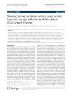
the area of interaction. The original idea for the AFM was to measure the van der Waals
interaction of an atom at the very end of the tip with atoms at a surface of a solid substrate.
To bring a single atom at a tip close to within angstrom distance toward a surface is only
possible if the surface is atomically flat (Fig. 1c), such as, for example, the crystalline
surface of mica. If the surface is rough on a nanometer scale (Fig.1b), groups of atoms can
interact and determine, according to their size, the possible resolution. With a roughness
at the micrometer scale (Fig. 1a) the macroscopic level is reached where instruments like
the surface profiler are able to measure surface roughness. A similarly important part
of the scanning probe microscope is the mechanism which moves the tip closer to the
surface and scans it across with precision fitting to the highest resolution. What enables
such precise manipulation is the property of some materials to change size proportional
1. Local Probe Techniques 3
Fig. 1 Scanning probe tip structures shown at different scales.
to an applied electric field. These materials can also generate an electric field if a force
is applied, an effect first described by Pierre and Jacques Curie in 1880 for quartz. The
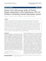
piezo-tube scanner is widely used to produce movements in all three directions easily and
consists of a thin-walled hard piezo-electric ceramic that is radially polarized. Electrodes
are attached to the internal and external faces of the tube. The external electrode is split
into quarters parallel to the axis as shown in Fig. 2. By applying a voltage between
the inner and all the outer electrodes, the tube expands or contracts and in this way
either moves a tip closer to a surface or retracts it from a surface, respectively. If the
voltage is applied just between the inner and one outer electrode, the tube will bend,
i.e., moving the tip along the surface, with a precision determined by both the noise of
the voltage source used and the overall mechanical stability. The disadvantage of these
piezo-tubes is that the tip is not scanned exactly parallel to the surface but is moved
on an arc, leading to an effect known as “eyeballing” when large scans are carried out.
Another problem of piezo-materials is the hysteresis, which like the arc motion must
be corrected by the electronic equipment controlling the movement by providing the
necessary voltage.
In the meantime, many other types of scanning probe microscopes using various
types of interactions have been developed and are too numerous to mention in this short
Fig. 2 Piezo-electric effect of quartz and the piezo-ceramic tube scanner with inner and segmented outer
electrodes used in scanning probe microscopes.
4 J. K. Heinrich H¨orber
introduction. I prefer, therefore, to name only one other: the scanning nearfield optical
microscope (SNOM), developed by Pohl et al. (1988), which is, as the name implies,
the near-field equivalent to the conventional optical microscope working in the farfield
of the radiation. The STM, on the other hand, can be seen as the nearfield equivalent
to the electron microscope. The optical microscope, like other types of microscopes
using radiation in the farfield range, is limited in its resolution by the wavelength of the
radiation. This limit, reported by Abbe in 1873, restricts the optimal resolution to several
hundred nanometers for using visible light. The only way of overcoming this limit is by
using nearfield effects observed within a wavelength from a radiation source. In high
resolution, the very small tip can be used again. The tip of a SNOM is, at least in many
instruments, a specially prepared end of an optical fiber, which acts as a light source. The
interaction of the electromagnetic nearfield at the tip with the surface determines how
much light is radiated from the source and how much is reflected back into the optical
fiber. In this way the aspects of the surface structure correlated to the interaction with
electromagnetic fields can be studied.
Many types of scanning probe microscopes have been developed and can be used not
only for measuring surface topologies but also for measuring various material properties
at or close to surfaces. This can be done in vacuum, in gas, or in liquids in a broad
temperature range with a resolution down to either the atomic or the molecular level.
In this way, it is the only type of microscopy that can complement optical microscopy
in biology on a smaller scale. Additionally, these instruments allow manipulations at
either the single-atomic or the molecular level, making experiments which no one ever
dreamed of 20 years ago possible. Experiments at the nanometer scale provide a com-
plete new insight into processes which, before the development of these instruments, were
accessible only by ensemble-average processes, where all of the elements can never be
identical, and all of the information concerning the behavior of individuals is lost. With
the available information on single components using scanning probe techniques we can
now learn how processes, which we were previously unaware of, are determined by the
properties of the single elements of such ensembles.
II. Scanning Tunneling Microscopy
It is of particular interest to understand the images of biological structures obtained
by the STM, as this technique allows imaging with a signal-to-noise ratio unequalled by
other techniques and under near-physiological conditions (H¨orber et al., 1988; H¨orber,
Schuler, Witzemann, Schr¨oter et al., 1991; H¨orber, Schuler, Witzemann, M¨uller et al.,
1991; Heckl et al., 1989; Ruppersberg et al., 1989; G¨obel et al., 1992; Maaloum et al.,
1994). This is an advantage that can only be exploited by having a deeper knowledge of
both the “tunneling” or electron transport mechanism and the environmental conditions
under which it takes place. Furthermore, a means to understand the nature of the images
produced, namely, a model that can be used as reference, is necessary. For this purpose, a
sample can, for instance, be imaged by scanning tunneling and electron microscopy, and
the results can then be compared to investigate the physical mechanism of image contrast
1. Local Probe Techniques 5
formation in the STM for biological samples. For example, the electron microscope can
produce a three-dimensional image of a helical structure, e.g., a bacteriophage tail. For
such experiments performed by my group, T5 tails were purified and adsorbed to glow
discharged indium tin oxide (ITO) surfaces in solution (Gu´enebaut et al., 1997). The
surface was washed with distilled water, which was removed partially by blotting, leaving
only a thin layer of aqueous solution. The STM used was a noncommercial “pocket-size”
type (Smith and Binnig, 1986), equipped not only with tungsten tips etched in KOH
by alternating currents but also with a patch-clamp amplifier allowing measurements
down to 0.5 pA with an equivalent noise current of 200 fA. Importantly, all the current
measurements were carried out in the picoampere range. The feedback circuit controlling
the movement of the tip in the z direction, which is the distance to the sample, was
equipped with a logarithmic amplifier to correct for the exponential behavior of the
current. However, the direct measurements of the current variation giving the constant
height images usually are not corrected for this exponential behavior. Therefore, the
logarithms of these images were extracted, before combining the left-to-right and right-
to-left scans, to produce a real-space representation of the specimen. Both scans can be
normalized by histogram equalization and, after combining the different scan directions,
they could be compared to the transmission electron microscope (TEM) reconstruction
of the phage tail structure. The time constant of the feedback used was the limiting factor
in the tip movement, and adding both right- and left-scan images significantly suppressed
the z feedback effect. With this setup, recording the feedback signal simultaneously with
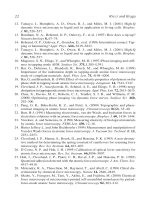
the current signal is alsopossible. Inprinciple, thiscombined constantheight andconstant
current imaging mode increases the height resolution of the instrument, showing the fine
structure on the top of the phage tails as a constant height image (Fig. 3).
Fig. 3 STM image of the tail of the bacteriophage T5 prepared on an ITO surface. The scan size is
18 × 18 nm
2
. The picture was taken with a 30-pA current at a 120-mV tip voltage within a thin layer of water.
6 J. K. Heinrich H¨orber
The general characteristics of the bacteriophage T5 tail make it an excellent test spec-
imen for comparing TEM and STM results. Bacteriophage T5 is a member of the T-odd
phage family having an icosahedral head with a diameter of 80 nm. Its noncontractile
flexible tail is 160 nm long and is composed of 120 copies of a 58 kDa protein. The
proteins are arranged as trimers, each trimer forming a ring with an external diameter of
11 nm. The superposition of 40 of these rings, with a 40-degree angular shift between
each stack, confers a helical symmetry to the tail. The tail model was calculated from
cryo-TEM images using helical reconstruction methods. The general dimensions of the
tail allowed for its easy identification in the STM images. These Bacteriophage T5 tail
images exhibit size features approaching 3 nm, which were used in comparison to the
reference obtained from electron microscopy data.
As for other biological materials observed using STM, the tail appears with a positive
contrast and exhibits complex features that prevent trivial interpretation of the images.
It is difficult to correlate these two observations with the classical concept of the elec-
tron “tunneling” mechanism between two conductors through an energetically forbidden
region. Nevertheless, it is clear from the many experiments performed thus far that it
is possible to image nonconductive molecular structures using the STM. The imaging
of cyanobiphenyl monomolecular layers of liquid crystals, where near-atomic details
were observed, confirmed the transfer of electrons through thin, nonconductive, and
organic materials (Smith et al., 1989, 1990). However, the mechanism by which this
phenomenon occurs through thicker nonconductive layers of organic material, either a
multilayered arrangement of small molecules or larger molecular structures, is still not
understood. The role of water, which is always present under ambient conditions (Freund
et al., 1999), while keeping biological samples under physiological conditions, remains
unknown.
By comparing TEM results to those of STM on these bacteriophage tails it became
clear that, although the STM images did not show the surface of tail structures, they,
however, could be directly compared to contrast-inverted TEM images. The actual sit-
uation for STM imaging such samples, i.e., the position of the tip with respect to the
sample, can be studied using current/distance measurements. It was found that phage
tails freshly adsorbed on ITO-coated glass retained a thin (50- to 100-nm) film of water.
While imaging, the tip was immersed several tens of nanometers into this film; at these
distances, currents of 5–50 pA were observed. In this situation (Fig. 4), the electrons had
to cross a water layer of up to several tens of nanometers in addition to the molecular
structure, but still could provide a resolution of 3 nm. The exponential distance depen-
dence of the measured current decays faster in water than through the macromolecules,
leading to a positive contrast. Without hypothesizing on the nature of the electron transfer
mechanism across biological material and through water, the observation that the protein
structure has less resistance to the current than to the surrounding aqueous solution is
very interesting. This produces a positive image of the specimen, while cryo-TEM, based
on high-energy electron scattering by the specimen, produces a negative image. A pos-
sible explanation might be that as denser protein structures are more ordered low-energy
electrons do not scatter as frequently.
1. Local Probe Techniques 7
Fig. 4 Scaled schematic drawing of the imaging situation as determined by current/distance measurements.
The diameter of the tail is 11 nm and the tip surface distance while imaging is about 60–70 nm above the
surface. The water is kept by cooling the sample as a thin layer of 100–200 nm on top of the sample.
If the physical basis for the use of the STM on biological structures can be identified,
then the STM can become an important complement to TEM in structural studies, as
completely different preparation methods are used and the samples remain hydrated
under close to physiological conditions.
III. Atomic Force Microscopy
A. Combination with Optical Microscopy
It has been shown in many experiments that the AFM can be used to study biological
structures under physiological conditions. It is even possible for the AFM to both image
living cells (H¨aberle et al., 1991) and study dynamic processes at the plasma membrane,
although such experiments are quite difficult, as the AFM cantilever is by far much
more rigid than cellular membrane structures (Schneider et al., 1997; Jena and Cho
in this book). The preparation of cells and the parallel optical observation, which are
necessary for having standard biological controls for cell activities available, present
other problems. To address these problems, in 1988 we initiated an IBM Physics project
in Munich to develop a special AFM built into an inverted optical microscope. This
instrument could make the first reproducible images of the outer membrane of a living
cell, fixed only by a pipette in its normal growth medium (H¨orber et al., 1992; Ohnesorge
et al., 1997). This pipette was moved by a conventional piezo-tube scanner. The detection
system, in principle, was a normal optical detection scheme using a glass fiber as a light
source and placed very close to the cantilever (Fig. 5). This configuration allowed a
very fast scanning speed for imaging cells in the variable deflection mode, as the parts
moving in the liquid are very small. Therefore, in contrast to the standard procedure
of imaging cells attached to a flat substrate on the scanning stage, neither significant
excitation of disturbing waves nor convection in the liquid occurs. Additionally, the
severe deformation of the cells was avoided, which normally occurs when they are
squeezed between a solid substrate and a cantilever. Since it was possible to keep the
8 J. K. Heinrich H¨orber
Fig. 5 A schematic drawing of the AFM built onto an inverted optical microscope with a patch-clamp
pipette as a sample holder. An optical fiber as a light source very close to the cantilever is used for the optical
detection of the cantilever deflection. The detection of the reflected light is done by a quadrant photo-diode
above the sample chamber.
cell alive and well for days while imaging, this made studies of live activities and
kinematics in addition to the application of other measuring techniques possible. With
this step in the development of scanning probe instruments, the capability of optical
microscopy to investigate the dynamics of biological processes of cell membranes under
physiological conditions could be extended into the nanometer range with the help of
the AFM.
In the initial experiments with the AFM, we observed the reaction of cultured monkey
kidney cells infected by orthopox viruses. We usually saw no reaction during the first
few minutes after adding the virus suspension to the fluid chamber where the cells were
kept in buffer solution. Yet in one case we observed a decaying protrusion after about
1 h. The size of the protrusion was comparable to that of a virus (200–300 nm), but
we observed an effect like this only once. The fact that we usually did not observe
the endocytosis of the virus might have been due to a shadowing by the lever and the
imaging tip, which prevented the penetration of viruses into this area. On the other hand,
at about the time when the virus would be expected to enter the cell (a few minutes after
adding viruses according to estimates of diffusion times in the surrounding liquid) we
noticed a strong softening of the cells, which was always accompanied by the danger
of the tip easily penetrating the membrane and the images losing considerable contrast.
One might imagine that a virus only locally modifies the membrane to enable its entry
into the cell. However, from the fact that the dramatic softening of the cell membrane
is always observed when viruses are added, we conclude that the cell membrane as a
whole is affected by the penetration or adhesion of the viruses. It is known that 4 to
1. Local Probe Techniques 9
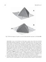
Fig. 6 Exocytotic process imaged by AFM 3 h after monkey kidney cells were infected by pox viruses. The
size of the structure seen is about 200 nm and similar to the size of viral particles.
6 h after infection the first viruses reproduced inside the cell and emerged from the cell
through the cell membrane. However, approximately 2.5 h after infection we observed
a series of processes occurring in our SFM images. Single clear protrusions became
visible and grew in size. The objects quickly disappeared and the original structures on
the cell surface were more or less restored. Such processes can occur several times in
the same area and last about 90 s for a small protrusion (about 20-nm lateral extent) and
up to 10 min for a larger one (cross section of about 100 nm). Each process proceeds
distinctly, apparently independently of the others, and is never observed with uninfected
cells and never prior to 2 h after infection.
The fact that the growing protrusions abruptly disappeared after a certain time led us
to believe that we observed an exocytodic process but not the virus release. First-progeny
viruses are known to appear 5–8 h after infection and they are clearly bigger than the
structures observed. It is alsoknown, however, that after2–3 h only the early stageof virus
reproduction is finished and the final virus assembly has just begun. Since the protrusions
are observed after this characteristic time span, we believe that they are related to the
exocytotic processes connected to the virus assembly. Significantly more than 6 h after
infection even more dramatic changes are seen in the cell membrane (Fig. 6). Large
protrusions, with cross sections of 200–300 nm, grow out of the membrane near deep
folds. These events occur much less frequently than those which occur after only 2 h.
These protrusions also abruptly disappear, leaving behind small scars on the cell surface.
Considering the timing and their size, we believe these protrusions are progeny viruses
exiting the cell. Assuming that approximately 20–100 viruses exit the living cell and that
roughly 1/40 of the cell surface is accessible to our SFM, one should be able to observe
one or two of these events for each infected cell. We actually observed two processes
exhibiting the correct size and timing during one 46-h experiment on a single infected
cell: one after 19 h and the other after 35 h. It is known from electron microscopy that
individual viruses exit the cell at the end of finger-like microvilli that are formed at the cell
membrane. Figure 7 actually shows a finger-like protrusion at whose end an exocytotic
process is observed. The release of the particle observed also occurs in a region where the
cell membrane is dominated by finger-like structures. This striking similarity to results
from electron microscopy made us believe that we indeed had imaged the exocytosis of
a progeny virus through the membrane of an infected live cell.
10 J. K. Heinrich H¨orber
Fig. 7 Sequence of images showing the escape of a viral particle at the end of a microvillus 19 h after
infection of the cells.
With the setup developed, it was finally possible to observe structures as small as
10–20 nm at high-imaging rates of up to one frame per second. This, in principle, gives
one access to processes besides endo- and exocytosis such as the binding of labeled anti-
bodies, pore formation, and the dynamics of surface structures in general. Nevertheless,
still after more than 10 years much work must be done to control the interaction between
tip and plasma membrane structures, which can be influenced quite strongly by the so-
called extracellular matrix of cells containing a broad variety of sugars and other polymer
structures.
As with the integrated tip of the cantilever, forces in the range of some 10 to 100 pN
are applied to the investigated cell membrane, and the mechanical properties of cell sur-
face structures dominate the imaging process. On the one hand, topographic and elastic
properties of the sample in the images are combined; on the other hand, additional infor-
mation is provided regarding cell membranes and their dynamics in various situations
during the life of the cell. To separate the elastic and topographic properties, additional
information is needed, which can be provided either by topographic data from electron
microscopy or by the use of AFM modulation techniques. The pipette–AFM concept
is very well suited for such modulation measurements, because, as mentioned earlier,
perturbation by the excitation of convection or waves in the solution are extremely small
compared to the normal situation in AFM measurements. Furthermore, the cells held
by a pipette are supposedly in a state much more comparable to the natural situation
than a cell adhering to a substrate. For a thorough analysis of a cell membrane elasticity
map, one would have to record pixel by pixel a complete frequency spectrum of the
cantilever response and derive image data from various frequency regimes. This would
require too much time for a highly dynamic system like a living cell. Nevertheless, we