BioMEMS and Biomedical Nanotechnology Volume I pdf
Bạn đang xem bản rút gọn của tài liệu. Xem và tải ngay bản đầy đủ của tài liệu tại đây (10.35 MB, 532 trang )
BioMEMS and Biomedical
Nanotechnology
Volume I
Biological and Biomedical Nanotechnology
BioMEMS and Biomedical
Nanotechnology
Mauro Ferrari, Ph.D., Editor-in-Chief
Professor, Brown Institute of Molecular Medicine Chairman
Department of Biomedical Engineering
University of Texas Health Science Center, Houston, TX
Professor of Experimental Therapeutics
University of Texas M.D. Anderson Cancer Center, Houston, TX
Professor of Bioengineering
Rice University, Houston, TX
Professor of Biochemistry and Molecular Biology
University of Texas Medical Branch, Galveston, TX
President, the Texas Alliance for NanoHealth
Houston, TX
Volume I
Biological and Biomedical Nanotechnology
Edited by
Abraham P. Lee
Biomedical Engineering
University of California, Irvine
L. James Lee
Chemical and Biomolecular Engineering
The Ohio State University
Abraham P. Lee
University of California, Irvine
Irvine, California
James Lee
Ohio State University
Columbus, Ohio
Mauro Ferrari
Ohio State University
Columbus, OH
Library of Congress Cataloging-in-Publication Data
Volume I
ISBN-10: 0-387-25563-X e-ISBN 10: 0-387-25842-6 Printed on acid-free paper.
ISBN-13: 978-0387-25563-7 e-ISBN-13: 978-0387-25842-3
Set
ISBN-10: 0-387-25661-3 e-ISBN:10: 0-387-25749-7
ISBN-13: 978-0387-25561-3 e-ISBN:13: 978-0387-25749-5
C
2006 Springer Science+Business Media, LLC
All rights reserved. This work may not be translated or copied in whole or in part without the written permission of
the publisher (Springer Science+Business Media LLC, 233 Spring Street, New York, NY 10013, USA), except for
brief excerpts in connection with reviews or scholarly analysis. Use in connection with any form of information
storage and retrieval, electronic adaptation, computer software, or by similar or dissimilar methodology now
known or hereafter developed is forbidden.
The use in this publication of trade names, trademarks, service marks and similar terms, even if they are not
identified as such, is not to be taken as an expression of opinion as to whether or not they are subject to
proprietary rights.
987654321 SPIN 11406068
springer.com
Dedicated to Richard Smalley (1943–2005), in Memoriam
To Rick,
father founder of nanotechnology
prime inspiration for its applications to medicine
gracious mentor to its researchers
our light—forever in the trenches with us
(Rick Smalley received the 1996 Chemistry Nobel Prize
for the co-discovery of carbon-60 buckeyballs)
Contents
List of Contributors xv
Foreword xix
Preface xxi
1. Biomolecular Sensing for Cancer Diagnostics Using Carbon Nanotubes 1
Jun Li and M. Meyyappan
1.1. Introduction 1
1.2. Carbon Nanotubes . 2
1.3. Carbon Nanotube Electrodes 3
1.3.1 Characteristics of a Good Electrode 3
1.3.2 Why Use Nanoelectrode? 4
1.3.3 Why Use Carbon Nanotubes? 5
1.3.4 Fabrication of CNT Nanoelectrodes 5
1.4. Preliminary Results 8
1.4.1 Electronic Nano-Chip Development 8
1.4.2 Electrochemical Properties of CNT Nanoelectrode Arrays 11
1.4.3 Functionalization of Oligonucleotide Probes 12
1.4.4 Electrochemical Detection of DNA Hybridization 14
1.5. Summary 16
Acknowledgements 17
References 17
2. Microspheres for Drug Delivery 19
Kyekyoon “Kevin” Kim and Daniel W. Pack
2.1. Introduction 19
2.2. Background 20
2.2.1 Factors Affecting Release Rates 20
2.2.2 Recent Applications of Controlled Release Microspheres 21
2.3. Fabrication of Polymer Micro- and Nanoparticles 24
2.3.1 Techniques for Fabricating Uniform Microspheres 25
2.3.2 Techniques for Fabricating Uniform Core-Shell Microparticles 29
2.3.3 Use of Electrohydrodynamic Spraying for Fabrication of Uniform
Micro and Nanospheres 33
2.4. Controlled Release from Precision Microspheres 35
2.4.1 In-vitro Release from Uniform Microspheres 36
viii CONTENTS
2.4.2 In-vitro Release from Mixtures of Uniform Microspheres 37
2.4.3 In vitro Release with Double-Wall Microspheres 39
2.4.4 Release of Macromolecules from Monodisperse Microspheres 40
2.5. Conclusions 41
References 42
3. Nanoscale Polymer Fabrication for Biomedical Applications 51
L. James Lee
3.1. Introduction 51
3.2. Potential Biomedical Applications of Polymer Nanostructures 52
3.2.1 Drug Delivery and Gene Therapy 52
3.2.2 Medical Diagnostics and Nanofluidics 53
3.2.3 Tissue Engineering and Bioreactors 54
3.3. Mold (Master) Making and Prototyping 55
3.3.1 Non-Cleanroom based Mold Making and Prototyping 55
3.3.2 Cleanroom based Mold Making 57
3.4. Nanoscale Polymer Replication 62
3.4.1 Soft Lithography 63
3.4.2 Nanoimprinting 63
3.4.3 Injection Molding at the Nanoscale 71
3.4.4 Other Technologies 73
3.5. Assembly and Bonding 80
3.6. Conclusions and Future Directions 83
References 89
4. 3D Micro- and Nanofabrication and Their Medical Application 97
E. Di Fabrizio, F. Perennes, F. Romanato, S. Cabrini, D. Cojoc, M. Tormen,
L. Businaro, L. Vaccari, R. Z. Proietti, and Rakesh Kumar
4.1. Introduction 97
4.2. 3D Micro and Nanofabrication 98
4.2.1 3D Fabrication by X-ray and Deep X-ray Lithography for
Biomedical Application 98
4.2.2 3D Microparts for Transdermal Drug Delivery System 102
4.3. Emerging Methods for 3D Micro and Nanofabrication 107
4.3.1 Two Photon assisted Microfabrication 108
4.3.2 Nanoimprint and Soft Lithography 112
4.3.3 Focused Ion Beam Lithography for 3 Dimensional Structures 115
4.4. Hybrid Lithography Approach 121
4.4.1 X-ray and Nanoimprint Lithography for 3D Patterning 121
4.4.2 Lithography at Interface-Binary Resist Process Combined with
Multiple Tilted XRL and EBL Lithography 123
4.5. 3D Trapping and Micro Manipulation by Means of Optical Tweezers 129
4.5.1 Optical Tweezers Enabled 3D Trapping and
Micromanipulation 129
4.5.2 3D Micromanipulation of Cells by Means of Optical Tweezers 133
4.6. Mems Devices for Biomedical Applications 136
CONTENTS ix
4.6.1 Self-standing Metallic Nanogap MEMS Structures for Nano
Trapping Application 137
Conclusions 138
References 139
5. Sacrificial Oxide Layer for Drug Delivery 145
Piyush M. Sinha and Mauro Ferrari
5.1. Introduction 145
5.2. Silicon Dioxide Fabrication 146
5.2.1 Thermally Grown Oxide 147
5.2.2 Deposited Silicon Dioxide 148
5.2.3 Thermally Grown Oxide vs Deposited Oxide 149
5.2.4 Silicon-On-Insulator (SOI) as Sacrificial Layer 149
5.3. Sacrificial Oxide Etching 150
5.3.1 Etch Mechanism 150
5.3.2 Etch Selectivity 152
5.3.3 Stiction . . . 152
5.3.4 On-Chip Packaging 153
5.4. Application of Sacrificial Oxide in Devices 153
5.4.1 Sacrificial Oxide for MEMS 154
5.4.2 Sacrificial Oxide in ICs 162
5.5. Summary 166
References 166
6. Carbon Nanotube Biosensors 171
Pingang He and Liming Dai
6.1. Introduction 171
6.2. The Structure and Chemical Reactivity of Carbon Nanotubes 172
6.3. Functionalization of Carbon Nanotubes 173
6.3.1 Non-covalent Functionalization 173
6.3.2 Chemically Covalent Modification 175
6.4. Fabrication of Carbon Nanotube Electrodes 178
6.4.1 Non-aligned Carbon Nanotube Electrodes 178
6.4.2 Aligned Carbon Nanotube Electrodes 182
6.5. Carbon Nanotube Biosensors 185
6.5.1 Protein and Enzyme Biosensors 185
6.5.2 DNA Sensors 191
6.6. Conclusion 198
Acknowledgements 198
References 198
7. Characterization Methods for Quality Control of Nanopore
and Nanochannel Membranes 203
Carlo Cosentino, Francesco Amato, and Mauro Ferrari
7.1. Introduction 203
7.2. Microscopy Observation 205
x CONTENTS
7.3. Bubble Point 207
7.4. Gas Permeability . . 210
7.5. Permoporometry. . 211
7.6. Thermoporometry . 212
7.7. Electrical Conductance 213
7.8. Ultrasonic Spectroscopy 214
7.9. Molecular Transport 216
7.9.1 Classical Transport Models 216
7.9.2 Diffusion Through Nanochannels 218
References 222
8. Magnetic Nanoparticles for MR Imaging 227
Lee Josephson
8.1. Introduction 227
8.2 A Brief History Of Polymer Coated Iron Oxide Nanoparticles
As Pharmaceuticals. 227
8.3 Magneto/optical Nanoparticles As Optical Probes 230
8.4 Magnetic Nanoparticles As Biosensors 231
8.5 Magnetic Nanoparticles For Cell Loading And Tracking By MRI 232
8.6 Molecularly Targeted Nanoparticle Based MRI Contrast Agents 234
8.7 The Future 235
References 235
9. Polymer Design for Nonviral Gene Delivery 239
Kam W. Leong
9.1 Introduction 239
9.1.1 Barriers for Nonviral Gene Transfer 240
9.2 Synthetic Polymeric Gene Carriers 243
9.2.1 Polyethyleneimine 243
9.2.2 Polylysine . . 243
9.2.3 Poly(α -(4-aminobutyl)-L-glycolic acid) 246
9.2.4 Polyamidoamine Dendrimer 247
9.2.5 Poly((2-dimethylamino)ethyl methacrylate) 248
9.2.6 Poly(β-amino ester) 248
9.2.7 Polyphosphazene 249
9.2.8 Cyclodextrin-containing Polycation 250
9.2.9 Polyphosphoester 251
9.3 Natural Polymeric Gene Carriers 254
9.3.1 Chitosan . . . 254
9.4 Biomaterials Approach to Gene Delivery 256
9.5 Summary 258
References 259
10. Dip-Pen Technologies for Biomolecular Devices 265
Debjyoti Banerjee
10.1 Introduction 265
CONTENTS xi
10.2 General Applications 268
10.3 Bio-molecular Patterning using Dpn 269
10.3.1 Nano-Pattering of Oligonucleotides Using DPN 270
10.3.2 Nano-Patterning of Protein and Petides Using DPN 276
10.3.3 Nano-Patterning of Composite Bio-Molecular Structures 291
10.4 Dpn Bio-Molecular Devices for Cell and Virus Capture 292
10.5 Using Microfluidics for Dpn Applications in Biomolecular Patterning 295
10.5.1 Analysis 296
10.5.2 Computational Fluid Dynamic (CFD) Simulation 297
10.5.3 Fabrication 298
10.5.4 Experimental Apparatus 299
10.5.5 Results and Discussion 299
10.6 Summary, Conclusion and Future Direction 302
References 303
11. Engineered Inorganic-Binding Polypeptides for Bionanotechnology 307
Candan Tamerler and Mehmet Sarikaya
11.1 Introduction 307
11.2 Selection of Inorganic Binding Polypeptides 309
11.3 Binding Affinity of Inorganic-Binding Polypeptides 312
11.3.1 Molecular Adsorption of GEPI 312
11.3.2 Physical Specificity and Molecular Modeling 314
11.4 Potential Applications of Molecular Biomimetics in Bio-And
Nanobiotechnology. 316
11.4.1 GEPI-Assisted Cell and Phage Sorting and Differentiation 317
11.4.2 Target Immobilization via Engineered Polypeptides as Molecular
Erector Films 318
11.4.3 Genetically Engineered Bifunctional GEPI-Alkaline Phosphatase
Molecular Construct: Expressing both Catalytic and
Inorganic-Binding Activity 320
11.4.4 Bionanofabrication: Silica Synthesis Using Inorganic
Binding Polypeptides 321
11.5 Future Prospects and Potential Applications in Nanotechnology 322
Acknowledgements 323
References 323
12. Dynamic Nanodevices Based on Protein Molecular Motors 327
Dan V. Nicolau
12.1 Introduction 327
12.2 Protein Molecular Motors—Biophysical Aspects 328
12.2.1 Rotary Motors 328
12.2.2 Linear Motors 329
12.2.3 Actin/Microtubule Polymerisation 333
12.3 Nanodevices Based on Protein Molecular Motors—Operational Aspects . . 333
12.3.1 Motility Assays and Single Molecule Techniques 333
12.3.2 Interaction of Motor Proteins with the Device Environment 336
xii CONTENTS
12.4 Design, Fabrication and Operation of Protein Molecular Motors-Based
Nanodevices 341
12.4.1 Lateral Confinement of Movement for Motile Elements 341
12.4.2 Control of Unidirectional Movement by External Means 343
12.4.3 Control of Unidirectional Movement by Self-Assembled Tracks 346
12.4.4 On-Off Control of the Operation of Protein Molecular Motors
Devices 347
12.5 Prototypes of Nanodevices Based on Protein Molecular Motors 349
12.5.1 Sensing Devices 350
12.5.2 Nanomechanical Devices 350
12.5.3 Information Storage and Processing 354
12.6 Perspectives 354
12.7 Conclusion 356
Acknowledgements 357
References 357
13. Nanodevices in Biomedical Applications 363
Bryan Ronain Smith, Mark Ruegsegger, Philip A. Barnes, Mauro Ferrari,
and Stephen C. Lee
13.1 Introduction 363
13.1.1 Defining Nanotechnology and Nanodevices 363
13.2 Opportunities for Biomedical Nanotechnology: Technological and
Biological 366
13.2.1 Device Assembly 366
13.2.2 Targeting: Delimiting Nanotherapeutic Action in
Three-Dimensional Space 373
13.2.3 Triggering: Spatially and Temporally Delimiting
Nanotherapeutic Action 374
13.2.4 Sensing Approaches 380
13.2.5 Imaging Using Nanotherapeutic Contrast Agents 383
13.3 Specific Therapeutic Applications of Hybrid Nanodevices 385
13.3.1 Hybrid Nanotherapeutic Devices in Oncology 385
13.3.2 Nanotherapeutics for Cardiovascular Applications 396
13.3.3 Hybrid Nanotherapeutics and Specific Host Immune Responses 388
13.4 Conclusions 389
Acknowledgements 390
References 390
14. Modeling Biomolecular Transport at the Nanoscale 399
A. T. Conlisk
14.1 Introduction 399
14.2 Background 402
14.3 Governing Equations for Synthetic Ion Channels in the Continuum
Regime: The Poisson-Nernst-Planck System 403
14.4 The One-Dimensional Poisson-Nernst-Planck Equations 406
14.5 Hindered Diffusion Concepts 408
14.6 Calculating the Electrical Potential 412
CONTENTS xiii
14.7 Ionic and Biomolecular Transport: Comparison with Experiment 416
14.8 Brownian Dynamics 423
14.9 Molecular Dynamics Simulations 427
14.10 Summary 431
Acknowledgements 432
References 433
15. Nanotechnology in Cancer Drug Therapy: A Biocomputational
Approach 435
Hermann B. Frieboes, John P. Sinek, Orhan Nalcioglu, John P. Fruehauf,
and Vittorio Cristini
15.1 Introduction 435
15.1.1 Challenges with Chemotherapy 435
15.1.2 Possibilities of Nanotechnology 436
15.1.3 Chemotherapy via Nanoparticles 436
15.1.4 Challenges of Nanotechnology 437
15.1.5 Biocomputation in Cancer Treatment 437
15.2 Issues with Chemotherapy: How Nanotechnology can Help and the
Role of Biocomputation 438
15.2.1 Drug Resistance 438
15.2.2 Drug Toxicity 439
15.2.3 Drug Targeting 439
15.2.4 Drug Transport 440
15.2.5 Drug Dosage and Scheduling 442
15.2.6 Drug Concentration 455
15.2.7 Drug Release 447
15.3 Biocomputation at the System Level 450
15.3.1 Modeling at the Nanoscale 450
15.3.2 Modeling at the Tumor Scale 452
15.3.3 Modeling of Cancer Therapy 453
15.4 Outlook on Modeling 456
References 456
16. Nanomechanics and Tissue Pathology 461
Jason Sakamoto, Paolo Decuzzi, Francesco Gentile, Stanislav I. Rokhlin,
Lugen Wang, Bin Xie, and Senior Author: Mauro Ferrari
16.1 Introduction 461
16.1.1 Background 461
16.1.2 The Diagnostic Conundrum 463
16.1.3 Oncologic Opportunity: Breast Cancer 463
16.1.4 Screening for Malignant Melanoma 465
16.2 The Classic Approach: Characterization-Mode Ultrasound and
Continuum Mechanics Model 467
16.2.1 Continuum Mechanics Description of Ultrasonic Wave
Propagation 467
16.3 An Introduction to “Doublet Mechanics” 471
16.3.1 Connotations and Interpretation of Doublet Mechanics 471
xiv CONTENTS
16.3.2 Microstrains and Microstresses: A Deeper Insight into Doublet
Mechanics 472
16.3.3 Comparison with Other Theories 473
16.4 Doublet Mechanics within the Linear Elastic Framework (Mathematical
Formulation of Doublet Mechanics) 474
16.4.1 Microstructure 474
16.4.2 Microstrains 474
16.4.3 Microstresses and Transition to Macrostresses 476
16.4.4 Linear Elastic Doublet Mechanics 478
16.5 Plane Waves Propagation within the Linear Elastodynamics of Doublet
Mechanics 479
16.5.1 Significance of the Analysis 479
16.5.2 Dynamic Scaling Equations 479
16.5.3 Plane Elastic Waves in Granular Media 480
16.5.4 Discussion 483
16.6 Reflection and Transmission of Plane Waves (Numerical Applications
of Doublet Mechanics to Malignant Tissue) 483
16.6.1 The Reflection Equations 484
16.6.2 Solution of the Equations: the Forward Problem 486
16.6.3 The Inverse Problem and the Doublet Mechanics Parameters
Identification 487
16.6.4 The Doublet Mechanics Approach: Final Marks 488
16.7 Experimental Practice 488
16.7.1 Characterization-Mode Ultrasound 488
16.7.2 Characterization-Mode Ultrasound System 489
16.7.3 The Model 489
16.7.4 Tissue Preparation 490
16.7.5 Experimental Findings: Breast Cancer detection 491
16.8 Nanomechanical Method for the Molecular Analysis of Breast Cancer 494
16.8.1 Introduction 494
16.8.2 The HER-2/neu Oncogene 494
16.8.3 HER-2/neu Exploitation 495
16.8.4 Ultrasound Interaction with Tissues with Targeted Nanoparticles 497
16.8.5 Preliminary Results: Randomly Distributed Particles in the Bulk 497
16.8.6 Preliminary Results: Randomly distributed particles upon
an interface 499
16.9 Future of Characterization-Mode Ultrasound 499
Acknowledgements 501
References 501
About the Editors 505
Index 507
List of Contributors
VOLUME I
Francesco Amato, Dept. of Experimental and Clinical Medicine, Universit`a degli Studi
Magna Graecia di Catanzaro, Catanzaro, Italy
Debjyoti Banerjee, Group Leader and Staff Mechanical Engineer, Applied Biosystems Inc.
(formerly Microfluidics Engineer, NanoInk Inc.)
Phillip A. Barnes, Biomedical Engineering Center, The Ohio State University, Columbus,
Ohio USA
L. Businaro, LILIT Group, National Nanotechnology Laboratory-TASC, Instituto
Nazionale per la Fisica della Materia, Basovizza (Trieste) Italy
S. Cabrini, LILIT Group, National Nanotechnology Laboratory-TASC, Instituto Nazionale
per la Fisica della Materia, Basovizza (Trieste) Italy
D. Cojoc, LILIT Group, National Nanotechnology Laboratory-TASC, Instituto Nazionale
per la Fisica della Materia, Basovizza (Trieste) Italy
A.T. Conlisk, Dept. of Mechanical Engineering, The Ohio State University, Columbus,
Ohio USA
Carlo Cosentino, Dept. of Experimental and Clinical Medicine, Universit`a degli Studi
Magna Graecia di Catanzaro, Catanzaro, Italy
Vittorio Cristini, Dept. of Biomedical Engineering/Mathetmatics, University of California,
Irvine, Irvine, California USA
Liming Dai, Dept. of Chemical and Materials Engineering, University of Dayton, Dayton,
Ohio USA
Paolo Decuzzi, CEMeC—Center of Excellence in Computational Mechanics, Dept. of
Experimental Medicine, University Magna Gracia at Catanzaro, Italy
E. Di Fabrizio, LILT Group, National Nanotechnology Laboratory-TASC, Instituto
Nazionale per la Fisica della Materia, Basovizza (Trieste) Italy
xvi LIST OF CONTRIBUTORS
Mauro Ferrari, Ph.D., Professor, Brown Institute of Molecular Medicine Chairman, De-
partment of Biomedical Engineering, University of Texas Health Science Center, Houston,
TX; Professor of Experimental Therapeutics, University of Texas M.D. Anderson Cancer
Center, Houston, TX; Professor of Bioengineering, Rice University, Houston, TX; Professor
of Biochemistry and Molecular Biology, University of Texas Medical Branch, Galveston,
TX; President, the Texas Alliance for NanoHealth, Houston, TX
Hermann B. Frieboes, Dept. of Biomedical Engineering, University of California, Irvine,
Irvine, California USA
John P. Fruehauf, Medicine—Hematology/Oncology, University of California, Irvine,
Irvine, California USA
Francesco Gentile, Dept. of Experimental Medicine, University Magna Gracia at
Catanzaro, Italy
Pingang He, Dept. of Chemistry, East China Normal University, Shanghai, China
Lee Josephson, Center for Molecular Imaging Research, Massachusetts General Hospital/
Harvard Medical School, Charlestown, Massachusetts USA
Kyekyoon “Kevin” Kim, University of Illinois at Urbana-Champaign, Illinois USA
Rakesh Kumar, LILIT Group, National Nanotechnology Laboratory-TASC, Instituto
Nazionale per la Fisica della Materia, Basovizza (Trieste) Italy
L. James Lee, Dept. of Chemical and Biomolecular Engineering, The Ohio State University,
Columbus, Ohio USA
Stephen C. Lee, Dorothy M. Davis Heart and Lung Research Institute, Dept. of Cellular
and Molecular Biology, The Ohio State University, Columbus, Ohio USA
Kam W. Leong, Dept. of Biomedical Engineering, Johns Hopkins School of Medicine,
Baltimore, Maryland USA
Jun Li, NASA Ames Research Center, Center for Nanotechnology, Moffett Field, California
USA
M. Meyyappan, NASA Ames Research Center, Center for Nanotechnology, Moffett Field,
California USA
Orhan Nalcioglu, Radiological Sciences and Tu & Yuen Center for Functional Onco-
Imaging, University of California, Irvine, Irvine, California USA
Dan V. Nicolau, Department of Electrical Engineering and Electronics, University of
Liverpool, Liverpool, UK
Daniel W. Pack, University of Illinois at Urbana-Champaign, Illinois USA
F. Perennes, Sincrotrone Trieste ELETTRA, Basovizza (Trieste) Italy
LIST OF CONTRIBUTORS xvii
R.Z. Proietti, LILIT Group, National Nanotechnology Laboratory-TASC, Instituto
Nazionale per la Fisica della Materia, Basovizza (Trieste) Italy
Stanislav I. Rokhlin, Nondestructive Evaluation Program, The Ohio State University,
Columbus, Ohio USA
F. Romanato, LILT Group, National Nanotechnology Laboratory-TASC, Instituto
Nazionale per la Fisica della Materia, Basovizza (Trieste) Italy
Mark Ruegsegger, Dorothy M. Davis Heart and Lung Research Institute, Cardiology
Divison, Biomedical Engineering Center, The Ohio State University, Columbus, Ohio USA
Jason Sakamoto, Biomedical Engineering, The Ohio State University, Columbus, Ohio
USA
Mehmet Sarikaya, Molecular Biology & Genetics, Istanbul Technical University, Maslak,
Istanbul, Turkey
John P. Sinek, Mathematics Dept., University of California, Irvine, Irvine, California USA
Piyush M. Sinha, Electrical and Computer Engineering, The Ohio State University,
Columbus, Ohio USA
Bryan Ronain Smith, Biomedical Engineering Center, The Ohio State University,
Columbus, Ohio USA
Candan Tamerler, Materials Science & Engineering, University of Washington, Seattle,
Washington USA
M. Tormen, LILIT Group, National Nanotechnology Laboratory-TASC, Instituto
Nazionale per la Fisica della Materia, Basovizza (Trieste) Italy
L. Vaccari, LILIT Group, National Nanotechnology Laboratory-TASC, Instituto Nazionale
per la Fisica della Materia, Basovizza (Trieste) Italy
Lugen Wang, Nondestructive Evaluation Program, The Ohio State University, Columbus,
Ohio USA
Bin Xie, Nondestructive Evaluation Program, The Ohio State University, Columbus, Ohio
USA
Foreword
Less than twenty years ago photolithography and medicine were total strangers to one
another. They had not yet met, and not even looking each other up in the classifieds. And
then, nucleic acid chips, microfluidics and microarrays entered the scene, and rapidly these
strangers became indispensable partners in biomedicine.
As recently as ten years ago the notion of applying nanotechnology to the fight against dis-
ease was dominantly the province of the fiction writers. Thoughts of nanoparticle-vehicled
delivery of therapeuticals to diseased sites were an exercise in scientific solitude, and grounds
for questioning one’s ability to think “like an established scientist”. And today we have
nanoparticulate paclitaxel as the prime option against metastatic breast cancer, proteomic
profiling diagnostic tools based on target surface nanotexturing, nanoparticle contrast agents
for all radiological modalities, nanotechnologies embedded in high-distribution laboratory
equipment, and no less than 152 novel nanomedical entities in the regulatory pipeline in
the US alone.
This is a transforming impact, by any measure, with clear evidence of further acceleration,
supported by very vigorous investments by the public and private sectors throughout the
world. Even joining the dots in a most conservative, linear fashion, it is easy to envision
scenarios of personalized medicine such as the following:
r
patient-specific prevention supplanting gross, faceless intervention strategies;
r
early detection protocols identifying signs of developing disease at the time when
the disease is most easily subdued;
r
personally tailored intervention strategies that are so routinely and inexpensively
realized, that access to them can be secured by everyone;
r
technologies allowing for long lives in the company of disease, as good neighbors,
without impairment of the quality of life itself.
These visions will become reality. The contributions from the worlds of small-scale tech-
nologies are required to realize them. Invaluable progress towards them was recorded
by the very scientists that have joined forces to accomplish the effort presented in this
4-volume collection. It has been a great privilege for me to be at their service, and
at the service of the readership, in aiding with its assembly. May I take this opportu-
nity to express my gratitude to all of the contributing Chapter Authors, for their in-
spired and thorough work. For many of them, writing about the history of their spe-
cialty fields of BioMEMS and Biomedical Nanotechnology has really been reporting about
their personal, individual adventures through scientific discovery and innovation—a sort
xx FOREWORD
of family album, with equations, diagrams, bibliographies and charts replacing Holiday
pictures
It has been a particular privilege to work with our Volume Editors: Sangeeta Bhatia,
Rashid Bashir, Tejal Desai, Michael Heller, Abraham Lee, Jim Lee, Mihri Ozkan, and
Steve Werely. They have been nothing short of outstanding in their dedication, scientific
vision, and generosity. My gratitude goes to our Publisher, and in particular to Greg Franklin
for his constant support and leadership, and to Angela De Pina for her assistance.
Most importantly, I wish to express my public gratitude in these pages to Paola, for her
leadership, professional assistance throughout this effort, her support and her patience. To
her, and our children Giacomo, Chiara, Kim, Ilaria and Federica, I dedicate my contribution
to BioMEMS and Biomedical Nanotechnology.
With my very best wishes
Mauro Ferrari, Ph.D.
Professor, Brown Institute of Molecular Medicine Chairman
Department of Biomedical Engineering
University of Texas Health Science Center, Houston, TX
Professor of Experimental Therapeutics
University of Texas M.D. Anderson Cancer Center, Houston, TX
Professor of Bioengineering
Rice University, Houston, TX
Professor of Biochemistry and Molecular Biology
University of Texas Medical Branch, Galveston, TX
President, the Texas Alliance for NanoHealth
Houston, TX
Preface
The growing demand for nanoscale structures and devices in the biomedical field presents
significant career opportunities for future generations. Various novel materials and technolo-
gies have been developed in recent years. There, however, lacks a comprehensive book to
systematically address this broad spectrum of new science and technologies. This volume
is intended to provide an introduction to nanoscale devices for biological and biomedi-
cal applications. Sixteen chapters are included in this volume experts in the field of the
nanobiotechnology have contributed to this work.
The volumeis dividedinto threeparts. Thefirst part,Synthetic Nanodevices for Biotechnol-
ogy and Biomedicine; covers the fabrication and characterization techniques of representative
nanoscale structures such as carbon nanotubes, micro/nanospheres and particles, nanopores
and nanochannels, and macro or microscale structures containing two-dimensional and three-
dimensional nanoscale features made of polymers, silicon and other materials. The applica-
tions of these nanostructures and devices for biosensing, drug delivery and bioseparation
are also introduced. The second part, Hybrid Synthetic and Biomolecular Nanodevices; fo-
cuses on the synthesis, interface structures, and medical applications of nanodevices made
of biomolecule-polymer and biomolecule-inorganics hybrids. Finally, the third part, Compu-
tation, Simulation, and Informatics for Bionanodevices, provides nanoscale fluid and solid
phase computation methodologies for selected biomedical applications.
We would like to thank all authors who devoted a great deal of time to make this
volume possible. We hope the collected efforts from these distinguished professionals will
present you a cohesive and balanced path into the intellectually exciting and fast evolving
nanobiotechnology field.
Abraham P. Lee
Biomedical Engineering, University of California at Irvine
L. James Lee
Chemical and Biomolecular Engineering, The Ohio State University
Mauro Ferrari
Professor, Brown Institute of Molecular Medicine Chairman
Department of Biomedical Engineering
University of Texas Health Science Center, Houston, TX
Professor of Experimental Therapeutics
University of Texas M.D. Anderson Cancer Center, Houston, TX
Professor of Bioengineering, Rice University, Houston, TX
Professor of Biochemistry and Molecular Biology
University of Texas Medical Branch, Galveston, TX
President, the Texas Alliance for NanoHealth, Houston, TX
1
Biomolecular Sensing for
Cancer Diagnostics Using
Carbon Nanotubes
Jun Li and M. Meyyappan
NASA Ames Research Center, Center for Nanotechnology, Moffett Field, CA 94035
1.1. INTRODUCTION
The field of biomolecule sensing in the medical field is broad and rapidly evolving. The
devices range in size from microns to centimeters across the sensing surface and rely on
electronic, optical or other form of signals. If the sensing technology utilizes toxic reagents,
then the use is limited to only in vitro application. In this chapter, biomolecule sensing using
carbon nanotubes (CNTs) is discussed with specific application to cancer diagnostics.
Beyond the expected size advantages of the CNT-based sensors, there are other benefits
as well. Conventional cytogenetic analysis and fluorescence in situ hybridization (FISH)
take about three weeks for completion of the analysis. Molecular diagnostic arrays by PCR
techniques take less time, still about a week. The sensitivity (i.e. ratio of the number of
positive cells detected to all cells) of FISH is 5–10% and conventional cytogenetics is about
5%. Most cytogenetics, FISH and molecular diagnostic testing procedures involve bone
marrow aspiration that causes pain. A biosensor that utilizes a nanoelectrode (such as CNT
based electrode), in principle, can overcome many of these limitations. The CNT-based
cancer diagnostics sensor discussed here can provide instantaneous results, facilitating
rapid turn around time and chemotherapy dosing regimens. The detection ability can be 1
positive cell in 1000–10000 cells. Current testing is targetted at in vitro application and may
be extended for in vivo diagnostics in the future, eliminating bone marrow aspiration.
The CNT based biosensor consists of a nanoelectrode array fabricated using conven-
tional microfabrication techniques. In this array, each nanotube electrode is functionalized
2 JUN LI AND M. MEYYAPPAN
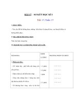
FIGURE 1.1. Schematic of a prototype catheter for cancer diagnostics.
with a probe molecule. The probe-target interaction is captured through the measurement
of electrochemical signals amplified by the use of metal-ion mediators.
A proposed design for a biosensor catheter for cancer diagnostics is shown in Fig. 1.1
and the expected operating principle is as follows. The working end of the catheter consists
of the carbon nanotube electrode functionalized with the probe molecules. The catheter is
inserted into a soft tissue area suspicious for cancer and a pair of external electrodes (shown
by the dark outline of the catheter), by applying a current to them, heats and lyzes the
cells. The DNA from cancerous cell diffuses towards the stationary probe molecules and
the hybridization is detected as an electrochemical signal.
In the sections below, discussion of carbon nanotubes and their interesting properties,
nanoelectrode fabrication, testing and characterization are discussed, as a progress report
in the fabrication of the biosensor Catheter.
1.2. CARBON NANOTUBES
For a detailed discussion on the properties, growth and applications of carbon
nanotubes, the reader is referred to [1]; here, only a brief overview is provided. A carbon
nanotube is an elongated fullerene molecule with diameter as small as 7
˚
A. Configura-
tionally, a nanotube is equivalent to a sheet of graphite rolled into a tube. The resulting
single-walled carbon nanotube (SWCNT) is denoted by its chiral vector (n, m) where n and
m are indices in the graphene sheet lattice vector. When (n-m)/3 is an integer, the resulting
BIOMOLECULAR SENSING FOR CANCER DIAGNOSTICS 3
tube is metallic; otherwise, it is a semiconductor. Besides this intriguing electrical property,
the SWCNT is mechanically very strong. It exhibits an Young’s modulus of over 1 TPa. The
strength/weight ratio of SWCNT is about 600 times higher than that of steel. The maximum
strain is about 10% which is higher than that of any other material. Thermal conductivity
along the axis of the tube is very high, exhibiting a value upto 3000 W/mK. The conductivity
and current carrying capacity are much higher than that of metals such as copper.
A MWCNT is a set of concentric cylinders with a central core where the wall separation
is close to 0.34nm. Some of the properties discussed above drop off from the values of
SWCNT, nevertheless high enough to create excitement in the research community for
a variety of applications. Both SWCNTs and MWCNTs were first produced by an arc
discharge synthesis [2]. But for most applications involving devices, electrodes, sensors
etc, chemical vapor deposition (CVD) has emerged as a powerful alternative [3]. CVD
allows in situ growth on a patterned substrate with possibility of subsequent processing
steps in an assembly-like fashion. Variations of CVD with the use of a glow discharge have
also emerged and this popular plasma enhanced CVD or PECVD has been used to grow
individual, free-standing, vertically- aligned multiwalled tubes [3]. For the most part, the
PECVD-grown nanostructures tend to have the hollow core periodically interrupted by a
bamboo-like closure. The resulting structure is somewhat inferior when it comes to electron
transport compared to an ideal MWCNT as electrons have to hop across these closures. In the
literature, these structures are called multiwalled carbon nanofibers (MWCNFs) or simply
carbon nanofibers (CNFs). In the remainder of this chapter, for generality, they are referred
to as MWCNTs.
While the various forms of CNTs are chemically inert, their ends and sidewalls are
amenable for attaching a variety of chemical groups. All these interesting structural, me-
chanical, electrical, thermal and other properties have led to an incredible array of application
development. SWCNT based diodes and transistors have been constructed showing inter-
esting electronic properties for memory and logic applications. Interconnects for wiring
electronic circuits using nanotubes have been investigated. Potential for near term appli-
cation with a mass market appeal exists with CNT based field emitters for flat panel TV
displays. On the structural side there is active research in developing high strength, low
weight polymer matrix, metal matrix and ceramic matrix composites with applications
in automotive, aerospace, and construction industries. The ability to functionalize CNTs
mentioned above opens up the possibilities to developing chemical sensors and biosensors.
The biosensors can serve the needs in biomedical, homeland security, and astrobiology
applications.
1.3. CARBON NANOTUBE ELECTRODES
1.3.1. Characteristics of a Good Electrode
Electrodes of all sizes using metals, various forms of carbon and other materials have
been around for a variety of applications. Typical expectations of a good electrode and
related issues are as follows:
– Appropriate level of conductivity for the chosen application
– The right size to meet the needs
– Ease of fabrication
4 JUN LI AND M. MEYYAPPAN
– Reliability: lifetime, wear characteristics
– Compatibility with the environment
– Signal processing issues: integrity, signal-to-noise, cross-talk in an ensemble
– Approach: electrical vs. electrochemical
– Integration into a functional system
1.3.2. Why Use Nanoelectrode?
The sensitivity of an electrode is mainly determined by its signal to noise ratio. The
noise is the background current mainly due to the capacitive charging/discharging current
at the electrode/electrolyte interface and thus proportional to the surface area (A) of the
electrode as given by:
i
n
∝ C
d
0
A (1.1)
where C
d
0
is the specific capacitance at the interface. In voltammetry measurements, the
magnitude of the peak current of the redox signal is the sum of two terms: a linear diffusion
as described in the Cottrell equation, and a nonlinear radial diffusion [4]:
i
1,peak
= nFAC
0
∗
D
0
πt
+ nFAC
0
∗
D
0
r
(1.2)
where i
l, pea k
is the diffusion-limited electrical current, n is the number of electrons involved
in the reaction with one electroactive species, F is the Faraday constant, C
o
is the elec-
troactive species concentration, D
o
is the diffusion coefficient, t is time, and r is the radius
of the electrode. Both terms are proportional to the concentration of the species present in
the solution. The first term is proportional to the electrode surface area and decays to zero
over time, whereas the second term is proportional to the inverse of the electrode radius and
represents a steady state current due to a constant flux of material to the surface. The ratio
of the second term to the first becomes larger as the radius is decreased. The second term
dominates the measured peak current, i
l, pea k
if the electrode size is less than 25 µm, which is
commonly referred as ultramicroelectrode (UME) [5]. In this regime, the magnitude of the
current decreases, but the signal-to-noise ratio is improved as the electrode size decreases,
according to:
i
l,peak
/i
n
∝ nFC
o
D
o
/r (1.3)
Clearly, the signal to noise will be improved by 1000 times if the electrode size is reduced
from 20 µmto20nm.
The response time of an electrode is also a function of the electrode dimension. The
cell time constant can be described as
τ = R
u
C
d
= rC
d
0
/4 κ (1.4)
where κ is the conductivity of the electrolyte. The electrode can respond 1000 times faster
when the size is reduced from microns to nanometers so that fast electrochemical techniques
can be applied. The electrochemical signal is defined by the total number of electrons that
can be transferred between the electroactive species and the electrode. For high sensitivity
BIOMOLECULAR SENSING FOR CANCER DIAGNOSTICS 5
analytical applications, this number is always limited. By employing fast electrochemical
techniques, the same amount of electrons can be transferred to the measuring circuit in a
much shorter time. As a result, the current, i.e. the real physical quantity being measured,
will be much larger and can be differentiated much easier from the background noise.
From the above discussion, it is clear that the performance of an electrode with respect
to temporal and spatial resolution scales inversely with the electrode radius. Therefore, the
sensitivity can be dramatically improved by reducing the size of the electrodes to nanoscale.
Indeed, a single redox molecule was detected using a Pt-Ir electrode with a diameter of 15 nm
[6]. With the diameter approaching the size of the target molecules, nanoelectrodes can also
interrogate biomolecules much more efficiently than conventional electrodes. There have
been strong efforts in developing nanoelectrode based chemical and biosensors since 1980s.
However, a reliable method to fabricate nanoelectrodes was lacking until the recent reports
of CNT nanoelectrode fabrication using microfabrication approaches discussed below.
1.3.3. Why Use Carbon Nanotubes?
The outside diameter of a MWCNT varies from a few nanometers to about
200 nanometers and the length varies from a few microns to hundreds of microns. The
physical dimension of MWCNTs is ideal for fabricating nanoelectrodes, with the dimen-
sion approaching the size of biomolecules. MWCNTs normally show highly conductive
metallic properties. The open end is an ideal electrode similar to graphite edge plane, while
the sidewall is very inert similar to graphite basal plane. The difference in electron transfer
rate (ETR) between the open end and the sidewall differs by 5 to 6 orders of magnitude [7].
This makes MWCNT an ideal nanoelectrode which can pick up the signal at the tip and
transfer it to the measuring unit connected at the other end with minimum interference by
the surrounding environment.
From an electrochemical point of view, CNTs possess great properties similar to com-
monly used carbon electrodes (particularly for biosensors) such as fast electron transfer rate,
wide potential window, flexible surface chemistry, and good biocompatibility. Only a few
materials can provide such properties, necessary for maximizing the signal from detecting
species while minimizing the noise from other species in the solution. For example, a metal
electrode of Pt or Au will electrolyze water before reaching the electropotential needed for
the detection of many biomolecules. This causes a large background current that masks out
the real signal. Such problems can be avoided by using carbon as the electrode material. The
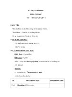
dangling carbon bonds at the open end of a CNT can form various oxides similar to the edge
of a graphite sheet, as shown in Fig. 1.2. Withelectrochemical etching or acid treatment, most
dangling bonds can be further converted into -COOH for highly selective functionalization
of biomolecules through the formation of amide bonds [8]. While it has been known for
sometime that a CNT, particularly a MWCNT, is ideal for biosensing, the major challenge
is how to fabricate and integrate it as a nanoelectrode. This is discussed in the next section.
1.3.4. Fabrication of CNT Nanoelectrodes
In principle, a single MWCNT can be grown on each individually addressed micro-
electrode indicated in Fig. 1.3 and used as a nanoelectrode. The microelectrode is more
precisely referred to as a microcontact since it only provides an electrical contact with the
MWCNTs. However, the use of individual CNTs is not reliable due to the large fluctuation
6 JUN LI AND M. MEYYAPPAN
Phenol
Carbonyl
Lactone
Carboxylic Acid
o-quinone
p-quinone
OH
O
O
O
H
O
C
O
O
OO
OH
FIGURE 1.2. The functional groups at the open end of a carbon nanotube.
in the detected signal. The signal is also very weak that can be easily masked out by the
electronic noises in the environment. These problems can be solved by using an array of
nanoelectrodes on each microcontact, as shown in Fig. 1.3. The small circles within each
microcontact in Fig. 1.3(a) represent a vertically aligned MWCNT. The side view indicates
that such a MWCNT array is embedded in an insulating matrix such as SiO
2
exposing only
the very end of MWCNTs to the solution. The other end of the MWCNT is attached to the
micron sized metal substrate and wired out to the measuring circuit. For a microcontact with
FIGURE 1.3. Schematic of a CNT nanoelectrode chip. (a) top view, (b) cross-sectional view, and (c) enlarged
cross-sectional view of MWCNT nanoelectrode array on a single microcontact spot. (d) illustration of the diffusion
layer around each MWCNT nanoelectrode.