Spinal Disorders: Fundamentals of Diagnosis and Treatment Part 12 potx
Bạn đang xem bản rút gọn của tài liệu. Xem và tải ngay bản đầy đủ của tài liệu tại đây (362.5 KB, 10 trang )
Corpectomy fusion technique. Spinal instability
after corpectomy or after vertebrectomy in the
lumbar spine often requires complex reconstruc-
tive procedures. The type and degree of instrumen-
tation depend strongly on the number of involved
levels and the retained functioning stabilizing
structures. Generally, after corpectomy anterior
support is mandatory and long-term stability can-
not be achieved with rod/pedicle screw instrumen-
tation alone. Furthermore, the combination with an
anterior tension band device still exhibits a certain
instability in extension and rotation. Therefore, from
the biomechanical perspective, substantial anterior
instability requires “front and back” instrumenta-
tion. In the cervical spine, however, single-level cage
stabilization is sufficiently supported by an anterior
tension band device. Multiple-level cervical corpec-
tomies are particularly unstable and anterior plating
may be insufficient; consequently additional pedi-
cle/lateral mass screw devices must be considered.
Anterior tension band technique. Anterior rods/
plates act as tension bands in extension and func-
tion as buttress plates in flexion. For the cervical
spine, the latest generation of “semi-constrained/
dynamic” plates allow locked angle-stable mono-
cortical screw fixation while axial compression of
the graft is permitted. This offers increased stability
combined with a minimized risk of stress-shielding.
In the lumbar spine, anterior rod/double-rod
instrumentation increases anterior stability after
cage or graft implantation especially in extension.
In flexion and lateral bending they are still inferior
to pedicle screw devices.
Biomechanics of the “adjacent segment”. Unphysi-
ologically long and stiff spinal segments increase
motion and intradiscal pressure in the adjacent
segments. However, it is still unclear if adjacent seg-
ment degeneration after spinal fusion is resulting
from the changed biomechanics or exhibits simply
the progression of the natural history.
Disc arthroplasty. Disc arthroplasty offers several
advantages such as preservation of segmental
motion, potential absence of adjacent segment
degeneration and no need for harvesting autolo-
gous bone graft. Current prostheses differ in bear-
ing materials (metal or polyethylene) and kinemat-
ics principles. Constrained prostheses have a fixed
center of rotation whereas unconstrained devices
allow translational movement and thus respect the
physiological helical axis of motion. Kinematics
studies have shown that both types successfully re-
establish almost the physiological range of motion.
Only a few data exist so far on the long-term radio-
logical and clinical outcome.
Posterior dynamic stabilization technique. Improv-
ing primary or iatrogenic spinal instability while
“unloading/protecting” certain spine elements
without performing a spinal fusion are the objec-
tives of posterior dynamic implants. All systems
successfully reduce segmental motion. However,
rotation is poorly controlled while the posterior
devices are particularly stiff in flexion. As it is
unknown how much stability is needed in which
particular entity of spine pathology combined with
the partially undefined clinical indications, an
assessment of this technique is currently impossi-
ble. Finally, only long-term prospective clinical trials
will give the necessary evidence for the efficacy of
this particular treatment method.
Key Articles
Cripton PA, Jain GM, Wittenberg RH, Nol te LP (2000) Load-sharing characteristics of
stabilized lumbar spine segments. Spine 25:170 – 179
Biomechanical cadaver study using pressure sensors, strain gauges and an optoelectronic
tracking system. Load-sharing between an internal fixator and anatomical structures was
assessed in asequential injury scenario. Applied loads were mostly supported by equal and
opposite forces between disc and fixator. Based on the results, the paper highlights the fact
that an anterior column insufficiency may lead to fixator overloads and implant failure.
Laxer E (1994) A further development in spinal instrumentation. Technical Commission
for Spinal Surgery of the ASIF. Eur Spine J 3:347 – 352
Introduction of the Universal Spine System with a single set of implants and instruments
for various spinal disorders and surgical approaches.
Spinal Instrumentation Chapter 3 85
Magerl FP (1984) Stabilization of the lower thoracic and lumbar spine with external
skeletal fixation. Clin Orthop Relat Res 125–141
Classic article introducing the concept of a new angle-stable transpedicular fixation
device which formed the basis for the development of second generation internal spinal
fixation devices.
Panjabi MM (1988) Biomechanical evaluation of spinal fixation devices: I. A conceptual
framework. Spine 13:1129 – 1134
Panjabi M, Abumi K, Duranceau J, Crisco J (1988) Biomechanical evaluation of spinal
fixation devices: II. Stability provided by eight internal fixation devices. Spine
13:1135 – 1140
Abumi K, Panjabi MM, Duranceau J (1989) Biomechanical evaluation of spinal fixation
devices. Part III. Stability provided by six spinal fixation devices and interbody bone
graft. Spine 14:1249 – 1255
These three publications are milestone papers as they introduced the basic concepts for
testing and evaluation of spinal implants. Guidelines for three categorical biomechanical
tests are stated: assessment of strength, fatigue and stability.
TsantrizosA,AndreouA,AebiM,SteffenT(2000) Biomechanical stability of five stand-
alone anterior lumbar interbody fusion constructs. Eur Spine J 9:14 – 22
The authors compared five different stand-alone cages with respect to stabilizing proper-
ties (kinematics) and pull-out strength using human specimens. The results demon-
strated a general stabilizing effect of all implants but load/displacement curves also sug-
gested micro-instability. Influencing factors of the cage design concerning dimensions,
height and wedge angle were pointed out.
References
1. Abumi K, Panjabi MM, Duranceau J (1989) Biomechanical evaluation of spinal fixation
devices. Part III. Stability provided by six spinal fixation devices and interbody bone graft.
Spine 14:1249–1255
2. Aebi M, Etter C, Kehl T, Thalgott J (1988) The internal skeletal fixation system. A new treat-
ment of thoracolumbar fractures and other spinal disorders. Clin Orthop Relat Res 227:
30–43
3. Aebi M, Etter C, Kehl T, Thalgott J (1987) Stabilization of the lower thoracic and lumbar
spine with the internal spinal skeletal fixation system. Indications, techniques, and first
results of treatment. Spine 12:544–551
4. Aebi M, Thalgott JS, Webb JK (1998) AO ASIF principles in spine surgery. Springer-Verlag,
Berlin Heidelberg New York
5. Albee FH (1972) The classic. Transplantation of a portion of the tibia into the spine for Pott’s
disease. A preliminary report. JAMA 57:885, 1911. Clin Orthop Relat Res 87:5–8
6. Anderson PA, Rouleau JP, Bryan VE, Carlson CS (2003) Wear analysis of the Bryan cervical
disc prosthesis. Spine 28:S186–194
7.
Arand M, Wilke HJ, Schultheiss M, Hartwig E, Kinzl L, Claes L (2000) Comparative sta-
bility of the “Internal Fixator” and the “Universal Spine System” and the effect of cross-
linking transfixating systems. A biomechanical in vitro study. Biomed Tech (Berl) 45:
311–316
8. Bagby GW (1988) Arthrodesis by the distraction-compression method using a stainless
steel implant. Orthopedics 11:931–934
9. Bastian L, Lange U, Knop C, Tusch G, Blauth M (2001) Evaluation of the mobility of adjacent
segments after posterior thoracolumbar fixation: a biomechanical study. Eur Spine J 10:
295–300
10. Battie MC, Videman T, Parent E (2004) Lumbar disc degeneration: epidemiology and
genetic influences. Spine 29:2679–2690
11. Benzel EC (2001) Biomechanics of spine stabilization, 1st edn. American Association of
Neurological Surgeons, Rolling Meadows, IL
12. Berlemann U, Cripton P, Nolte LP, Lippuner K, Schlapfer F (1995) New means in spinal pedi-
cle hook fixation. A biomechanical evaluation. Eur Spine J 4:114–122
13. Boos N, Webb JK (1997) Pedicle screw fixation in spinal disorders: a European view. Eur
Spine J 6:2–18
14. Boucher HH (1959) A method of spinal fusion. J Bone Joint Surg Br 41B:248–259
15. Burns BH (1933) An operation for spondylolisthesis. Lancet 224:1233–1239
86 Section Basic Science
16. Cain CM, Schleicher P, Gerlach R, Pflugmacher R, Scholz M, Kandziora F (2005) A new
stand-alone anterior lumbar interbody fusion device: biomechanical comparison with
established fixation techniques. Spine 30:2631–2636
17. Carlisle E, Fischgrund JS (2005) Bone morphogenetic proteins for spinal fusion. Spine J
5:S240–249
18. Chang BS, Brown PR, Sieber A, Valdevit A, Tateno K, Kostuik JP (2004) Evaluation of the
biological response of wear debris. Spine J 4:239S–244S
19. Chow DH, Luk KD, Evans JH, Leong JC (1996) Effects of short anterior lumbar interbody
fusion on biomechanics of neighboring unfused segments. Spine 21:549–555
20. Cotrel Y, Dubousset J (1984) A new technic for segmental spinal osteosynthesis using the
posterior approach. Rev Chir Orthop Reparatrice Appar Mot 70:489–494
21. Cripton PA, Jain GM, Wittenberg RH, Nolte LP (2000) Load-sharing characteristics of stabi-
lized lumbar spine segments. Spine 25:170–179
22. Cunningham BW (2004) Basic scientific considerations in total disc arthroplasty. Spine J
4:219S–230S
23. Cunningham BW, Gordon JD, Dmitriev AE, Hu N, McAfee PC (2003) Biomechanical evalua-
tion of total disc replacement arthroplasty: an in vitro human cadaveric model. Spine 28:
S110–117
24. de Kleuver M, Oner FC, Jacobs WC (2003) Total disc replacement for chronic low back pain:
background and a systematic review of the literature. Eur Spine J 12:108–116
25.
Dekutoski MB, Schendel MJ, Ogilvie JW, Olsewski JM, Wallace LJ, Lewis JL (1994) Comparison
of in vivo and in vitro adjacent segment motion after lumbar fusion. Spine 19: 1745–1751
26. DiAngelo DJ, Foley KT, Morrow BR, Schwab JS, Song J, German JW, Blair E (2004) In vitro
biomechanics of cervical disc arthroplasty with the ProDisc-C total disc implant. Neurosurg
Focus 17:E7
27. Dick W, Kluger P, Magerl F, Woersdorfer O, Zach G (1985) A new device for internal fixation
of thoracolumbar and lumbar spine fractures: the ’fixateur interne’. Paraplegia 23:225–232
28. Dvorak M, MacDonald S, Gurr KR, Bailey SI, Haddad RG (1993) An anatomic, radiographic,
and biomechanical assessment of extrapedicular screw fixation in the thoracic spine. Spine
18:1689–1694
29. Eggli S (1994). Steifigkeitsanalyse von transpedikulären multisegmentalen Fixationssyste-
men der Wirbelsäule. Medizinische Fakultät, Universität Bern, Bern
30. Epari DR, Kandziora F, Duda GN (2005) Stress shielding in box and cylinder cervical inter-
body fusion cage designs. Spine 30:908–914
31. Ferguson SJ, Tolkmitt F, Nolte L-P (2004) Kinematic analysis of intervertebral disc prosthe-
ses. Proceedings of the 14th Conference of the European Society of Biomechanics. ’s Herto-
genbosch, The Netherlands
32. Freudiger S, Dubois G, Lorrain M (1999) Dynamic neutralisation of the lumbar spine con-
firmed on a new lumbar spine simulator in vitro. Arch Orthop Trauma Surg 119:127–132
33. Fritzell P, Hagg O, Wessberg P, Nordwall A (2002) Chronic low back pain and fusion: a com-
parison of three surgical techniques: a prospective multicenter randomized study from the
Swedish lumbar spine study group. Spine 27:1131–1141
34. Gaines RW, Jr. (2000) The use of pedicle-screw internal fixation for the operative treatment
of spinal disorders. J Bone Joint Surg Am 82-A:1458–1476
35. Gardner A, Pande KC (2002) Graf ligamentoplasty: a 7-year follow-up. Eur Spine J 11 Suppl
2:S157–163
36. Graf H (1992) Lumbar instability. Rachis 412:123–137
37. Greene DL, Crawford NR, Chamberlain RH, Park SC, Crandall D (2003) Biomechanical
comparison of cervical interbody cage versus structural bone graft. Spine J 3:262–269
38. Guidera KJ, Hooten J, Weatherly W, Highhouse M, Castellvi A, Ogden JA, Pugh L, Cook S
(1993) Cotrel-Dubousset instrumentation. Results in 52 patients. Spine 18:427–431
39. Halvorson TL, Kelley LA, Thomas KA, Whitecloud TS, 3rd, Cook SD (1994) Effects of bone
mineral density on pedicle screw fixation. Spine 19:2415–2420
40. Harrington PR (1962) Treatment of scoliosis. Correction and internal fixation by spine
instrumentation. J Bone Joint Surg Am 44-A:591–610
41. Hibbs RA (1964) The classic: the original paper appeared in the New York Medical Journal
93:1013, 1911. I. An operation for progressive spinal deformities: a preliminary report of
threecasesfromtheserviceoftheorthopaedichospital.ClinOrthopRelatRes35:4–8
42. Hilibrand AS, Robbins M (2004) Adjacent segment degeneration and adjacent segment dis-
ease: the consequences of spinal fusion? Spine J 4:190S–194S
43. Holdsworth FW (1964) Fractures and dislocations of the lower thoracic and lumbar spines,
with and without neurological involvement. Curr Pract Orthop Surg 23:61–83
44. Huang RC, Girardi FP, Cammisa FP, Jr., Wright TM (2003) The implications of constraint in
lumbar total disc replacement. J Spinal Disord Tech 16:412–417
45. Isomi T, Panjabi MM, Kato Y, Wang JL (2000) Radiographic parameters for evaluating the
neurological spaces in experimental thoracolumbar burst fractures. J Spinal Disord 13:
404–411
Spinal Instrumentation Chapter 3 87
46. Jeanneret B (1996) Posterior rod system of the cervical spine: a new implant allowing opti-
mal screw insertion. Eur Spine J 5:350–356
47. Jost B, Cripton PA, Lund T, Oxland TR, Lippuner K, Jaeger P, Nolte LP (1998) Compressive
strength of interbody cages in the lumbar spine: the effect of cage shape, posterior instru-
mentation and bone density. Eur Spine J 7:132–141
48. Kandziora F, Pflugmacher R, Schaefer J, Scholz M, Ludwig K, Schleicher P, Haas NP (2003)
Biomechanical comparison of expandable cages for vertebral body replacement in the cer-
vical spine. J Neurosurg 99:91–97
49. Kandziora F, Schleicher P, Scholz M, Pflugmacher R, Eindorf T, Haas NP, Pavlov PW (2005)
Biomechanical testing of the lumbar facet interference screw. Spine 30:E34–39
50. Kettler A, Wilke HJ, Dietl R, Krammer M, Lumenta C, Claes L (2000) Stabilizing effect of
posterior lumbar interbody fusion cages before and after cyclic loading. J Neurosurg 92:
87–92
51. Laxer E (1994) A further development in spinal instrumentation. Technical Commission for
Spinal Surgery of the ASIF. Eur Spine J 3:347–352
52. Lindsey DP, Swanson KE, Fuchs P, Hsu KY, Zucherman JF, Yerby SA (2003) The effects of an
interspinous implant on the kinematics of the instrumented and adjacent levels in the lum-
bar spine. Spine 28:2192–2197
53. Lowe TG, Hashim S, Wilson LA, O’Brien MF, Smith DA, Diekmann MJ, Trommeter J (2004)
A biomechanical study of regional endplate strength and cage morphology as it relates to
structural interbody support. Spine 29:2389–2394
54. LundT,OxlandTR,JostB,CriptonP,GrassmannS,EtterC,NolteLP(1998)Interbodycage
stabilisation inthe lumbar spine: biomechanical evaluation of cage design, posterior instru-
mentation and bone density. J Bone Joint Surg Br 80:351–359
55. Magerl FP (1984) Stabilization of the lower thoracic and lumbar spine with external skeletal
fixation. Clin Orthop Relat Res 189:125–141
56. Markwalder TM, Wenger M (2003) Dynamic stabilization of lumbar motion segments by
use of Graf’s ligaments: results with an average follow-up of 7.4 years in 39 highly selected,
consecutive patients. Acta Neurochir (Wien) 145:209–214; discussion 214
57. McLain RF,Sparling E, Benson DR (1993) Early failure of short-segment pedicle instrumen-
tation for thoracolumbar fractures. Apreliminary report. J Bone Joint Surg Am75:162–167
58. Montesano PX, Magerl F, Jacobs RR, Jackson RP, Rauschning W (1988) Translaminar facet
joint screws. Orthopedics 11:1393–1397
59. Morgenstern W, Ferguson SJ, Berey S, Orr TE, Nolte LP (2003) Posterior thoracic extrapedi-
cular fixation: a biomechanical study. Spine 28:1829–1835
60. Mummaneni PV, Rodts GE, Jr. (2005) The mini-open transforaminal lumbar interbody
fusion. Neurosurgery 57:256–261
61. Nibu K, Panjabi MM, Oxland T, Cholewicki J (1997) Multidirectional stabilizing potential of
BAK interbody spinal fusion system for anterior surgery. J Spinal Disord 10:357–362
62. Nydegger T, Oxland TR, Hoffer Z, Cottle W, Nolte LP (2001) Does anterolateral cage inser-
tion enhance immediate stabilization of the functional spinal unit? A biomechanical investi-
gation. Spine 26:2491–2497
63. Oda I, Abumi K, Sell LC, Haggerty CJ, Cunningham BW, McAfee PC (1999) Biomechanical
evaluation of five different occipito-atlanto-axial fixation techniques. Spine 24:2377–2382
64. Oxland TR, Grant JP, Dvorak MF, Fisher CG (2003) Effects of endplate removal on the struc-
tural properties of the lower lumbar vertebral bodies. Spine 28:771–777
65. Panjabi MM (1988) Biomechanical evaluation of spinal fixation devices: I. A conceptual
framework. Spine 13:1129–1134
66. Pflugmacher R, Schleicher P, Schaefer J, ScholzM, Ludwig K, Khodadadyan-Klostermann C,
Haas NP, Kandziora F (2004) Biomechanical comparison of expandable cages for vertebral
body replacement in the thoracolumbar spine. Spine 29:1413–1419
67. PhillipsFM,CunninghamB,CarandangG,GhanayemAJ,VoronovL,HaveyRM,Patward-
han AG (2004) Effect of supplemental translaminar facet screw fixation on the stability of
stand-alone anterior lumbar interbody fusion cages under physiologic compressive prelo-
ads. Spine 29:1731–1736
68. Pitzen T, Wilke HJ, Caspar W, Claes L, Steudel WI (1999) Evaluation of a new monocortical
screw for anterior cervical fusion and plating by a combined biomechanical and clinical
study. Eur Spine J 8:382–387
69. Polly DW, Jr., Klemme WR, Cunningham BW, Burnette JB, Haggerty CJ, Oda I (2000) The
biomechanical significance of anterior column support in a simulated single-level spinal
fusion. J Spinal Disord 13:58–62
70. Puttlitz CM, Rousseau MA, Xu Z, Hu S, Tay BK, Lotz JC (2004) Intervertebral disc replace-
ment maintains cervical spine kinetics. Spine 29:2809–2814
71. Putzier M, Funk JF, Schneider SV, Gross C, Tohtz SW, Khodadadyan-Klostermann C, Perka
C, Kandziora F (2006) Charit´e total disc replacement – clinical and radiographical results
after an average follow-up of 17 years. Eur Spine J 15:183–195
72. Rathonyi GC, Oxland TR, Gerich U, Grassmann S, Nolte LP (1998) The role of supplemental
88 Section Basic Science
translaminar screws in anterior lumbar interbody fixation: a biomechanical study. Eur
Spine J 7:400–407
73. Reidy D, Finkelstein J, Nagpurkar A, Mousavi P, Whyne C (2004) Cervical spine loading
characteristics in a cadaveric C5 corpectomy model using a static and dynamic plate. J Spi-
nal Disord Tech 17:117–122
74.
RichterM,WilkeHJ,KlugerP,ClaesL,PuhlW(1999)Biomechanicalevaluationofanewly
developed monocortical expansion screw for use in anterior internal fixation of the cervical
spine. In vitro comparison with two established internal fixation systems. Spine 24:207 –212
75. Roberts DA, Doherty BJ, Heggeness MH (1998) Quantitative anatomy of the occiput and the
biomechanics of occipital screw fixation. Spine 23:1100–1107; discussion 1107–1108
76. Rohlmann A, Bergmann G, Graichen F, Mayer HM (1998) Influence of muscle forces on
loads in internal spinal fixation devices. Spine 23:537–542
77. Rohlmann A, Bergmann G, Graichen F, Mayer HM (1995) Telemeterized load measurement
using instrumented spinal internal fixators in a patient with degenerative instability. Spine
20:2683–2689
78. Rohlmann A, Bergmann G, Graichen F, Neff G (1999) Braces donot reduce loads on internal
spinal fixation devices. Clin Biomech (Bristol, Avon) 14:97–102
79. Rohlmann A, Bergmann G, Graichen F, Weber U (1997) Comparison of loads on internal
spinal fixation devices measured in vitro and in vivo. Med Eng Phys 19:539–546
80. Rohlmann A, Calisse J, Bergmann G, Weber U (1999) Internal spinal fixator stiffness has
only a minor influence on stresses in the adjacent discs. Spine 24:1192–1195; discussion
1195–1196
81. Rohlmann A, Graichen F, Weber U, Bergmann G (2000) 2000 Volvo Award winner in biome-
chanical studies: Monitoring in vivo implant loads with a telemeterized internal spinal fixa-
tion device. Spine 25:2981–2986
82. Roy-Camille R, Roy-Camille M, Demeulenaere C (1970) [Osteosynthesis of dorsal, lumbar,
and lumbosacral spine with metallic plates screwed into vertebral pedicles and articular
apophyses]. Presse Med 78:1447–1448
83. Samartzis D, Shen FH, Lyon C,Phillips M, Goldberg EJ, An HS (2004) Does rigid instrumen-
tation increase the fusion rate in one-level anterior cervical discectomy and fusion? Spine J
4:636–643
84. Sato M, Ochi T, Nakase T, Hirota S, Kitamura Y, Nomura S, Yasui N (1999) Mechanical ten-
sion-stress induces expression of bone morphogenetic protein (BMP)-2 and BMP-4, but not
BMP-6, BMP-7, and GDF-5 mRNA, during distraction osteogenesis. J Bone Miner Res
14:1084–1095
85. Schmidt R, Wilke HJ,Claes L, Puhl W, Richter M(2005) Effect ofconstrained posterior screw
and rod systems for primary stability: biomechanical in vitro comparison of various instru-
mentations in a single-level corpectomy model. Eur Spine J 14:372–380
86. Schmoelz W, Huber JF, Nydegger T, Dipl I, Claes L, Wilke HJ (2003) Dynamic stabilization
of the lumbar spine and its effects on adjacent segments: an in vitro experiment. J Spinal
Disord Tech 16:418–423
87. Seitsalo S, Osterman K, Hyvarinen H, Schlenzka D, Poussa M (1990) Severe spondylolisthe-
sis in children and adolescents. A long-term review of fusion in situ. J Bone Joint Surg Br
72:259–265
88. Senegas J (2002) Mechanical supplementation by non-rigid fixation in degenerative inter-
vertebral lumbar segments: the Wallis system. Eur Spine J 11 Suppl 2:S164–169
89. Shono Y, Kaneda K, Abumi K, McAfee PC, Cunningham BW (1998) Stability of posterior
spinal instrumentation and its effects on adjacent motion segments in the lumbosacral
spine. Spine 23:1550–1558
90. Singh K, Vaccaro AR, Kim J, Lorenz EP, Lim TH, An HS (2003) Biomechanical comparison
of cervical spine reconstructive techniques after a multilevel corpectomy of the cervical
spine. Spine 28:2352–2358; discussion 2358
91. Smith GW, Robinson RA (1958) The treatment of certain cervical-spine disorders by ante-
rior removal of the intervertebral disc and interbody fusion. J Bone Joint Surg Am 40-
A:607–624
92. Spivak JM, Chen D, Kummer FJ (1999) The effect of locking fixation screws on the stability
of anterior cervical plating. Spine 24:334–338
93. Steffen T, Tsantrizos A, Aebi M (2000) Effect of implant design and endplate preparation on
the compressive strength of interbody fusion constructs. Spine 25:1077–1084
94. Strauss PJ, Novotny JE, Wilder DG, Grobler LJ, Pope MH (1994) Multidirectional stability of
the Graf system. Spine 19:965–972
95. Szpalski M, Gunzburg R, Mayer M (2002) Spine arthroplasty: a historical review. Eur Spine
J 11 Suppl 2:S65–84
96. Totoribe K, Chosa E, Tajima N (2004) A biomechanical study of lumbar fusion based on a
three-dimensional nonlinear finite element method. J Spinal Disord Tech 17:147–153
97. Tsantrizos A, Andreou A, Aebi M, Steffen T (2000) Biomechanical stability of five stand-
alone anterior lumbar interbody fusion constructs. Eur Spine J 9:14–22
Spinal Instrumentation Chapter 3 89
98. Tsantrizos A, Baramki HG, Zeidman S, Steffen T (2000) Segmental stability and compres-
sive strength of posterior lumbar interbody fusion implants. Spine 25:1899–1907
99. Vaccaro AR, Lim MR, Lee JY (2005) Indications for surgery and stabilization techniques of
theoccipito-cervicaljunction.Injury36Suppl2:B44–53
100. Valdevit A, Kambic HE, McLain RF (2005) Torsional stability of cross-link configurations:
a biomechanical analysis. Spine J 5:441–445
101. van Ooij A, Oner FC, Verbout AJ (2003) Complications of artificial disc replacement: a
report of 27 patients with the SB Charite disc. J Spinal Disord Tech 16:369–383
102. Vender JR, Rekito AJ, Harrison SJ, McDonnell DE (2004) The evolution of posterior cervi-
cal and occipitocervical fusion and instrumentation. Neurosurg Focus 16:E9
103. White AA, Panjabi MM (1990) Clinical biomechanics of the spine, 2nd edn. JB Lippincott
Co, Philadelphia
104. Whitesides TE, Jr. (2003) The effect of an interspinous implant on intervertebral disc pres-
sures. Spine 28:1906–1907; author reply 1907–1908
105. Wilke HJ, Kavanagh S, Neller S, Claes L (2002) [Effect of artificial disk nucleus implant on
mobility and intervertebral disk high of an L4/5 segment after nucleotomy]. Orthopade
31:434–440
106. Wilke HJ, Kemmerich V, Claes LE, Arand M (2001) Combined anteroposterior spinal fixa-
tion provides superior stabilisation to a single anterior or posterior procedure. J Bone Joint
Surg Br 83:609–617
107. Wiseman CM, Lindsey DP, Fredrick AD, Yerby SA (2005) The effect of an interspinous pro-
cess implant on facet loading during extension. Spine 30:903–907
90 Section Basic Science
4
Age-Related Changes of the Spine
Atul Sukthankar, Andreas G. Nerlich, Günther Paesold
Core Messages
✔
The spinal column degenerates far earlier than
other musculoskeletal tissues
✔
Age-related changes of the spine are not syn-
onymous with painful alterations
✔
Time course and probability of early disc
degeneration are largely determined by
genetic disposition
✔
Theintervertebraldiscisthelargestavascular
structure of the human body resulting in large
diffusion distances to allow for disc nutrition
✔
Compromised disc nutrition is a key factor for
disc degeneration
✔
Changes in the matrix components of the inter-
vertebral disc, especially the proteoglycans,
determine age-related changes of the disc
✔
Orientation and misalignment of the facet
joints correlate with development of early
osteoarthritis of the joint
✔
Changes in bone architecture of the vertebral
bodies and formation of osteophytes alter
mechanical properties of the spinal column
✔
Changes in matrix molecules and fiber orienta-
tion in ligaments alter behavior of the liga-
ments
✔
Age-related changes of the three joint complex
lead to disc herniation, osseous and ligamen-
tous stenosis
Epidemiology
Musculoskeletal impair-
ments are a predominant
health problem in the aging
population
Musculoskeletal impairments are prevalent and symptomatic health problems in
individuals of middle and old age. Naturally, aging of an individual is accompa-
nied by decreasing strength, pain and restricted movement. As a consequence,
increasing age is concomitant with limited abilities for work and leisure activi-
ties. Regular physical activities are important to maintain optimal mobility and
general health. Age-related changes in the musculoskeletal system occur due to
alteration in a multitude of tissues, such as bone and soft tissue including mus-
cles, articular cartilage, intervertebral discs, tendons, ligaments and joint cap-
sules [40]. In addition, a decrease in musculoskeletal function increases proba-
bility and severity of soft tissue and skeletal damage due to trauma and also
enhances the likelihood of complications during surgery.
The number of people over
65 years will double within
25 years
Considering estimations that predict a doubling of the number of people over
65 years of age during the next 25 years, patients suffering from musculoskeletal
impairments will increase significantly [79]. In the USA, musculoskeletal and
associated conditions in the elderly caused direct costs of US $51 billion in 1992
[158]. These facts impressively underline the impact on healthcare systems that
age-related alterations of the musculoskeletal system will have in the future.
Basic Science Section 91
ab
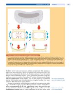
Case Introduction
This spinal specimen shows the extreme course of the result of aging on the lumbar spine. A sagittal section through the
lumbar spine (L3–S1) of an 8-year-old individual (
a) demonstrates that the nucleus pulposus can be clearly distinguished
from the anulus fibrosus. The cartilage endplates are composed of a thick layer of hyaline cartilage. The disc height is
somewhat less than the vertebral body height. The vertebral bodies demonstrate rounded edges. On the contrary, the
parasagittal section (
b) of a 77-year-old individual demonstrates that the disc space has completely collapsed. Anterior
or posterior displacement of the vertebral bodies is visible at all levels. The cartilaginous endplates are partially resorbed
and exhibit severe sclerotic alterations. The vertebral bodies exhibit severe bridging osteophyte formation. Despite
these dramatic changes there is no close link between these alterations and pain.
General Age-Related Changes
Various mechanisms on a cellular and systemic level have been identified to con-
tribute to age-related changes in the musculoskeletal system [45].
At the cellular level:
cellular senescence, leading to a decreasing ability of somatic cells to repli-
cate, repair, and maintain tissue
apoptosis (programmed cell death), leading to decreased cell numbers in
the affected tissue
accumulation of post-translational modifications of matrix proteins, lead-
ing to altered properties of the extracellular matrix
increasing generation of oxidative stress due to generation of reactive oxy-
gen species (ROS), leading to cell damage
genetic predisposition, leading to premature aging or phenotypic changes
in the musculoskeletal system
At the systemic level:
Systemic and cellular factors
contribute to musculo-
skeletal age-related changes
declining levels of trophic hormones, leading to altered tissue environment
and response of tissue to use and injury
general age-related changes,suchasadecreaseinreactiontime,proprio-
ception, vision, hearing, pulmonary and cardiovascular function, leading to
decreased mobility and therefore affecting the musculoskeletal system
socioeconomic and psychosocial factors alsocontribute,mainlybyinflu-
encing the individual variation regarding the age-related impairment of
mobility
The diversity of contributing factors on cellular and systemic levels underlines
the multifactorial nature of age-related changes that will finally lead to alter-
92 Section Basic Science
ations of the local environment within the affected tissue. These local alterations
can then directly affect the function of the respective tissue. Although the result,
i.e. altered tissue function, can be observed and analyzed, the exact relationships
and interactions between cellular and systemic changes are not yet clear.
The spine is most frequently
affected by
age-related alterations
Although any part of the musculoskeletal system can be affected by age-
related alterations, lower extremities and especially the lumbar spine are the
most frequently reported locations of musculoskeletal impairment (
Case Intro-
duction
). Between 70% and 85% of the population in Western industrialized
countries will experience back pain at least once during their lives, underlining
the impact of age-related alterations to the spine [33, 35, 86, 151, 152]. These epi-
sodes of back pain often lead to sickness leave and sometimes to chronic disabili-
ties (approx. 10%) causing an enormous socioeconomic burden on society [80].
In this context, it is important to notice that normal age-related degenerative
changes and pathological degeneration leading to back pain have to be distin-
guished. Several studies have shown that between 7% and 72% of individuals
that exhibit signs of disc degeneration never experienced relevant low back pain
[15, 115, 155].
Among age-related alterations of the spine, the so-called “degenerating spon-
dylosis” or spinal osteoarthritis is the most common and is probably inevitable
Degenerative spondylosis
is inevitable with aging
with increasing age. This alteration is radiologically characterized by osteophy-
tes (bone spurs) arising from the margin of the vertebral body and is usually
accompanied by disc space narrowing. The term “spondylosis” was historically
an effort to distinguish between degenerative changes in the spine and those in
synovial joints (osteoarthritis) such as facet joints [145]. However, it has been
shown that pathological changes in the spine and osteoarthritis of the synovial
joints coexist and in most cases are interrelated [145]. Autopsy studies by
Schmorl and Junghanns [64] reported evidence of spondylosis in 60% of women
and 80% of men by the age of 49 years, and in 95% of both sexes by the age of
70 years.
Functional Spine Unit
The motion segment is the
functional unit of the spine
The spine is a multi-segmented column, which provides stability and mobility to
the body at each segmental level and gives protection to the nerve roots and the
spinal cord. The smallest anatomical unit of the spine which exhibits the basic
functional characteristics of the entire spine is called the “motion segment”or
“functional spine unit”(
Fig. 1). It was first described by Schmorl and Junghanns
[64]. Each motion segment consists of two adjacent vertebrae, separated dorsally
by the zygapophyseal joints or facet joints and anteriorly by the interposed inter-
vertebral disc. The vertebrae are further connected by spinal ligaments, joint
capsules and segmental muscles. The spinal ligament complex consists of the
interspinous, supraspinous intertransverse, yellow, anterior and posterior longi-
tudinal ligaments. In contrast to the extrinsic muscles, the intrinsic muscles span
over two vertebrae and consist of splenius, erector spinae, transversospinal and
segmental muscles. Spine motion, stability and equilibrium are achieved by the
antagonist action of the powerful flexor and extensor muscle groups.
The motion segment
is a three joint complex
The normal spinal function largely depends on the integrity of these compo-
nents and their coordinated interplay. Kirkaldy-Willis [71] introduced the term
“the three joint complex” to highlight the importance of a normal interaction of
the three joints in a segment, i.e. the intervertebral disc and the two facet joints.
Any alterations in one of these components will result in a disturbance of their
interplay with subsequent dysfunction, finally leading to back pain, deformity
and neurological compromise.
Age-Related Changes of the Spine Chapter 4 93
a
b
Figure 1. Functional spinal unit
Schematic representation of a functional spinal unit (motion segment) in a the cervical and b lumbar spine.
94 Section Basic Science


