Spinal Disorders: Fundamentals of Diagnosis and Treatment Part 16 pps
Bạn đang xem bản rút gọn của tài liệu. Xem và tải ngay bản đầy đủ của tài liệu tại đây (560.85 KB, 10 trang )
that operate in sensory pathways to generate those neural signals that we ulti-
mately interpreted as pain [9, 18, 55, 112].
Epidemiology of Chronic Pain
Chronic pain
is very common
Epidemiological studies show a prevalence of chronic pain from 24 %to46 % in
the general population [31, 102]. Elliott et al. [31] showed that about 15% of
patients suffer from the worst degree of pain. The most frequently reported
forms of pain in this study are back pain and arthritic pain. In a 1-year follow-up
study, 79% of patients reporting chronic pain at the baseline investigation still
suffered from pain at the end of the study [31]. During this period the average
annual incidence was about 8.3%, whereas the recovery rate was about 5.4%
[31]. Chronic pain is localized in 90 % of patients to the musculoskeletal system.
Axial pain is very frequent
(85%) and strongly tends
to chronify
The incidence of musculoskeletal pain is reported to vary from 21% for shoul-
der pain up to 85% for low back pain in the industrialized nations [3, 10, 24, 42].
Thereportedlifetimeprevalenceofbackpainis84%[15]andthatofneckpain
67% [20]. Dorsal (thoracic) pain is much less frequent. The 1-year prevalence of
dorsal pain was 17% compared to 64% for neck and 67% for low back pain in a
Finnish study [85]. In a primary care setting, most patients improve considerably
during the first 4 weeks after seeking treatment. Sixty-six to 75% continue to
experience at least mild back pain 1 month after seeking care. At 1 month,
approximately 33% report continuing pain of at least moderate intensity,
whereas 20–25% report substantial activity limitations. After more than 1 year,
approximately 33% of patients report intermittent or persistent pain of at least
moderate intensity, 14% continue to report back pain of severe intensity, and
20% report substantial activity limitations [118]. The patient population suffer-
ing from chronic back pain has been found to be responsible for an enormous
part of the cost of the health care system (intake of analgesics, medical consulta-
tions, hospitalizations, requirement for diagnostic and therapeutic procedures)
[82] (see also Chapter
6 ).
Definition and Classification
The manifestation of pain is largely variable but we define all sensations that hurt
or are unpleasant as pain. The Taxonomy Committee of the In ternational Asso-
ciation for the Study of Pain (IASP) [50] has provided a definition, which is
widely used today (
Table 1).
Table 1. Definition of pain
“Pain is an unpleasant sensory and emotional experience associated with actual or poten-
tial tissue damage, or described in terms of such damage”.
The IASP task force [50] stresses the fact that the inability to communicate ver-
bally does not exclude that an individual is experiencing pain and requires
appropriate pain-relieving treatment. Furthermore, the task force highlights that
Pain is always subjectivepain is always subjective. Each individual learns the application of the word
through experiences related to injury in early life. Accordingly, pain is that expe-
rience we associate with actual or potential tissue damage. It is also always
unpleasant and therefore has an emotional experience. However, many people
report pain in the absence of tissue damage or any likely pathophysiological
cause. This latter pain cannot be differentiated from pain due to tissue damage if
Pathways of Spinal Pain Chapter 5 125
we consider the subjective report. If these individuals regard their experience as
pain and if they report it in the same ways as pain caused by tissue damage, it
should be accepted as pain [50].
Temporal Course
From a temporal perspective [50, 101], pain can be differentiated as:
acute pain (<4 weeks)
subacutepain(4weeksto3months)
chronic pain (>3–6 months)
Chronic pain induces
molecular and cellular
changes in the nervous
system
Acute pain is caused by an adequate stimulation of nociceptive neurons. This
pain typically results from soft tissue injury or inflammation and has a protective
role by enabling healing and tissue repair [81, 122]. Subacute pain is often less
intense and follows the acute phase. It is regarded as organic pain from tissue
healing and remodeling. It usually lasts up to 12 weeks but usually not longer. In
contrast, chronic pain has lost its protective role. In retrospect, it is often difficult
to identify the noxious stimulus or tissue damage in patients presenting with
chronic pain which originally causes the pain. Chronic pain induces biochemical
and phenotypic changes in the nervous system that escalate and alter sensory
inputs, resulting in physiologic, metabolic and immunologic alterations that
threaten homeostasis and contribute to illness and death [81].
Contemporary Pain Classification
A timely distinction of pain is given by Clifford Woolf [106, 123], who suggests
differentiating (
Fig. 1):
nociceptive pain
inflammatory pain
neuropathic pain
functional pain
Nociceptive Pain
Nociceptive pain is a vital physiologic sensation which occurs in situations like
trauma or surgery [123]. Acute nociceptive pain is elicited by noxious stimula-
tion of normal tissue sufficiently intense to damage tissue. It has the important
function of protecting tissue from further damage by, e.g. eliciting withdrawal
reflexes.
Inflammatory Pain
Adaptive pain
is a physiologic
protection mechanism
In the case of tissue damage that occurs despite an intact nociceptive defensive
system, the role of the nociceptive system switches from preventing noxious
stimulation to promoting healing of the injured tissue. Inflammatory pain is
characterized by an increased sensitivity to stimuli, which does not cause pain
under normal conditions. This protects the individual from further damage to
the injured part until the healing and repair process is completed. Inflammatory
pain normally decreases during the healing process. An exception is inflamma-
tory pain states due to surgery or chronic diseases such as rheumatoid arthritis.
In these cases, pain management has to be conceptualized that decreases or nor-
malizes pain sensitivity without impairing the warning system of nociceptive
pain [59, 61, 106, 123, 125, 126].
126 Section Basic Science
ab
cd
Figure 1. Classification of pain
Redrawn from Woolf [123] (with permission from ACP).
Neuropathic Pain
Neuropathic pain
is the result of direct damage
or disease of neurons
In contrast to nociceptive pain, which is provoked by noxious stimulation of the
sensory endings in the tissue, neuropathic pain is the result of a direct damage or
disease of neurons in the periphery or central nervous system and seems not to
have any beneficial effect. Therefore, peripheral neuropathic pain syndromes are
differentiated from central pain. Neuropathic pain normally is felt as abnormal,
because it is not related primarily to a signal of tissue damage. It often occurs
spontaneously in a continuous or episodic form and is associated with other sen-
sory abnormalities. Neuropathic pain often has a burning or electrical character
Allodynia and hyperalgesia
are found in neuropathic
pain
and might be combined with allodynia and/or hyperalgesia. This type of pain
often shows a chronic course and in most cases is difficult to treat. Neuropathic
pain can have a variety of causes, e.g. [27, 106, 123, 128, 134]:
nerve root injury (traumatic, compression syndrome)
spinal cord injury
brain lesions
diabetic polyneuropathy
AIDS polyneuropathy
postherpetic
Pathways of Spinal Pain Chapter 5 127
Functional Pain
No morphological correlate
can be found in functional
pain
This form of pain occurs due to an abnormal responsiv eness or function of the
nervous system. In the clinical examination, no neurological or peripheral
abnormalities can be found. The physiological basis of functional pain is an
increased sensitivity or hyperresponsiveness of the sensory system that amplifies
symptoms. Syndromes which belong to this class of pain are, e.g. [106, 123]:
fibromyalgia
irritable bowel syndrome
non-cardiac chest pain
tension headache
Pathways of Pain
The physiologic processes [61, 81,123] involved in pain sensation include (Fig. 2):
transduction of noxious stimuli (thermal, mechanical and chemical) into
electrical activity at the peripheral terminal of nociceptor sensory fibers
conduction of the resulting sensory input to the central terminal of nociceptors
transmission and modulation of the sensory input from one neuron to
another
projection to the brain stem, thalamus and cortex
perception ofthesensoryinputatthesomatosensorycortex.
Figure 2. Pathways of pain
128 Section Basic Science
Transduction
Nociception can be defined as the detection of noxious stimuli and the subse-
quent transfer of encoded information to the brain while pain is a perceptual
processthatarisesinresponsetosuchactivity[61].Nociceptionismediatedby
activation of peripheral sensory-nerve terminals located in, e.g. the skin, deep
fascias, muscles, and joints. These terminals are called primary sensory neurons
There are three types of
nociceptor: mechanical,
thermal, and chemical
or nociceptors. We can differentiate three types of noxious stimuli which are tar-
geted by the receptor of nociceptors, i.e.:
mechanical (pressure and mechanical stress)
thermal (hot/cold)
chemical
Primary sensory neurons can be excited by noxious heat, intense pressure or
irritant chemicals, but not by innocuous stimuli such as warm or light touch [55].
The conversion of a noxious thermal, mechanical, or chemical stimulus into elec-
trical activity in the peripheral terminals of nociceptor sensory fibers is
described as transduction [123].
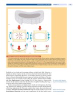
Mechanical stress resulting from direct pressure, tissue deformation or osmo-
larity changes can activate nociceptors allowing for the detection of touch, deep
pressure, distension of a visceral organ, destruction of bone or swelling [55]
(
Fig. 3a). These stimuli are mediated by mechanosensory transducers such as ion
channels of the degenerin family (mammalian degenerin, MDEG) or acid-sens-
ing ion channel 2 (ASIC2) [39, 55]. Mechanical stimulation can release ATP from
the cell activating G-protein-coupled ATP receptors (P2Y) or ATP-gated ion
channels (P2X) [55, 83]. Noxious heat can be detected by the vanilloid receptor
(TRPV1,formerlyalsocalledVR1)andthevanilloid receptor-like (TRPV2, for-
merly called VRL-1) channel, which belong to the larger family of transient
receptor potential (TRP) channels. The core membrane structure of the recep-
tors resembles that of voltage-gated potassium or cyclic nucleotide-gated chan-
nels [55, 83]. The TRPM8 receptor, a distant relative of TRPV1, has been identi-
fied as detecting noxious cold [75, 88]. Nociceptors uniquely express two voltage-
ab
Figure 3. Nociceptive transduction and transmission
a Nociceptive transduction (ASIC acid sensitizing ion channel, TRP transient receptor potential channels, MDEG mamma-
lian degenerin channel, P2X ATP-gated ion channel).
b Nociceptive transmission (AMPA -amino-3-hydroxy-5-methyl-4-
isoxazolepropionic acid receptors). Redrawn from Woolf [123] (with permission from ACP).
Pathways of Spinal Pain Chapter 5 129
gated sodium channels (Na
v
1.8 and Na
v
1.9), which could become the target for
selective anesthetics blocking only pain but leaving innocuous sensation, motor
and autonomic output intact [123].
Conduction
Conduction is the action
potential passage from the
peripheral to the central
nociceptor terminal
Conduction is the passage of action potentials from the peripheral terminal
along axons to the central terminal of nociceptors in the spinal cord [123]. Dorsal
root ganglion (DRG) cell bodies give rise to three different fiber types [55, 61]:
Ctypefibers
A fibers
A fibers
C type fibers are unmyelinated fibers ranging in diameter from 0.4 to 1.2 μm and
have a velocity of 0.5–2.0 m/s. These fibers present the thermosensitive receptors
reacting to temperature (heat/cold), mechanoreceptors of low threshold and spe-
cific receptors for algogenic substances [2, 55, 78].
A␦ fibers are lightly myelinated ranging in diameter from 2.0 to 6.0 μm and
have a velocity of 12–30 m/s. These fibers are classified into two subgroups. Type
I presents high-threshold mechanoreceptors and they respond weakly to chemi-
cal and thermal stimuli. Type II corresponds mainly to mechanothermal recep-
tors for high temperatures and intense cold [2, 55, 78].
A fibers are myelinated with a diameter of more than 10 μm and a velocity of
30–100 m/s. These fibers mediate the sensations of touch and mild pressure, as
well as the sensation of joint positions (proprioception) and vibration [2, 55, 78].
Their activation contributes to mechanisms of segmental suppression in the spi-
nal cord.
Activation of C type fibers and A fibers leads to burning sensations and
twinges. Under pathological conditions, signs of neuropathic pain, e.g. dysesthe-
sia and paresthesia, can result from activation of A fibers. Pathologic pain sen-
sation can manifest as hyperalgesia mediated by C fibers and A fibers. Under
pathologic conditions, activation of low threshold mechanoreceptors (A fibers)
can evoke allodynia (touch evoked pain) [2, 55, 78].
Transmission and Modulation
Transmission is the first
synaptic transfer
Transmission is the synaptic transfer of sensory input from one neuron to
another [123].
The sensory input
is modulated in the
dorsal horn
The primary sensory neurons terminate in the dorsal horn in a highly orga-
nized fashion, innervating both intrinsic dorsal horn interneurons and projec-
tion neurons. The dorsal horn is the first site of synaptic transmission (or inte-
gration) in the nociceptive pathway and is subject to considerable local and
descending modulation [18].
Dorsal Horn Cytoarchitecture
The dorsal horn exhibits
a distinct cytoarchitecture
The gray matter of the spinal cord can be divided into ten laminae.Ofthese,lami-
nae I (marginal layer), II (substantia gelatinosa), III, IV (nucleus propius), V and
VI(deeplayers)comprisethedorsalhorn[78].Thelaminaeformcolumns
extending along the spinal cord [81, 99]. Within the columns, a large number of
second-order excitatory and inhibitory interneurons receive multiple inputs
from surrounding columns and send outputs to the brain and to the anterior
horn [81]. The neuronal network of the dorsal horn hence serves as a gate con-
trolling propagation of nociceptive signals to higher brain areas [132].
130 Section Basic Science
Figure 4. Cytoarchitecture of the dorsal horn
The cytoarchitecture of the dorsal horn is very complex [2, 78, 81, 99, 127]. Sim-
plified, large myelinated low-threshold A afferents terminate in laminae III and
IV, lightly myelinated high-threshold A fibers synapse at laminae I and V, and
non-myelinated high-threshold C fibers terminate in lamina II but also terminate
with some fibers in laminae I and V [111, 127] (
Fig. 4).
There are three distinct
neuron types within
the dorsal horn
Within the dorsal horn three distinct types of neurons can be identified
according to the type of afferents and their response pattern to nociceptive input
[78]:
nociceptive-specific (SN) neurons
multireceptorial or wide-dynamic range (WDR) neurons
non-nociceptive neurons
Nociceptive-specific (NS) neurons are located in the substantia gelatinosa but
can also occur in layers (laminae V and VI) under physiologic conditions. They
are exclusively activated by high intensity noxious stimuli mediated by C and A
fibers [78].
Multireceptorial or wide-dynamic range (WDR) neurons respond to thermal,
mechanical and chemical stimuli via C, A and A fibers. These neurons are
foundtoalesserdegreeintheventralhorn(VH).WDRneuronspresentacon-
siderable convergence from cutaneous, muscle and visceral input. This type of
neuron is the major type of neuron that encodes stimulus intensity [26]. Addi-
tionally, these neurons participate mainly in the C-fiber-mediated processes of
sensitization and amplification of prolonged pain [78].
Non-nociceptive (N-NOC) neurons are activated by innocuous stimuli such
as low intensity mechanical, thermal and proprioceptive stimuli, mediated by
A and A fibers. They are found predominately in laminae II, III and IV [78].
These neurons act indirectly in segmental suppression mechanisms [2]. The dif-
ferent types of neurons are connected via second order excitatory and inhibitory
interneurons. These interneurons receive multiple inputs from other columns
and send information and impulses to the brain [81]. After modulation and
modification of the nociceptive stimulus within the dorsal horn, the informa-
tion is transmitted to the CNS. Afferents of the spinal cord dorsal horn neurons
form so called spinal tracts that transmit nociceptive informations to the CNS.
Pathways of Spinal Pain Chapter 5 131
Plasticity or modifiability of synaptic transfer in the dorsal horn is a key feature
of its function and integral to the generation of pain and pain hypersensitivity
[18].
The major synapses responsible for transmission are located in the dorsal
horn of the spinal cord in lamina I (marginal zone) and lamina II (substantia
gelatinosa). These impulses are conveyed to the thalamus, the main region for the
integration of brain input [37]. The transfer of nociceptive stimuli is mediated by
direct monosynaptic contact or through multiple excitatory or inhibitory inter-
neurons. Transmission of nociceptive stimulus is inhibited by descending path-
ways of the brain stem and midbrain and collateral influences within the dorsal
horn [37, 106].
Modulation of Sensory Inputs
Transmission of the peripheral nociceptive signals to the brain undergoes vari-
ous modulatory influences in the dorsal horn by descending pathways [9, 37, 78].
Many neurotransmitters have been identified which mediate this modulation [9,
37] (
Table 2).
The sensory input is
modulated by inhibitory
and excitatory mechanisms
Modulation can be described as the process in which pain transmission is
modified or altered – “gated” – before being transmitted to the CNS. Nociceptive
impulses are modulated in two ways, i.e. by:
excitatory (facilitatory) mechanisms
inhibitory mechanisms
Inhibitory Mechanisms
The majority of the
inhibitory mechanism
is GABA-dependent
Inhibitory mechanisms can originate from local (segmental) inhibitory inter-
neurons or from descending antinociceptive pathways. The majority of local
inhibitory neurons in the spinal cord release glycine and/or -aminobutyric acid
(GABA). The descending inhibition pathways originate at the level of the cortex
and thalamus, and descend via the brain stem (periaqueductal gray) and the dor-
sal columns to terminate at the dorsal horn of the spinal cord. These descending
pathways modulate nociceptive transmission through the release of serotonin (5-
HT) and/or norepinephrine [37, 78]. Inhibition can be postsynaptic or presynap-
tic. Postsynaptic inhibition results from a hyperpolarization of the cell mem-
brane and/or from the activation of a shunting conductance, which impairs prop-
Table 2. Neurotransmitters
Peptides Non-peptides
Opioid peptides
-endorphin
enkephalins
dynorphins
Non-opioid peptides
substance P
somatostatin
neurotensin
cytokines (IL-1 ,IL-6,TNF- )
calcitonin gene related peptide (CGRP)
galanin
neuropeptides Y
nerve growth factor (NGF)
cholecystokinin (CCK)
purines
nociceptin
Monoamines
norepinephrine
serotonin (5-HT)
Amino acids
inhibitory amino acids (GABA, glycine)
excitatory amino acids (aspartate, glutamate)
Nitric oxide (NO)
132 Section Basic Science
agation of excitatory postsynaptic potentials along the dendrite of neurons [132].
Presynaptic inhibition occurs at axoaxonic synapses of GABAergic neurons with
primary sensory nerve terminals [37].
Excitatory Mechanisms
Glutamate plays a pivotal
role as an excitatory
transmitter
The excitatory transmitter glutamate is released by primary afferent fibers and
plays a pivotal role in the spinal mechanisms of nociceptive transmission [9].
Synaptically released glutamate acts on kainate and AMPA ( -amino-3-hydroxy-
5-methyl-4-isoxazolepropionic acid) receptors, being responsible for a fast syn-
aptic transmission at the first synapse in the dorsal horn (
Fig. 3b). Transient and
non-injurious noxious stimuli result in stable AMPA receptor-mediated synaptic
signals which are finally perceived as a transient localized pain [123]. Glutamate
can also act on N-methyl-
D-aspartate (NMDA) receptors, but this receptor is
blocked under resting conditions by extracellular magnesium ions [81]. Depolar-
ization of the postsynaptic neuron, e.g. through intense AMPA receptor activa-
tion,removesthismagnesiumblock.Inaddition,activatorsofproteinkinaseC
can reduce the sensitivity of NMDA receptors to magnesium, possibly contribut-
ing to spinal hypersensitivity and amplification of peripheral inputs. The activa-
tion of the NMDA receptors also leads to an entry of calcium, which is a key event
in the generation of long lasting potentiation of synaptic transmission (LTP). In
addition, calcium activates various enzymes such as nitric oxide (NO) synthase
and phospholipases [9], which can also augment pain sensitivity.
Wind-up is an activity-
dependent phenomenon
responsible for increasing
pain in response to
repeated stimuli
Closely timed repeated stimulation of C fibers results in an increased response
even though the amplitude of the input signal remains unchanged. This activity-
dependent phenomenon known as wind-up is responsible for the increasing
pain experienced in response to closely repeated stimulation of the skin by nox-
ious heat [72, 123].
Pain Projection
Nociceptive information
is projected to supraspinal
structures via afferent
bundles
Subsequent to pain transmission and modulation within the dorsal horn, noci-
ceptive information is projected to the supraspinal structures via afferent bun-
dles (
Fig. 5). These bundles can be differentiated into several tracts with special
functions [2]:
spinothalamic tract involved in sensory-discriminative components and
motivational-affectiveaspectsofpainaswellastheaffectivecomponentsof
painful experience
spinoreticular tract involved in the motivational-affective aspects and neu-
rovegetative responses to pain
spinomesencephalic tract involved in somatosensory processing, activation
of descending analgesia, inducing aversive behaviors in response to nocicep-
tive stimuli as well as autonomic, cardiovascular, motivational and affective
responses
spinoparabrachial tract involved in autonomic, motivational, affective regu-
lation and in the neuroendocrine responses to pain
spinohypothalamic tract involved in neuroendocrine autonomic, motiva-
tional, affective and alert responses of somatic and visceral pain
spinocervical tract involved in the sensory-discriminative components and
motivational-affective and autonomic responses of pain, and plays a role in
sensory integration and modulation of afferent inputs
postsynaptic pathways of spinal column involved in the sensory-discrimina-
tive components and motivational-affective aspects of pain
Pathways of Spinal Pain Chapter 5 133
Figure 5. Afferent pathways
Pain Perception
Thalamus and
somatosensory cortex
are the main structures
of pain perception
The spinal projection pathwa ys project to the reticular formation of the brain
stem and surrounding nuclei before converging in the thalamus, the main struc-
ture for reception, integration and nociceptive transfer of nociceptive stimuli
before transmission to the somatosensory cortex. However, only a small propor-
tion of all the sensory input from the spinal cord arrives at the thalamus because
of local processing, modulation, and controlling [123]. The somatosensory cor-
tex in turn projects to adjoining cortical association areas, predominately the
limbic system. The limbic system includes [81]:
cingulate gyrus (behavior and emotion)
amygdala (conditioned fear and anxiety)
hippocampus (memory)
hypothalamus (sympathetic autonomic activity)
134 Section Basic Science


