Spinal Disorders: Fundamentals of Diagnosis and Treatment Part 30 pps
Bạn đang xem bản rút gọn của tài liệu. Xem và tải ngay bản đầy đủ của tài liệu tại đây (262.81 KB, 10 trang )
Table 5. Indications for provocative discography
Differentiation of symptomatic and asymptomatic disc alterations
Disc degeneration
Annular tears (high intensity zones)
Endplate changes (modic changes)
Minor disc protrusions with questionable nerve root compromise
Technique
Inject an MRI normal disc
as a negative control
Discography should be performed by a spine specialist or a dedicated radiologist
with experience of the diagnostic assessment of spinal disorders. It is mandatory
that the patient is awake during the procedure to allow for communication about
the injection response. However, mild sedation is helpful during the procedure.
Lumbar Discography
In lumbar discography the posterolateral approach is widely accepted as the
technique of choice. A double needle technique (with a short 18-gauge external
and an internal 22-gauge needle) is widely recommended [48, 116]. In patients
withunilateralpain,theneedleisintroducedfromthecontralateralsidetodis-
tinguish between iatrogenic and genuine pain. The needle position is verified
underfluoroscopyintwoplanes.Afteraccurateneedlepositioning,contrast
medium containing an iodine concentration of 300 mg/ml is injected into each
disc by using a 5-ml syringe. The amount of contrast agent injectable before leak-
age usually ranges from 0.8 ml to 3.0 ml before leakage [10]. Non-ionic contrast
agent is injected with a 5-ml syringe until firm resistance to the injection is felt,
untilseverepainisprovoked,oruntilcontrastmediumisseentoleakoutofthe
Pain provocation should
be graded as concordant
or non-concordant
disc into the spinal canal. During discography, the patient is asked to grade the
pain provoked on a visual analogue scale. The type of pain should be graded
according to the Dallas Discogram Description [97] as follows:
no sensation
pressure
dissimilar pain
similar pain, or
exact pain reproduction
Discogenic pain is based
on the provocation of
concordant pain
Pain sensation occurring during discography is defined as concordant if the
patient had exact pain reproduction or felt similar pain. Accordingly, non-con-
cordant pain is defined as pressure, dissimilar pain sensation, or no pain provo-
cation. Evaluation of disc morphological characteristics is performed with con-
ventional radiographs by using the classification of Adams etal.[1].Theclassifi-
cation includes five stages of disc degeneration distinguished by their morpho-
logical appearance on discograms:
cotton ball (Type I)
lobular type (Type II)
irregular (Type III)
fissured (Type IV)
ruptured (Type V)
Types I and II are interpreted as non-degenerative discs and Types III–V as
degenerative discs.
It has been very helpful to include an MRI normal disc as an internal control.
In our practice, we only regard concordant pain predictive of discogenic pain
when the injection of the control level does not provoke pain [129].
272 Section Patient Assessment
Thoracic Discography
Thoracic discography is performed under CT guidance on an outpatient basis.
The patient is placed in a prone position on the CT table. Following a scout film
Thoracic discography
should only be done under
CT guidance
of the thoracic spine the level of interest is scanned with a section thickness of
3 mm. After choosing the target thoracic disc, the CT-table position is adjusted.
The side opposite, if present, is chosen as the injection side, so as not to provoke
patient pain while advancing the needle. Under CT guidance a 25-gauge needle is
advanced into the target disc. After positioning of the needle in the center of the
disc, contrast medium (iopamidol, 1.5 cc) is injected and a CT discogram scan
performed. The patient is questioned about the pain provoked during injection
as mentioned above.
Cervical Discography
For this procedure, the patient lies supine with the neck in slight extension. The
neck is draped in a sterile fashion. By using a 22-gauge needle, through an ante-
romedial approach (medial to the m. sternocleidomastoideus), the needle is
advanced to the center of the disc under biplanar fluoroscopic control.Thetra-
chea and esophagus remain medially and the carotid artery is palpated and dis-
placedlaterally.Theamountofcontrastagentinjectedusuallyrangesfrom
0.3 ml to 1.0 ml. The pain response is assessed similarly to the lumbar proce-
dure.
Complications
Any needle technique carries with it the risk of infection, which appears to be
most relevant in cases of cervical and lumbar discography. The reported rate for
discitis after lumbar discography is in the order of magnitude of 0.25% [130].
Further complications are reported such as retroperitoneal hemorrhage, allergic
reaction, subarachnoidal bleeding, nerve root sheath injuries, or annular or end-
Therateofpost-discography
discitis ranges between
0.16% and 0.37 %
plate injections due to incorrect needle placement. Of 807 injected cervical discs,
Grubb et al. [47] had a rate of discitis of 0.37% corresponding to 1.7% patients
with discitis treated. In Zeidmann’s [136] review of 4400 diagnostic cervical dis-
cography cases, discitis occurred in 7 cases (0.16%).
Diagnostic Efficacy
In 1948 Lindblom [50] introduced discography as a morphological test to replace
or add information to myelography. Today the role of discography is related to a
Diagnostic accuracy is diffi-
cult to determine because
a gold standard is lacking
pain provocation test. The assessment of the diagnostic accuracy of provocative
discography for discogenic LBP is problematic since no gold standard is avail-
able. A reasonable practical approach is to include an adjacent normal disc level
as internal control [129]. Thus, a positive pain response would include an exact
pain reproduction at the target level and no pain provocation or only pressure at
the normal disc level. However, careful interpretation of the findings is still man-
datory with reference to the clinical presentation.
Lumbar Discography
In a prospective, controlled study, Walsh et al. [123] studied ten asymptomatic
volunteers and seven symptomatic patients with low back pain by lumbar discog-
raphy. In the asymptomatic individuals, the injection produced minimum pain
in 5 (17%) of the 30 discs and in 3 moderate to bad pain. The false-positive rate
Spinal Injections Chapter 10 273
a
b
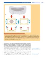
Figure 4. CT discography
Axial CT discogram showing contrast medium distribution within the intervertebral disc. a Sagittal view of CT/discogram
showing contrast medium extension to the margin of the disc.
b Corresponding MRI of the disc
of 0% and a specificity of 100% led the authors to conclude that discography is a
highly reliable and specific diagnostic test for the evaluation of low back pain dis-
orders [123]. In 1999, Caragee et al. [24] reported on patients with no history of
The diagnostic value
of discography remains
amatterofdebate
low back pain, who underwent posterior iliac crest bone graft. These patients
often experienced concordant pain on lumbar discography. However, this study
can be criticized because asymptomatic patients cannot perceive concordant dis-
cogenic pain. In 2000, Carragee repeated provocative discography in 26 older
subjects without history of low back pain [23]. They concluded that the rate of
false-positive discography may be low in subjects with normal psychological
testing and without chronic pain. Furthermore, Caragee and colleagues [23] per-
formed provocative discography in 20 asymptomatic patients who underwent
single level discectomy for sciatica. Forty percent injections were positive in discs
that had previous surgery.
Patientswithlowbackpainwhohadlumbarfusionsurgerybasedonpositive
discograms have been shown to have only moderate results. Complete pain relief
was achieved only in a few cases. Successful clinical results ranged between
86.1% and 46%. This indicates that confounding factors other than morphologi-
cal alterations may play a more important role in predicting surgical outcome
(see Chapter
7 ).
CT discography (
Fig. 4)representsafurtherstepintheapplicationofdiscog-
raphy and evaluation of the structure of the disc. The debate as to whether CT/
discography is superior to MRI because there is a theoretical advantage of CT/
discography over MRI in demonstrating the internal architecture of the disc has
not been conclusively answered. But, CT discography was found to have a higher
accuracy than pain provocation and plain discography, 87% vs 64% vs 58%
respectively [54, 55].
Thoracic Discography
Thoracic discography performed by experienced radiologists with CT guidance
is quite safe with a very low rate of complications. Similar to lumbar discography,
274 Section Patient Assessment
it seems to be accurate in distinguishing painful symptomatic discs from asymp-
tomatic discs. Wood et al. performed four-level thoracic discography in ten
asymptomatic volunteers and compared the discograms with MRI studies. Three
of the 40 discs were reported as intensely painful, all exhibiting prominent end-
plate infractions typical of Scheuermann’s disease. Of the 40 discs studied, only
13 were judged to be normal morphologically on discography versus 20 on MRI.
The remaining 27 discs were abnormal, exhibiting endplate irregularities, annu-
lar tears, and/or herniations. Wood et al. studied concomitantly thoracic disco-
grams of ten adults with chronic thoracic pain. In this group 48 discs were ana-
lyzed, of which 24 were concordantly painful and 17 had non-concordant pain or
pressure. On MRI, 21 of the 48 discs appeared normal, whereas on discography
only 10 were judged as normal. The authors concluded that thoracic discography
detects pathologies which may not be seen on MRI [134].
Cervical Discography
Results of cervical
discography must be
interpreted carefully
Ohnmeiss et al. [82] studied 269 discs in patients with neck, shoulder and arm
pain by cervical discography. Comparing the pain responses during disc injec-
tion with radiological images, they found positive pain provocation in 234 radio-
graphically abnormal discs (77.8%). They pointed out that it is important not
just to assess pain intensity but to interpret the provoked pain in terms of its sim-
ilarity to clinical symptoms. Grubb et al. [47] reviewed their 12-year experience
with 807 injected cervical discs and found a 50% concordant pain response rate.
They concluded that cervical discography provokes concordant pain in multiple
discs and conclusions about which disc should be treated must be drawn cau-
tiously.
So far, provocative discography appears to be the only diagnostic test available
to differentiate symptomatic and asymptomatic disc degeneration allowing for a
direct relation of a radiological image to the patient’s pain [49, 129].
Facet Joint Blocks
Neck pain and low back
pain may be caused by
osteoarthritis of the facet
joints
Since the first report by Ghormley [44], facet joints have been recognized as a
predominant source of back pain. Their prevalence as a cause of low back pain
has been reported to vary greatly and to range from 7.7% to 75% depending on
the diagnostic criteria [21, 37, 53, 75–77, 99–104, 106]. Mooney and Robertson
[75] demonstrated that low back pain and referred pain could be provoked by
injection of hypertonic saline into the facet joints. Many authors today believe
that the diagnosis of a facet joint syndrome can be based on pain relief by an
intra-articular facet joint injection of an anesthetic or pain provocation by hyper-
tonic saline injection [25, 64, 70, 76].
Today, facet joint blocks are used as a diagnostic and/or therapeutic means to
eliminate pain presumably arising from the facet joints.
Indications
Similarly to disc degeneration, a differentiation of a symptomatic and asymp-
tomatic facet joint osteoarthritis based on imaging studies alone is not possible.
Therefore, facet joint blocks alleviating the patient’s symptoms presumably
resulting from alteration of the facet joints are the only modality to differentiate
symptomatic from asymptomatic states (
Table 6).
Spinal Injections Chapter 10 275
Table 6. Indications for facet joint blocks
differentiating symptomatic from asymptomatic facet joint alterations
short- to medium-term relief of back pain in patients with previous positive diagnostic
blocks
Technique
Lumbar Facet Joint Blocks
The blocks are performed under fluoroscopic guidance with the patient lying
prone. In order to visualize the lumbar joints either the patient is rotated and
supported in an oblique prone position or the X-ray beam is tilted accordingly.
The angulation is usually between 30° and 40°. After disinfection the skin over
the target joint is anesthetized with 2–3 ml of lidocaine. A spinal needle
(22 gauge) is then inserted in a lateromedial direction (parallel to the X-ray
beam) towards the joint. In obese patients, a double-needle technique is
employed where a 22-gauge needle is passed through a shorter 18-gauge needle.
Correct needle placement
should be documented by
contrast agent injections
Depending on the specific situation, either the mid point or rather the cranial or
caudal part of the joint is targeted. A minimal quantity of contrast medium
(<0.3 ml) is then injected under fluoroscopy to confirm the correct needle posi-
tion (
Fig. 5). If an intra-articular application is not possible, a periarticular injec-
tion is performed. Needle placement and contrast distribution are documented
by standard radiographs. Subsequently, 1.0 ml of a mixture of local anesthetics
(Carbostesin or bupivacaine and steroids, e.g. 40 mg triamcinolone) is injected.
The patients are kept under surveillance for at least 15 min.All patients should be
asked to assess the amount of pain prior to and 15–30 min after the injection
using a visual analogue scale. Further follow-up information on the course of
pain relief is helpful in interpreting the results.
Spondylolysis Block
A special type of lumbar facet joint block is injection into the spondylolysis. This
can be accomplished by injecting the facet joint located superior to the spondylo-
lysis using the same technique as outlined above. Since the facet capsule is often
connected to the spondylolysis zone, a filling can be observed which can extend
to the inferior facet joint (
Fig. 6).
Figure 5. Lumbar facet joint infiltration
Fluoroscopically guided lumbar facet infiltration docu-
menting the right position of the needles with correct
arthrography of the joint.
276 Section Patient Assessment
Figure 6. Spondylolysis block
A correct spondylosis block is performed by injecting the
facet joints at the level of L4/5. Contrast medium is extend-
ing through the lysis into the facet joint L5/S1.
Cervical Facet Joint Blocks
We prefer the posterior approach for the cervical facet joints C3/4 to C6/7. The
entry point lies two segments below the target joint. The patient is positioned
prone on the fluoroscopic table. A spinal needle (22 gauge) is passed through the
posterior neck muscles until it strikes the back of the target joint. For safety rea-
CT guided cervical facet
blocks are relatively safe
sons, the CT guided fluoroscopy can be used (Fig. 7).Theaccurateplacementof
the needle is confirmed by injection of 1 ml of contrast medium. Thereafter, the
steroid and anesthetic agent can be injected. Similarly to the lumbar spine, pain
relief is recorded prior to and 15–30 min after the injection using a visual ana-
logue scale.
Complications
Although complications are possible with any invasive procedure, reports on
series of thousands of facet joint injections reveal that they are relatively safe[68].
Any needle technique carries with it the risk of infection, which appears to be of
Complications of facet joint
blocks are rare
little relevance in cases of cervical and lumbar facet blocks. Complications are
reported such as retroperitoneal hemorrhage, allergic reaction, and nerve root
sheath injuries. There were some adverse effects like headache, nausea and pares-
thesiae, which are transient [70]. Obviously,side effects related to the pharmacol-
ogy of the anesthetic agent and corticosteroids are possible.
Spinal Injections Chapter 10 277
Figure 7. CT-guided facet block
CT guidance for cervical facet joint blocks is preferred
because of the spatial relationships to the spinal cord to
avoid neurological damage. Image showing correct nee-
dle placement at the level of C5/6. Note the correct arthro-
graphy on both sides.
Diagnostic and Therapeutic Efficacy
Lumbar Facet Joint Blocks
Facet joint blocks tackle
symptomatic facet joint
osteoarthritis
Some authors suggest that a facet joint syndrome can be diagnosed based on pain
relief by an intra-articular anesthetic injection or provocation of the pain by
hypertonic saline injection followed by subsequent pain relief after injection of
anesthetics [25, 64, 70, 76]. Jackson et al. [53] investigated clinical predictors
indicative of the injection response but had to conclude that there were no clear
clinical findings. Similarly, Revel et al. [89] did not find any difference in the fre-
quency of the 90 variables examined between the responder and non-responder
groups. Uncontrolled diagnostic facet joint blocks are reported with a false-pos-
itive rate of 38% and a positive predictive value of 31% [100]. It therefore is man-
datory to perform repetitive infiltrations to improve the diagnostic accuracy, e.g.
with two different local anesthetics as suggested by Schwarzer et al. [100]. Drey-
fuss [37] has concluded that there are no convincing pathognomonic, non-inva-
sive radiographic, historical, or physical examination findings that allow one to
definitively identify lumbar facet joints as a source of low back pain and referred
lower extremity pain.
Facet joints are innervated
polysegmentally making
interpretation of the pain
response difficult
According to a randomized double blind study by Marks etal. [70], intra-artic-
ular blocks are as effective as blocks of the medial branch of the dorsal ramus.
One problem of interpreting the response to a facet joint block is related to the
finding that facet joints are innervated by two to three segmental posterior
branches, making a diagnosis of the affected joint difficult. The evaluation of the
diagnostic accuracy of joint injections to diagnose a symptomatic facet joint is
difficult in the absence of a true gold standard.
Even less information is available on the therapeutic efficacy of facet joint
blocks in relieving pain attributed to facet joints [21]. Carette et al. [21] selected
110 out of 190 patients who experienced pain relief of more than 50% after an
intra-articular facet joint block with 2 ml lidocaine for a double blinded ran-
domized control trial comparing methylprednisolone versus isotonic saline
injection. They showed an immediate average pain reduction in the study
groupof76%vs79%intheplacebogroup.At6monthsfollow-up,however,the
patients in the study group reported a significantly higher pain relief (46% vs
15%).
278 Section Patient Assessment
Table 7. Therapeutic efficacy of facet joint blocks
Author/year Study design Technique Indication Patients Follow-up Outcome
Carette et al.
1991 [21]
randomized
double-blind
intra-articular lum-
bar facet block
saline vs steroid
low back
pain
49 vs 48 1, 3 and
6m
early benefit 42% vs 33 %,
after 6 months 46 % vs
15 %
Marks et al.
1992 [70]
randomized,
double blind
facet joint vs facet
nerve
lumbar or
lumbosa-
cral pain
42 vs 44 1 and 3 m no significant difference
Lilius et al.
1989 and
1990 [62, 63]
randomized,
not blinded
(1) intracapsular
steroid + bupiva-
caine, (2) pericap-
sular steroid +
bupivacaine, (3)
intracapsular saline
low back
pain
28 vs 39 vs
42
60 min, 3 m 64% benefit in all groups,
36% at 3 months, no sig-
nificant differences
between groups
Lynch 1986
[66]
controlled, not
randomized
2 levels intra-/
extracapsular vs
extracapsular
low back
pain
50 vs 15 6 m positive effect in all
treated patients
Revel et al.
1998 [88]
randomized,
double blind
intra-articular lido-
caine vs saline
low back
pain with
7 inclusion
criteria
43 vs 37 30 min significantly greater pain
relief in lidocaine group,
92%ofrespondersto
facet injection had 5 out
of 7 facet criteria
Gorbach et al.
2005 [46]
cohort, pro-
spective
intra-articular ste-
roid + bupivacaine
or mepivacaine
low back
pain
1 level: 29 15–30 min
= immedi-
ate
74% immediate pos.
effect (> 50%) pain relief,
57 % short term pos.
effect, 33 % medium term
pos. effect
2 levels: 13 >1 w = short
term
>3 m = me-
dium term
Note: w = weeks, m = months
Spondylolysis Block
There are no reports on the therapeutic value of pars infiltration. But, clinicians
who use pars infiltration preoperatively for patient selection have described
thatpatientswithpainreliefaremorelikelytobepainfreeafterlumbarfusion.
Patients without pain relief after pars infiltration could have other sources of
pain. Suh et al. reported that patients selected with positive pars infiltration
weremorelikelytohavepainrelief,tobefunctional,andtoreturntowork
[115].
Cervical Facet Joint Block
The result of facet joint
blocks is difficult to predict
So far, the accuracy and reliability of cervical facet blocks has not been demon-
strated.
Few data also exist about the therapeutic efficacy of therapeutic cervical
facet joint injections. One observational study found no benefit of cervical
intracapsular steroid injections in patients with chronic pain after whiplash
injury [2].
Spinal Injections Chapter 10 279
Sacroiliac Joint Blocks
The sacroiliac joints are
helpful in the diagnosis of a
symptomatic sacroiliac joint
Alterations of the sacroiliac (SI) joints remain a diagnostic and therapeutic
obstacle. Every joint can cause pain; therefore it is highly likely that pain can also
result from the SI joint [98]. Pain from the SI joint has been referred to the region
medial to the posterior superior iliac spine called the sacral sulcus. The pain can
also radiate into the groin, abdomen and thigh, which makes it difficult to distin-
guish SI joint pain from disc disease or facet arthropathy [41, 42]. The clinical
diagnosis is difficult to make since none of the clinical signs and tests has proven
to be predictive. Imaging is not very helpful in diagnosing painful SI joint
arthropathy in patients without inflammatory sacroiliitis [118]. A diagnostic
anesthetic block of the sacroiliac joint is a possibility for identifying this struc-
ture as a relevant source of pain [96]. Slipman et al. [109] suggested that the pain-
ful sacroiliac joint is caused by a mild synovial irritation, which is not detectable
on imaging. Other researchers assume that there is a chemical irritation of the
nerves innervating the joint by mediators from the joint fluid [41].
Therefore, the rationale for SI joint blocks is to support the clinical diagnosis
of an SI joint pathology.
Indications
Indications for sacroiliac joint blocks include the diagnostic work-up for patients
with low back and buttock pain radiating into the posterior thigh. Therapeutic
infiltrations have not been reported to be of long-lasting success and are there-
fore not very helpful.
Technique
This joint is for most of its extent inaccessible to needles due to the rough corru-
gated interosseous surfaces of the sacrum and the ileum. However, Bogduk et al.
[7] have described puncturing the joint from its inferior end where the joint
appears below the interosseous ligament and reaches the dorsal surface of the
sacrum deep to the gluteus muscles. The accurate method of sacroiliac joint
injection usually requires fluoroscopy or computed tomographic control [38, 39,
50, 108].
We describe here the technique which has been helpful in our service. With the
patient lying prone the entry point of the joint lies at the lower end of the joint
CT fluoroscopy facilitates
correct needle placement
and is identified with fluoroscopic aid. CT guidance is necessary in patients with
a complex orientation of the sacroiliac joint (
Fig. 8). In some patients even the
intra-articularaccesscanbeimpossible,alsoduetofusionofthejoint.Afterster-
ile skin preparation and draping, a 25-gauge needle (22 gauge) is introduced
through the skin directed to the posterolateral aspect of the sacrum and then
readjusted to enter the slit of the joint above the inferior edge. Once the needle is
in position, contrast medium is injected to confirm the correct position. Subse-
quently steroids and anesthetic agents can be injected for diagnostic and thera-
peutic purposes.
Complications
Complications due to sacroiliac joint injections are rare. Extravasation of anes-
thetic agent around the sciatic nerve can cause temporary numbness in up to 5%
of patients. If the needle is advanced too inferiorly, contact with the sciatic nerve
is possible [118].
280 Section Patient Assessment
ab
Figure 8. Sacroiliac joint block
Images showing correct needle placement (a) and art-
hrography of the sacroiliac joint (
b).
Diagnostic Efficacy
Sacroiliac joint infiltration
allows for the diagnosis of a
painful joint
Literature on sacroiliac joint injections and their impact on diagnosis and impact
is sparse [98]. No prospective or controlled evaluation of the technique has been
published. A few retrospective studies exist on the efficacy of sacroiliac joint
injections.
In the report by Maugurs et al. [72], 86% of patients had good pain relief after
sacroiliac joint injection after 1 month, which decreased to 58% after 6 months.
In the study by Bollow et al. [8], 92% of the 66 investigated patients had pain
relief. In Fortin’s study, 88% of 16 patients with non-inflammatory sacroiliac
joint syndrome had a decrease in pain after injection of anesthetic agent [41].
Slipmanetal.[108]selected31patientswithpaininthesacralsulcus,positive
stress test and relief of pain after a first sacroiliac injection with anesthetic agent.
After a second injection with an additional steroid mixture the patients had a sig-
nificant decrease in pain scores and improved functional status after a follow-up
of 94 weeks.
Today low back pain from the sacroiliac joint is best diagnosed when there is
relief of pain after injection of anesthetic agent. There is no gold standard for ver-
ifying the presence of sacroiliac joint pain to which the results of sacroiliac diag-
nostic block can be compared. Thus, there are no reliable data on the sensitivity
and specificity of this test [96].
Contraindications for Spinal Injections
There are few contraindications for spinal injections, which must be considered
before performing an infiltration. Alteration of the normal anatomy, e.g. pro-
nounced degenerative abnormalities, or after major surgery to the spinal canal,
where the positioning of the needle could be technically impossible, is per se not
a contraindication.
However, it is apparent that such injections can only be performed in patients
with normal hemostasis and without known allergic reactions. History taking on
potential allergic reactions is mandatory and laboratory screening strongly rec-
Spinal Injections Chapter 10 281


