Spinal Disorders: Fundamentals of Diagnosis and Treatment Part 33 pps
Bạn đang xem bản rút gọn của tài liệu. Xem và tải ngay bản đầy đủ của tài liệu tại đây (142.55 KB, 10 trang )
Several clinical tests can be applied to distinguish between these disorders.
In polyneuropathy the most specific finding is a pattern of loss of reflexes and
sensory deficit in a distal and sock like distribution (below the knee and/or in the
area covered by socks) of impaired light touch sensation and reduction of proprio-
ception. The latter is clinically tested by passively moving the foot or toes up and
down and asking the blindfolded patient to describe the direction of movement.
The impairment of dorsal column function is clinically tested by Romberg’s
test. This test is named after the German neurologist Moritz Heinrich Romberg
(1795–1873).
Romberg’s test is performed in two stages:
First, the patient stands with feet together, eyes open and hands by the sides.
Second, the patient closes the eyes while the examiner observes for a full
minute.
Because the examiner is trying to elicitwhether the patient falls when theeyes are
closed, it is advisable to stand ready to catch the falling patient. For large patients,
a strong assistant is recommended. Romberg’s test is positive if, and only if, the
following two conditions are both met:
The patient can stand with the eyes open; and
The patient falls when the eyes are closed.
The test is not positive if either:
The patient falls when the eyes are open; or
The patient sways but does not fall when the eyes are closed.
Maintaining balance while standing in the stationary position relies on intact
sensory pathways, sensorimotor integration centers and motor pathways.
The main sensory inputs are:
joint position sense (proprioception), carried in the dorsal columns of the
spinal cord
vision
Crucially, the brain can obtain sufficient information to maintain balance if
either the visual or the proprioceptive inputs are intact. Sensorimotor integra-
tion is carried out by the cerebellum. The first stage of the test (standing with the
eyes open) demonstrates that at least one of the two sensory pathways is intact,
and that sensorimotor integration and the motor pathway are intact. In the sec-
ond stage, the visual pathway is removed by closing the eyes. If the proprioceptive
Romberg’s test is not a test
of cerebellar function
pathway is intact, balance will be maintained. But if proprioception is defective,
both of the sensory inputs will be absent and the patient will sway then fall. Rom-
berg’s test is not a test of cerebellar function, as it is commonly misconceived.
Patients with cerebellar ataxia will generally be unable to balance even with the
eyes open: therefore, the test cannot proceed beyond the first step and no patient
with cerebellar ataxia can correctly be described as Romberg’s positive.Rather,
Romberg’stestissensitivetoanaffectionoftheproprioceptionreceptorsand
pathways caused by sensory peripheral neuropathies (such as polyneuropathy)
or disorders of the dorsal columns of the spinal cord.
Unterberger’s test identifies
labyrinth dysfunction
Unterberger’s stepping test is a simple means of identifying labyrinth dys-
function, which can induce vertigo and dysbalance during walking and standing.
During the clinical testing the patient is asked to perform stationary stepping for
1 min with their eyes closed and the arms lifted in front. A positive test is indi-
cated by rotational movement of the patient towards the side of the lesion.
Cerebellar dysfunct ion is clinically searched for by the heel-to-knee test and
the finger-to-nose test. These tests assess dysmetric and ataxic lower and upper
302 Section Patient Assessment
limb control, which is independent from the impairment of the deep sensory sys-
tem (proprioception). Patients move the right heel to the left knee and then move
The finger-to-nose and
heel-to-knee tests screen
for cerebellar dysfunction
theheelwithcontacttotheskinalongthetibiabonetotheankle,orpointwith
the tip of the index finger to the tip of the nose (with eyes closed and then
opened). The performance of a dysmetric and ataxic movement indicates a cere-
bellar dysfunction which is not completely corrected with open eyes.
Bowel and Bladder Dysfunction
In spinal disorders, bowl and bladder dysfunction are frequently underestimated
and patients do not report these problems immediately because they do not real-
ize there is any connection with their spinal problems. Patients have to be specifi-
cally asked for changes in:
frequency of micturition
urgency of voiding
any kind of urine or bowel incontinence
Asking about frequency addresses the question of whether a patient has to visit
A detailed history is needed
for bladder dysfunction
thebathroommorefrequentlythantheyusedto.Urgency describes whether a
patient is able to withhold voiding after the first desire to void or has to visit the
bathroom very quickly to avoid incontinence. Incontinence can describe a stress
incontinence where a physical activity (lifting a heavy object or coughing) that
increases the intra-abdominal pressure induces a non-voluntary urine loss or a
neurogenic bladder dysfunction with non-voluntary urine loss due to uncon-
trolled bladder activity (hyperreflexive detrusor). Besides these questions the
neurological examination of sacral segments is indispensable. After testing the
perianal sensitivity for light touch and pinprick (segments S4/S5), the sacral
reflexes, bulbocavernosus reflex (BCR) and anal reflex (AR) have to be examined
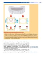
[5, 104]. Both the BCR and the AR represent the sacral segments S2–S4
(
Fig. 4).
Suspected bladder dysfunc-
tion should be investigated
by urodynamic assessment
It is most important to acknowledge that the function of the bladder (detrusor
muscle) cannot be clinically assessed. The clinical diagnosis of urine retention
along with the possibility of overflow as a typical finding in an areflexive bladder
cannot be reliably distinguished from a reflex bladder activity with incontinence
by clinical inspection. Only a full urodynamic examination is able to diagnose in
detail the bladder function (areflexive versus hyperreflexive detrusor, bladder
capacity and compliance) and interaction with the sphincter functions (detrusor
sphincter dyssynergia) [29, 76, 103]. The latter test should be considered when
the clinical examination shows a pathological finding (sacral motor and reflex
disturbance) or the patient describes pathological micturition behavior.
Disorders of the Autonomic System
Deterioration of autonomous column and sympathetic fibers which are con-
ducted through the spinal cord becomes obvious in changed hidrosis. Patients
may report skin areas with increased (wheat) or reduced (dry skin) sweating
(hidrosis). However, these symptoms have to be specifically explored because
patients usually do not report these alterations spontaneously. Areas of reduced
The spoon test indicates
areas of altered hidrosis
sweating can be tested by the so-called spoon test: A teaspoon is lightly stroked
over the skin. On the line of demarcation between the normal (wheat) and
impaired (dry) skin region, the spoon has a reduced friction as the skin with
reduced hidrosis shows a lower adhesion [15, 20, 22, 74, 96, 97, 109, 121].
Neurological Assessment in Spinal Disorders Chapter 11 303
Spinal Cord Injury
SCI is assessed according
to the ASIA protocol
For spinal cord injury (SCI), the Standard for Neurological Classification of SCI
(
Fig. 2) as developed by the American Spinal Injury Association (ASIA)provides
a standardized assessment protocol that can be applied in patients with acute and
chronic traumatic SCI [67–69].
The ASIA protocol allows important information to be obtained about the
level and extent of lesions in a reasonably short time [35, 67, 68]. It is important
to acknowledge that assigning one key muscle and one dermatome (defined by a
specific point) to represent a single spinal nerve segment is a simplification.
However, it could be shown that the ASIA testing allows for a reliable assessment
of the level and extent of lesions [73]. The neurological level refers to the lowest
segment of the spinal cord with normal sensory and motor function. Differentia-
tion between complete (ASIA A) and incomplete SCI (ASIA B–E) is given by the
absence (complete) or preservation (incomplete) of any sensory and motor func-
tion in the lowest sacral segment (S4/S5).
The ASIA protocol
is not approved
for non-traumatic SCI
In the ASIA protocol, appreciation of pinprick (algesia) and of light touch
(esthesia) is scored semiquantitatively on a three point scale (absent, impaired,
normal). The dermatomal key points defined by ASIA help to perform the sen-
sory examination in a standardized form. The involvement of sacral segments is
of predictable value for neurological outcome [125].
However, the ASIA protocol is not a suitable tool with which to guide the diag-
nosis of disorders affecting extraspinal neuronal structures, e.g. polyneuropathy,
plexus lesions or other peripheral neurological lesions. Furthermore, it does not
enable central lesions of spinal cord and brain disorders to be distinguished.
A pitfall in the diagnostic assessment of SCI is exhibited by the syndrome of
spinal shock. This initial state of transient depression of spinal cord function
below the level of injury is associated with loss of:
all sensorimotor functions
flaccid paralysis
bowel and bladder dysfunction
abolished tendon reflexes
Spinal shock can last from several days to weeks. The sacral reflexes [bulbocaver-
nosus (BCR) and anal (AR) reflexes] can be reliably assessed within 72 h after
injury and can be applied to search for an involvement of the conus medullaris
and cauda equina [5, 123] (
Fig. 4).
The neurophysiological examination enables valid information to be
obtained about the functional deficit of the spinal cord at an early time point after
SCI (see Chapter
12 ) [26, 55].
Spinal Cord Syndrome
Impairment of the intraspinal neural structures, i.e. the myelon and cauda
equina, results in typical clinical syndromes. These syndromes may occur with
any cause of an incomplete spinal cord lesion and describe by clinical means the
primarily affected areas of the spinal cord (
Table 3).
Brown-S´equard sy ndrome (spinal hemisyndrome). This is caused by the
deterioration of only half of the spinal cord and results in ipsilateral propri-
oceptive and motor loss and contralateral loss of pain and temperature per-
ception (dissociated sensitive disorder).
central cord syndrome. This lesion affects the central gray structures of the
spinal cord with deterioration of alpha-motoneurons and the crossing
304 Section Patient Assessment
Table 3. Spinal cord injury syndromes
Syndrome Paresis Reflexes Sensory function Vasomotor
dysfunc-
tion
Bladder/
bowel
Frequent cause
Tendon
tap
Babinski AR and
BCR
Deep
pressure
Pain
Complete lesion
spinal shock flaccid – +/– + – – + flaccid trauma
C1–T1 spastic tetra ++ +/– + – – + spastic trauma
T2–T12 spastic para ++ +/– + – – + spastic trauma, tumor
conus spastic and/
or flabby
(+)–(+)–––– spastic/
flaccid
trauma
cauda flaccid – – –––– flaccid trauma, disc her-
niation
Incomplete lesion
Brown-
S´equard
syndrome
spastic
hemiparesis
++ ipsi-
lateral
+ipsi-
lateral
+–ipsi-
lateral
– contra-
lateral
+/– –/spastic trauma
central cord
syndrome
spastic tetra
(flaccid pare-
sis of upper
limbs)
++ + + +/– – + spastic trauma, cervical
stenosis, syrinx,
disc herniation,
OPLL
anterior cord
syndrome
flaccid paresis – +/– + + – – spastic ischemia
posterior cord
syndrome
spastic or no
paresis
+/++ +/– + – + – spastic vitamin B
12
defi-
ciency syndrome
+ positive, ++ increased, – abolished
segmental spinothalamic fibers. The syndrome occurs most frequently in the
cervical region.
anterior cord syndrome. This syndrome refers to the disturbance of the
anterior spinal artery with consecutive affection of the anterior part (bilat-
eral) of the cord. Thus, there is loss of motor function and of sensitivity to
pain and temperature (ventrolateral column).
posterior cord syndrome. This syndrome occurs relatively seldom in trauma
andismorefrequentlyseeninnon-traumaticdisorders(suchasB
12
defi-
ciency). It produces primarily proprioceptive impairment as a result of
impaired posterior column.
conus medullaris syndrome. As a result of a compromise of the conus
medullaris (sacral spinal enlargement approximately at the spinal level L1–
L2 vertebrae) and/or cauda equina (lumbar nerve roots within the spinal
canal), a distinct pattern of bladder-bowel dysfunction and lower limb
impairment can be observed. Frequently a clear distinction between conus
medullaris and/or cauda equina lesion cannot be achieved. A pure cauda
equina lesion presents a remaining areflexive bladder dysfunction with loss
of sacral reflexes (BCR and AR) and saddle anesthesia. The lower limbs
show a flaccid paresis and in time a severe muscle atrophy. A conus medulla-
ris lesion can present a mixture of flaccid and spastic symptoms of both the
bladder and lower limbs depending on the localization within the conus.
Impotence accompanies both syndromes. The extent of symptoms depends
on the degree of damage (complete or incomplete) of the conus medullaris
and cauda equina.
Neurological Assessment in Spinal Disorders Chapter 11 305
Differential Diagnosis
Differentiation of Central and Peripheral Paresis
Spasticity differentiates cen-
tral and peripheral lesions
The neurological examination should not only confirm if there is any neurologi-
cal deficit but provide a somatotopic assessment of the location of the lesion. A
frequent problem is the differentiation between (
Table 4):
central paresis (spastic paresis)
peripheral paresis (flaccid paresis)
Differentiation between
spastic and flaccid paresis
allows the distinction
of central from peripheral
lesions
The differentiation into spastic and flaccid paresis is one of the most significant
factors for distinguishing between central and peripheral lesions.
A flaccid paresis indicates reduced or abolished muscle tone, while spastic pare-
sis is described by increased muscle tone with resistance to passive extension, brisk
jerks and cloni. The muscle resistance is especially present in fast passive extension
andatthestartofmovement.Inthepresenceofspasticity,themuscletoneshould
be assessed by the adapted Ashworth score (
Table 5
) [93, 110, 111].
Differentiation of Radicular and Peripheral Nerve Lesions
If a peripheral lesion is assumed, differentiation of a radicular and peripheral
nerve lesion is required. Differences in the dermatomal area of the roots and
peripheral nerves as well as differences in the key muscles may be helpful. How-
ever, the sensory examination can be very challenging particularly in elderly and
young patients, as well as in patients with impaired consciousness and psychiat-
ric disorders. Also the muscle strength testing depends on the cooperation of the
patient and is influenced by pain. The somatotopic relation between nerve root
and peripheral nerve is summarized in
Tables 6 and 7. Because of the similarity
of symptoms, the clinical differentiation between some radicular syndromes and
peripheral or plexus lesions can be difficult.
Table 4. Clinical differentiation of central and peripheral paresis
Central paresis Peripheral paresis
brisk tendon reflexes, muscle cloni diminished or absent tendon reflexes
uni- or bilateral increased stretch reflexes and enlarged reflex zones reduced or absent polysynaptic reflexes
pathological reflexes (Babinski sign, Gordon and Oppenheimer
reflexes), uni- and/or bilateral
no evidence of pathological reflexes
increased muscle tone flaccid muscle tone
para- or hemi-like distribution of motor deficit distribution related to peripheral nerve inner-
vation
spinal lesions from C1 to L1 (conus medullaris) lesions below L2
Table 5. Assessment of spasticity
Ashworth score Degree of muscle tone
0 no increase in muscle tone
1
slight increase in muscle tone, manifested by a catch and release or
by minimal resistance at the end of the range of motion when the
affected part(s) is moved in flexion or extension
2
slight increase in muscle tone, manifested by a catch, followed by
minimal resistance throughout the reminder (less than half) of the
ROM
3
more marked increase in muscle tone through most of the ROM, but
affected part(s) easily moved
4
considerable increase in muscle tone passive, movement difficult
5
affected part(s) rigid in flexion or extension
306 Section Patient Assessment
Table 6. Peripheral and segmental innervation of upper extremity muscles
Peripheral innervation Segmental
innervation
Muscles of the shoulder
trapezius
accessory n. C3–4
latissimus dorsi
thoracodorsal n. C6–8
rhomboids
dorsal scapular n. C5
levator scapulae
dorsal scapular n. C3–5
serratus posterior
(superior and inferior)
thoracic n.s T1–12
deltoideus
axillary n. C5–6
supraspinatus
suprascapular n. C4–6
infraspinatus
suprascapular n. C4–6
teres minor
axillary n. C5–6
teres major
subscapular n. C5–6
subscapularis
subscapular n. C5–6
Muscles of the arm
biceps brachii
musculocutaneous n. C5–7
brachialis
musculocutaneous n. C5–7
coracobrachialis
musculocutaneous n. C5–7
triceps brachii
radial n. C7–8
anconeus
radial n. C7–8
pronator teres
median n. C6–7
flexor carpi radialis
median n. C6–7
palmaris longus
median n. C6–7
flexor digitorum superficialis
median n.
C7–T1
flexor carpi ulnaris
ulnar n. C8–T1
flexor digitorum profundus
ulnarn.(ulnarside)
median n. (radial side)
C8–T1
flexor pollicis longus
anterior interosseous branch of median n. C8 –T1
pronator quadratus
anterior interosseous branch of median n. C8 –T1
brachioradialis
radial n. C5–6
extensor carpi radialis longus
radial n. C6–7
extensor carpi radialis brevis
radial n. C6–7
extensor digitorum
deep branch of radial n. C6–8
extensor digiti minimi
deep branch of radial n. C6–8
extensor carpi ulnaris
deep branch of radial n. C6–8
extensor pollicis longus
deep branch of radial n. C6–8
extensor indicis longus
deep branch of radial n. C6–8
abductor pollicis longus
deep branch of radial n. C6–8
extensor pollicis brevis
deep branch of radial n. C6–8
supinator muscle
deep branch of radial n. C6
Muscles of the hand
palmaris brevis
superficial branch of ulnar n. C8–T1
abductor pollicis brevis
median n. C8–T1
opponens pollicis
median n. C8–T1
flexor pollicis brevis
median n. (superficial head)
ulnar n. (deep head)
C8–T1
adductor pollicis
deep palmar branch of ulnar n. C8–T1
lumbricales
median n. (1
st
and 2
nd
)
ulnarn.(3
rd
and 4
th
)
C8–T1
abductor digiti minimi
deep palmar branch of ulnar n. C8–T1
flexor digiti minimi brevis
deep palmar branch of ulnar n. C8–T1
opponens digiti minimi
deep palmar branch of ulnar n. C8–T1
palmaris brevis
deep palmar branch of ulnar n. C8–T1
interosseous
deep palmar branch of ulnar n. C8–T1
According to Sobotta [113]
Neurological Assessment in Spinal Disorders Chapter 11 307
Table 7. Peripheral and segmental innervation of lower extremity muscles
Peripheral innervation Segmental
innervation
Muscles of the hip and thigh
iliopsoas
muscular branch of the lumbar plexus L1–4
sartorius
femoral n. L2–3
quadriceps
femoral n. L2–4
pectineus
femoral n. L2–4
adductor longus
anterior branch of obturator n. L2–4
adductor brevis
anterior branch of obturator n. L2–4
gracilis
anterior branch of obturator n. L2–4
obturator externus
anterior branch of obturator n. L3–4
adductor magnus
posterior branch of obturator n.
tibial part of sciatic n.
L2–4
L4–S1
gluteus maximus
inferior gluteal n. L5–S1
gluteus medius
superior gluteal n. L4–S1
gluteus minimus
superior gluteal n. L4–S1
tensor fascia lata
superior gluteal n. L4–S1
piriformis
1
st
and 2
nd
sacral n.s S1–2
obturatus internus
n. to obturator internus L5–S2
gemelli
n. to obturator internus L5–S2
quadratus femoris
n. to quadratus femoris L5–S2
Muscles of the leg
biceps femoris
tibial portion of the sciatic n. (long head)
peroneal portion of the sciatic n. (short
head)
S1–3
L5–S2
semitendinosus
tibial portion of the sciatic n. L5–S2
semimembranosus
tibial portion of the sciatic n. L5–S2
tibialis anterior
deep peroneal n. L4–S1
extensor hallucis longus
deep peroneal n. L4–S1
extensor digitorum longus
deep peroneal n. L4–S1
triceps surae
tibial n. S1–2
soleus
tibial n. S1–2
plantaris
tibial n. S1–2
popliteus
tibial n. L4–S1
tibialis posterior
tibial n. L5–S1
flexor digitorum longus
tibial n. L5–S1
flexor hallucis longus
tibial n. L5–S1
peroneus longus
superficial peroneal n. L4–S1
peroneus brevis
superficial peroneal n. L4–S1
Muscles of the foot
extensor digitorum brevis
deep peroneal n. L5–S1
extensor hallucis brevis
deep peroneal n. L5–S1
abductor hallucis
medial plantar n. L5 –S1
flexor hallucis
medial plantar n. L5 –S1
adductor hallucis
lateral plantar n. S2–3
abductor digiti minimi
lateral plantar n. S2–3
flexor digiti minimi
lateral plantar n. S2–3
opponens digiti minimi
lateral plantar n. S2–3
flexor digitorum brevis
medial plantar n. L5 –S1
quadratus plantae
lateral plantar n. S2–3
interossei
lateral plantar n. S1–2
According to Sobotta [113]
308 Section Patient Assessment
Radiculopathies
The clinical presentations of the radicular syndromes are summarized in Table 8.
The exact differentiation between radicular and peripheral nerve damage may
demand neurophysiological studies, i.e. EMG to show denervation of root- and/
or nerve-specific muscles as well as neurography to exclude conduction delay of
the peripheral nerve. Entrapment syndromes are an important differential diag-
nosis of radicular lesions. Knowledge of the characteristic symptoms is manda-
tory (
Table 9).
C5 Radiculopathy
In contrast to an isolated lesion of the musculocutaneous nerve,aC5lesion
causes not only a paresis of the biceps muscle, but also of the scapular muscle
Table 8. Radicular syndromes and differential diagnosis
Root Dermatome Muscle Reflex Important differential diagnoses
C1–4 neck and collar neck muscles – lung carcinoma
diaphragm (parado-
xic abdominal mus-
cle movements)
neuritis of brachial plexus
lymphoma
thymome
C5
lateral shoulder deltoid muscle biceps reflex frozen shoulder
Erb’s palsy
neuralgic amyotrophy of the shoulder
palsy of axillary nerve
C6
lateral arm and
thumb
extensors of hand,
flexors of elbow
biceps reflex carpal tunnel syndrome
brachioradial
reflex
radial nerve palsy
musculocutaneous nerve palsy
C7
dorsum of shoulder
and arm into the
long finger
triceps, wrist flexors,
finger extensors
triceps reflex palsy of posterior interosseus nerve,
brachial plexus paralysis (middle part)
C8–T1
medial arm into
ulnar two digits
intrinsic hand
muscles
Trömner’s reflex palsy of anterior interosseus nerve
brachial plexus paralysis (Klumpke type)
thoracic outlet syndrome
ulnar palsy
L2
inguinal ligament iliopsoas cremaster reflex femoral palsy
hip osteoarthritis
pelvic disorder (i.e. psoas muscle)
L3
medial femoral and
knee
femoral adductors,
vastus medialis of
quadriceps muscle
adductor reflex paralysis of obturator nerve
pelvic disorder (aseptic necrosis of
symphysis)
hip osteoarthritis
L4
lateral femoral and
medial shank
vastus lateralis of
quadriceps muscle
patellar reflex paralysis of femoral nerve
L5
lateral shank tibialis anterior
muscle
tibialis posterior
reflex
peroneal paralysis
S1
dorsal shank, along
heel into fifth digit
of foot
gastrocnemius
muscle
Achilles tendon
reflex
tibial paralysis
tarsal tunnel syndrome
S2
dorsal femoral ischiocrural muscles biceps femoris
reflex
sciatic pain syndrome
S3
proximal medial
femoral
bulbocavernosus
muscle and anal
sphincter
bulbocavernosus
and anal reflex
palsy of cutaneus posterior femoral
nerve (sacral plexus)
S4–5
perineum bulbocavernosus
muscle and anal
sphincter
bulbocavernosus
and anal reflex
palsy of clunium medii
palsy of anococcygei nerves (coccygeal
plexus)
Neurological Assessment in Spinal Disorders Chapter 11 309
Table 9. Frequent entrapment syndromes
Syndrome Findings
Carpal tunnel syndrome pain of hand and forearm, frequently at night (antebrachialgia nocturna)
hypesthesia of digits 1 to 3 including the radial side of digit 4
paresis and atrophy of the thenar muscles
positive Tinnel sign over the carpal tunnel
Sulcus ulnaris syndrome
numbness of digits 4 and 5
paretic intrinsic hand muscles and hypothenar muscles
positive Tinnel sign over the ulnar sulcus
Thoracic outlet syndrome
paresis of the intrinsic hand muscles
worsening of symptoms by elevating the shoulder
frequently associated with cervical rip or ligamental hypertrophy
pain of hand and forearm
Fibularis syndrome
paretic foot elevation
numbness of the dorsal foot
often history of repeating pressure over the fibular caput
Tarsal tunnel syndrome
paresis of short foot muscles
numbness of the plantar foot
atrophy of abductor hallucis muscle
group (supra- and infraspinatus, teres major and minor muscles). The sensory
deficits of a C5 radiculopathy are located at the posterolateral upper arm while
the musculocutaneous nerve also innervates the ventral aspects (see Chap-
ter
8 ).
C6 Radiculopathy
The sensory deficits in a C6 lesion may mimic median nerve lesion. However, in
median nerve lesion neither is the biceps tendon reflex (BTR) diminished nor the
biceps muscle paretic. Similarly, the middle finger is typically not involved in a
C6 hypesthesia but in a median nerve lesion.
C8/T1 Radiculopathy
This radiculopathy must be distinguished from an ulnar nerve lesion.InC8/T1
radiculopathy, the ulnar side of the forearm is hypesthesic and all intrinsic hand
muscles are affected. The ulnar nerve is mostly compressed within the sulcus,
resulting in paresis of the hypothenar and only those intrinsic hand muscles
innervated by the ulnar nerve. The sensory deficit affects the two ulnar fingers.
L3/4 Radiculopathy
In a neuropathy of the femoral nerve and in L3/4 radiculopathy, the patellar ten-
don reflex (PTR) is reduced or abolished with a predominant weakness of the
quadriceps muscles. However, detailed testing in femoral nerve neuropathy
shows a sensory deficit restricted to the ventral aspect of the thigh with paralysis
of hip flexion (iliopsoas muscle) while in L3/4 radiculopathy the sensory deficit
is extended to the medial site and below the knee with weakness of the thigh
adduction (adductor muscles).
L5 Radiculopathy
ParesisoffootelevationcanbeduetoaL5radiculopathyand/oralesionofthe
peroneal nerve (see Chapter
8 ,
Case Introduction
). Clinical differentiation is
310 Section Patient Assessment
possible by proving the hip abduction, which is also affected in a L5 radiculopa-
thy with weakness of the gluteal muscles (gluteus medius, tensor fasciae latae).
S1 Radiculopathy
In suspected S1 radiculopathy, damage of the tibial ner ve, e.g. tibial tunnel syn-
drome or partial sciatic lesion, has to be excluded. While S1 radiculopathy is sig-
naled by diminished Achilles tendon reflex and weak foot extension, the tibial
nerve affection involves the toe and ankle extensor muscles while the peroneal
nervelesionshowsparesisofthetoeandankleflexormuscles.
Differential Diagnosis of Spinal Cord Compression Syndromes
This group of syndromes is due to obliteration of the spinal canal resulting in
compression of the neural structures. Both cervical and lumbar stenosis fre-
quently originate from degenerative (secondary) changes of the spine. Also a
congenitally narrow spinal canal (primary spinal canal stenosis) can be present,
which exposes the patient to an increased risk of compression syndromes and a
greater danger of neuronal damage in minor spine trauma. In Asian people (e.g.
Japanese individuals), an ossified posterior longitudinal ligament (OPLL) can
cause spinal cord compression, which is only rarely described in Caucasian peo-
ple. Although all compression syndromes present with distinct symptoms, dif-
ferential diagnosis from other disorders is mandatory in equivocal cases
(
Table 10).
Table 10. Spinal cord compression syndromes
Compression syndrome Symptoms Differential diagnosis
Cervical stenosis clumsy painful hands multiple sclerosis
disturbed fine motor skills Myelitis
imbalance of gait B
12
hypovitaminosis
numb feet spinal tumors
urinary urgency polyneuropathy (PNP)
arteriovenous malformations
Thoracic stenosis
lower limb sensory deficit disc herniation (often calcified)
thoracic sensory level OPLL
spastic paraparesis arteriovenous malformations
bladder-bowel dysfunction spinal tumors
Lumbar stenosis
tired legs and weakness on walking vascular claudication
lumbar pain on walking spinal metastasis
pain relief during sitting, lying and forward
bending
polyneuropathy
Cauda equina syndrome
severe leg pain
cauda equina radiculitis (Elsberg’s syndrome)
flaccid paraparesis lesion of pelvic plexus
sensory loss of legs
urinary and bowel incontinence
saddle anesthesia
Miscellaneous Differential Diagnoses
Neurovascular Disorders
Girdle-like pain may be an
initial symptom of a spinal
ischemic or hemorrhagic
disorder
Non-traumatic acute paraplegia may be due to spinal ischemic or hemorrhagic
disorders. Typically,the first symptom is girdle-like pain in the dermatome refer-
ring to the involved level. Thereafter, motor paresis and sensory deficits appear,
mostly within minutes to a few hours. A very special but not so uncommon disor-
Neurological Assessment in Spinal Disorders Chapter 11 311


