Spinal Disorders: Fundamentals of Diagnosis and Treatment Part 42 doc
Bạn đang xem bản rút gọn của tài liệu. Xem và tải ngay bản đầy đủ của tài liệu tại đây (260.12 KB, 10 trang )
a
b
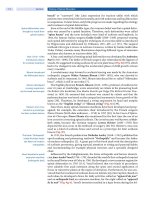
Figure 2. Eye and face protection
Details of the eye (a) and face protection (b) in a patient having anterior C-spine surgery due to trauma. The eyes are cov-
ered with cream and seal and are then padded to avoid damage by pressure or sharp objects. Nasogastric tube is in place.
After deployment of the surgical retractors in anterior cervical spine surgery, the
pressure inside the endotracheal tube cuff frequently reaches 40–50 mmHg. It
should be rechecked in order to maintain it between 15and 20 mmHg; this is even
more important if the anesthetist is using N
2
O in the gas mixture due to its fast dif-
fusion into the cuff. These marked increases in the cuff pressure along with
lengthy total intubation time are frequently reported to elevate tracheal and pha-
ryngeal morbidity such as hoarseness and vocal cord palsy [3]. Once the surgical
team finishes positioning the patient, it is wise to confirm that the endotracheal
tube hasnot moved and that bilateral ventilation and breath sounds are adequate.
It is also a good time to verify that the bronchial blocker is still in the right
place if one lung ventilation is desired.
Antibiotic Prophylaxis
Postoperative infections in spine surgery are primarily monomicrobial, although
in about half of infected patients more than one organism can be identified. The
Routine antibiotic
prophylaxis today is
standard in spinal surgery
bacteria most commonly cultured from wounds are Staphylococcus aureus and
epidermidis [17]. Postoperative infections occur in 0.3–9% of patients undergo-
ing spine surgery [75]. Increased risk of spine postoperative infections has been
associated with:
staged procedures
blood loss in excess of 1000 ml
surgery longer than 4 h
smoking
diabetes
malnutrition
obesity
immunocompromised patients
alcoholism
posterior approach
postoperative incontinence
Intraoperative Anesthesia Management Chapter 15 393
cancer surgery
extended preoperative hospitalization
intraoperative hypothermia
For the antibiotic proph ylaxis to be effective, a drug with bactericidal activity
against the most common infecting organisms must be present in the tissues at
risk from the moment of the incision and for the duration of the surgery. Cefazo-
lin’s spectrum is sufficiently broad to be effective but limited enough to avoid
resistance and superinfection. Cefazolin’s penetration into the subcutaneous tis-
sue and the intervertebral disc is adequate if serum concentration is maintained.
In most hospitals, cefazolin is the agent of choice because it has an optimal anti-
Redose antibiotics in cases
with prolonged surgery
and/or substantial
blood loss
microbial coverage, is relatively nontoxic and inexpensive, and hasexcellent pen-
etration into the tissues at risk. The agent should be started within 30 min before
skin incision. A blood loss greater than 1500 ml or a duration of surgery exceed-
ing 4 h warrants redosing of the antibiotic, which should only be given for 24 h
perioperatively. The responsibility for the prudent administration of prophylac-
tic agents has therefore moved to the domain of the anesthesiologist. These prac-
tices will result in the most efficacious and judicious use of antibiotics [14]:
maintaining therapeutic concentrations when appropriate
avoiding excessive cost
minimizing emergence of resistant microbial pathogens
Although adverse reactions are actually rare, patients with a history of these
events should receive an alternative antibiotic; vancomycin or clindamycin are
second line choices in this setting. In selecting the antibiotic, local patterns of
pathogens from infection control data should play a role. Hospitals with a high
prevalence of resistant microbes, such as the methicillin-resistant S. aureus
(MRSA), may consider using alternative agents. Most procedures with the
implantation of foreign material warrant prophylaxis. Foreign bodies not only
allow more efficient colonization, but also protect the organisms from systemic
antibiotics, making these complications extremely difficult to treat. Due to the
high rate of infection without prophylaxis, the severe associated morbidity, and
the lack of effective therapy, prophylaxis is indicated in any spinal procedure
where the intervertebral disc is manipulated. The use of antimicrobial prophy-
laxis in spinal surgery can reduce the number of both superficial and deep wound
infections. The benefits of this intervention include less patient pain and discom-
fort, shorter hospital stays, and fewer expenses.
Patient Positioning
Correct patient positioning
is mandatory for
a successful outcome
Patient position for surgery depends on the level of the spine to be operated on
and the kind of intervention to be performed. In some procedures (such as ante-
roposterior lumbar surgery) the patient is repositioned while asleep to complete
the operation. It is not clear whether positioning a patient with an unstable cervi-
cal spine is safer awake or asleep. In elderly patients with severe cervical spondy-
losis, positioning with the neck in extension may result in spinal cord compres-
sion between the ligamentum flavum and posterior vertebral body osteophytes.
Cervical approaches can be done with the patient prone or supine. Thoracolum-
bar surgery might require lateral decubitus to gain access to the intrathoracic
spine as well as the upper lumbar section. Most scoliosis procedures are done
with the patient in the prone position.
Attentionmustbegiventoprotect:
bony prominences and joints (elbows, anterior superior iliac spines, facial/
forehead area, knees and ankles/feet)
394 Section Peri- and Postoperative Management
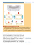
Figure 3. Position on the Jackson table
Observe the abdomen hanging free of pressure. The arms rest without axillary or elbow pressure and at a 90-degree
angle in the shoulders and elbows. Elbows are padded and the head is in neutral position with eyes, mouths and nose
in the hole of the foam holder with no pressure. The warming blower is in place over the lower limbs.
blood vessels (carotid/jugular, femoral, axillary artery)
nerves (ulnar, femoral, femorocutaneous, sciatic, peroneal, brachial plexus)
A 90° angle between the trunk and arms and between arms and forearms is rec-
ommended in the prone position. The abdomen must hang free [58] to decrease
The abdomen must hang
free with the patient
in the prone position
pressureontheinferiorvenacavaandsubsequentlyreduceepiduralveinpres-
sure and bleeding (
Fig. 3
).Theexternalgenitalsshouldbeunloadedofany
pressure or traction. In the prone position the eyes and nose should remain
free of pressure. A small risk of corneal abrasion exists if the patient wakes up
too actively in a WUT and the cornea remains uncovered afterwards in the
face-down position. The prone position might represent an advantage from a
respiratory point of view in patients properly positioned with a free-hanging
abdomen due to functional improvement in residual capacity and oxygenation
[59].
Sequential anteroposterior spinal access presents a challenge to keep themon-
itoring and lines in place when flipping from one position to the other. Coordina-
tion and communication are required since this is a combined effort of many
people in the OR. Jackson tables provide some advantages; however, precautions
must be taken to minimize compression and traction of linesand anatomic struc-
tures. Cervical spine procedures call for a thorough final check of lines and tubes
before prepping and draping. The endotracheal tube, nasogastric tube and tem-
perature probe have to be secured.
Skin Preparation. Current evidence based preoperative recommendations do not
endorse shaving the skin. If hair requires removal, it should be done by clipping
with an electrical device not by shaving (in fact shaving might lead to higher
Intraoperative Anesthesia Management Chapter 15 395
operative site infection rates than no hair removal or clipping) and the best tim-
ing is immediately before bringing the patient into the theater (not in the OR).
The patient’s skin should be physically scrubbed and cleaned before the applica-
tion of antiseptic [2, 35, 40].
Ischemic Optic Neuropathy
Perioperative increased
intraocular pressure
may lead to ischemic
optic neuropathy
Increases in intraocular pressure with ischemic optic neuropathy have been
linked to blindness after the patient has been in the face-down position in spine
surgery [72]. Ocular perfusion pressure (OPP) relates directly to mean arterial
pressure (MAP) and inversely to intraocular pressure (IOP), venous pressure in
the eye and central venous pressure. In patients free of ocular pathology under-
going spine surgery in the prone position, Cheng et al. [11] found a change in the
IOP from 19±1 mmHg preinduction/supine, to 13±1 mmHg 10 min postinduc-
tion/supine, to 27±2mmHg prone/before surgery, to 40±2 mmHg prone/end of
surgery, to 31±2 mmHg after returning the patient to the face-up position. They
also described a moderate correlation (r
2
=0.6)betweenthetimespentinthe
prone position and the elevation of the IOP. To minimize the chances of visual
troubles, a neutral-head or slight head-up position is recommended along with
equilibrated fluid balance and a MAP of notbelow 60 mmHg (eye perfusion pres-
sure = MAP – [CVP + IOP]). The most common cause of amaurosis after spine
surgery is anterior or posterior ischemic optic neuropathy (ION). Less common
causes are central retinal artery or vein occlusion and occipital lobe infarct. Risk
factors for ION are diabetes mellitus, hypertension, head-down position, smok-
ing, and the combination of intraoperative anemia and hypotension [62]. We
favor the use of the Mayfield head clamp for posterior cervical spine procedures
because pressure on eyes, nose, and chin can be avoided. Post spine surgery
blindness is an important topic that led The American Society of Anesthesiology
to evaluate this theme through the ASA Postoperative Visual Loss Registry. Pre-
liminary results have been published. Established in July 1999, the registry col-
lects information anonymously (http:depts.washington.edu/asaccp) to identify
risk factors to prevent this complication in the future [41, 43].
Maintenance of Anesthesia
Maintenance of anesthesia is intended to provide good surgical (a dry field, good
neuromonitoring, adequate muscle relaxation when needed) and anesthetic con-
ditions (amnesia, nociceptive suppression, temperature preservation, hemody-
namic andorgan function stability). These goals canbe achieved with total intra-
venous anesthesia (TIVA) or a gas/opioid approach. TIVA with target controlled
infusions (TCIs) has come into fashion in many places of the world except in
North America, because of its minimal interference with intraoperative neuro-
monitoring, smooth and fast anesthesia and quick control of the level of anes-
thetic depth. However, a low dose (0.3–0.5 minimum alveolar concentration or
MAC-awake) of desflurane or sevoflurane with remifentanil [4] can actually be as
good as or better than TIVA for neuromonitoring without the effect of propofol
Blood preservation
is important
on platelet function. Blood preservation is a primary goal in major spine surgery.
Propofol is known to decrease platelet function in studies describing the inhibi-
tory effect of propofol on human platelet aggregation [12, 49]. Because patients
often use prophylactic doses of aspirin or nonsteroidal anti-inflammatory drugs
(NSAIDs) for pain control preoperatively, the use of continuous infusions of pro-
Preoperative NSAID intake
substantially increases
bleeding and should be
stopped beforehand
pofol is a theoretical risk for more bleeding. If a WUT is required, patients on
low-dose desflurane or sevoflurane can be weaned faster and tend to respond ear-
lier to commands from the anesthetist than those on propofol. Remifentanil is an
396 Section Peri- and Postoperative Management
ultrashort acting and potent opiate that is completely metabolized and elimi-
nated from the circulation in 3–6 min by plasma esterases. It makes a perfect
match with thelow-dose gases technique. In continuous infusion, it not only pro-
vides excellent analgesia, but it also allows for quick changes in thedepth of anes-
thesia for WUT and it is a versatile tool for induction of controlled hypotension.
It has been our experience that for thoracolumbar and lumbar spine surgery the
use of intrathecal single shotmorphine (0.3–0.6 mgpreservative-free) before the
induction of anesthesia greatly contributes to intraoperative and early postoper-
ative stability and smooth WUT. Using this approach for the last 5 years we have
had no infections attributed to the technique and both surgeons and patients
appreciate it in equal measure. The same result is achieved with high thoracic
epidural analgesia (catheter at C6–T5) for thoracolumbar procedures where a
thoracotomy and chest drain are required. Any choice of maintenance drugs
must aim to give a stable depth or level of anesthesia. Neuromuscular relaxant
drugs should be used to facilitate airway control and then only as necessary
according to the surgical conditions.
Muscle relaxants do not
interfere with SSEPs
Muscle relaxants are generally not recommended when MEPs are being mon-
itored; however, if surgical conditions mandate some muscle relaxation while
monitoring MEPs, a low-dose continuous infusion of intermediate-acting mus-
cle relaxants (rocuronium, cisatracurium, etc.) titrated to keep 3 out of
4 twitches (3/4 TOF) from the nerve stimulator can be used without impairing
the MEP monitoring [38]. After the intubation dose of the muscle relaxant wears
off, MEPs should begin to get a baseline recording (unless baselines for SSEPs
andMEPswereobtainedbeforemusclerelaxationwasinduced).Then,thetitra-
tion of the muscle relaxant infusion should proceed. A theoretical advantage of
having some degree of muscle relaxation in major posterior procedures is better
abdominal decompression as opposed to the abdominal tightness of an unre-
laxed patient.
Intraoperative Monitoring Techniques
Advanced Monitoring of Vital Functions
Advanced monitoring of vital cardiopulmonary functions is suggested only in
patients with systemic pathology or those scheduled to have major spine proce-
dures. A central venous catheter is often inserted to measure central venous pres-
sure (CVP), administer volume and have separate lines for drugs. In anterior
lumbar spine surgery, monitoring hemoglobin saturation and plethysmographic
curves from the ipsilateral toes to the surgical access to the spine are recom-
mended (
Fig. 4). This simple measure can provide early warning of vascular com-
pression with retractors [33].
Cardiovascular System
Consider cardiac compromise
in patients with Duchenne’s
muscular dystrophy
Cardiac compromise may be a direct result of the underlying pathology, for
exampleinpatientswithDuchenne’smusculardystrophyorfromunrelatedcar-
diovascular disease such as hypertension or coronary artery disease. Cardiac
dysfunction may also result from severe scoliosis or kyphosis, which causes dis-
tortion of the mediastinum, and cor pulmonale secondary to chronic hypoxemia
and pulmonary hypertension. A direct arterial blood pressure line will be
required in the case of major surgery, patients with preoperative cardiopulmo-
nary pathologies or other anesthetic considerations (
Table 2).
An arterial catheter is usually inserted in the radial or femoral arteries for this
purpose.
Intraoperative Anesthesia Management Chapter 15 397
Figure 4. Plet hysmography of the toe
Simultaneous monitoring of the Hbsat and plethysmography in the toe and finger to detect arterial compression in the
anterior lumbar approach.
Table 2. Indications for direct arterial pressure monitoring
Preoperative conditions Surgical indications
coronary artery disease long operations (requiring blood sampling)
other cardiac conditions limiting heart function expected major blood loss
uncontrolled hypertension controlled hypotension to be used
severe peripheral vascular disease postoperative mechanical ventilation
advanced chronic obstructive pulmonary disease
With the patient in the prone position, the CVP may be a misleading indicator of
right and left ventricular end diastolic volume [71]. In a study in pediatric
patients scheduled for scoliosis surgery, the CVP rose from 9 to 18 mmHg on
turning patients from the supine to the prone position. The increase seems to
correlate with the pulmonary artery pressure (PAP). The left ventricular end dia-
stolic diameter measured by transesophageal echocardiography (TEE) fell from
37 to 33 mm, indicating a transient and positional diastolic ventricular dysfunc-
tion. Pulmonary artery catheters are controversial because they do not decrease
perioperative mortality and can cause significant morbidity. In healthy adults
Prone patient position
reduces cardiac function
[73], the face-down position reduces the cardiac index (15–25%) and increases
systemic vascular resistance possibly due to a decrease in venous return and ven-
tricular compliance. These changes are more pronounced with propofol-based
anesthesia than with gas. The main take-home message from this study is that
greater changes should be expected in individuals with established preoperative
cardiorespiratory pathology. Near infrared spectroscopy, a novel technology
with potential application in spine surgery patients undergoing controlled hypo-
tensive anesthesia (CHA), is enjoying a period of intense interest and research
[29]. This is a noninvasive device for following brain Hb-oxygen mixed satura-
tion in the territories supplied by the anterior and middle cerebral arteries. With
398 Section Peri- and Postoperative Management
CHA a small risk of brain hypoperfusion in the presence of unrecognized carotid
stenosis exists. This method has been extensively used in cardiac anesthesia to
reduce postoperative strokes and provides a transcranial reading of brain tissue
O
2
sat that is made up of 75% venous blood and 25% arterial blood, allowing the
anesthesiologist to adjust the brain blood flow and oxygenation to a safe level.
Maintenance Fluids
The type and volume of fluid maintenance will vary depending upon the magni-
tude of blood loss, the preoperative intravascular filling status, the systemic pre-
operative condition of the individual and the length of the procedure. Patients
scheduled for discectomy or simple hardware removal with minimal blood loss
can receive “normal” saline or balanced solutions (lactated Ringer’s, Hartmann’s
solution, etc.). Those that will be fast-tracked in day-surgery programs should
have (under normal conditions) no bladder catheter and crystalloid volumes
below 1000–1500 ml perioperatively. For major operations, fluid therapy should
be guided by theCVP andblood loss, and the latter replaced with the appropriate
Fluid therapy should
be guided by CVP
solution/blood product. Balanced crystalloid solutions are recommended to
avoid hyperchloremic acidosis induced by the so-called “normal” saline due to
the high content of chloride in it [8]. Preoperative fasting is usually replaced in
the first hour of surgery with 10 ml/kg of Ringer’s lactate solution. Recent publi-
cations [28] have raised concern about the potential harm of overloading
patients with fluids; therefore fluid volume therapy must follow a rational indica-
tion to replace preoperative negative balance, intraoperative maintenance, intra-
operative blood loss and postoperative requirements.
Bladder catheters are routinely inserted before procedures lasting for more
than 3 h to preclude bladder distension and to monitor urine output. Large blood
volume changes and the frequent use of vasoactive drugs make their use manda-
tory to observe urine output in these situations. Foley catheters are also recom-
mended to be inserted in elderly male patients who suffer from prostate hyper-
plasia and patients with urinary incontinence.
Body Temperature
Mild perioperative hypothermia (reductions of core body temperature of 1–2°C)
is associated with [64]:
increased postoperative cardiac complications
impaired hemostasis
impaired neutrophil function
wound area hypoxia
increased postoperative protein wasting
altered pharmacodynamics of muscle relaxants
delayed discharge from recovery room
increased infectious complications [24]
A temperature probe should be placed, particularly in juvenile and infantile
patients undergoing scoliosis surgery as well as in patients expecting to have
large blood volume changes. Body temperature decreases very quickly in uncov-
ered and anesthetized children and elderly patients; the main mechanisms are
redistribution of heat from the core compartment to the periphery along with
decreased heat production. Routine use of air-warming blan kets and intrave-
nous blood/liquid warming systems is recommended. Unless they are warmed,
each unit of blood or 1000 ml of crystalloid solution at room temperature will
reduce body temperature by 0.25°C. Patients that are only partially paralyzed
Intraoperative Anesthesia Management Chapter 15 399
produce more heat compared with those fully paralyzed. Temperature monitor-
ing must be used when neurophysiologic monitoring is planned since a normal
temperature is a requirement for successful WUT and neurophysiologic record-
ing. Although malignant hyperthermia nowadays is a very rare condition, its
incidence is increased in patients with scoliosis because of their association with
neuromuscular pathology.
Monitoring Depth of Anesthesia (Consciousness)
Since the introduction of anesthesia almost 150 years ago, the depth of anesthesia
has been monitored through surrogate variables (heart rate, arterial pressure,
eye behavior, etc.). Today, the level of consciousness at induction, steady-state
and wake-up phase can be monitored directly. The anesthesiologist uses these
tools in spine surgery to keep patients at an appropriate level of anesthesia, to
prevent recall of intraoperative events and to facilitate WUT performance (see
below). Bispectral Index (BIS)and other techniques (auditory evoked potentials,
entropy, etc.) have been evaluated and validated to correlate with consciousness
during anesthesia with propofol, isoflurane or sevoflurane [7]. The BIS is a pro-
Monitoring the level
of consciousness during
the anesthesia is necessary
cessed presentation of the EEG as a numerical rating from 100 (fully awake) to 0
(isoelectric EEG,total suppression of brain activity). Numbers between 45and 60
are desirable as indicators of an appropriate consciousness level for surgery. The
interaction of gases and propofol on thepharmacodynamic effects of opioids and
the BIS has been studied recently [52]. Bear in mind that the other components
of anesthesia (autonomic response, muscular relaxation, nociception, etc.) are
monitored with other instruments.
Neuromuscular monitoring
assesses the level
of muscular relaxation
Neuromuscular monitoring is performed in order to evaluate muscular relax-
ation during the intubation phase as well as during the surgical period and prior
to the WUT andextubation. The train-of-four (TOF) is a simple way for the anes-
thesiologist to assess neuromuscular relaxation in anesthetized patients. It con-
sists of a barrage of four electrical impulses delivered transcutaneously over the
ulnar nerve at 2 Hz to activate the adductor pollicis. Three responses in the TOF
are normally observed when there are over 75% of the neuromuscular receptors
free of a muscle relaxant effect. Patients monitored for MEP and/or nerve root
integritymusthaveatleast3/4twitchesintheTOF.
Intraoperative Blood Preserving Techniques
Use blood preserving
techniques
Blood product transfusions are frequently required in major spinal surgery.
Transfusionthresholdsforredbloodcellscommonlyusedareahemoglobincon-
centration of 7–9 g%, compensatory tachycardia and an increasing lactate blood
level. Patients with cardiopulmonary diseases and patients actively bleeding are
considered for transfusion in the upper threshold margin. Complications of
transfusions include transfusion transmitted infections (1:1900000 transfused
units for HIV, 1:1600000 for hepatitis C, 1:220000 for hepatitis B),bacterial con-
tamination (1:1000 or 2000 for platelet concentrates), immunosuppression,
transfusion-related acute lung injury, transfusion reactions (cutaneous, cardio-
vascular, respiratory) and graft-versus-host reaction. The Cumulative Serious
Hazards of Transfusions (SHOT) survey in the United Kingdom over 6 years
describes 35 reports of transfusion transmitted infections of which 21 were bac-
terial with 6 fatalities. Of these, 17/21 were due to platelets and also 5/6 deaths
were related to platelets. The SHOT report will not pick up viral complications as
they are often more chronic and may develop outside of the considered “win-
dow” for reporting [5].
400 Section Peri- and Postoperative Management
Transfusions increase
the risk of postoperative
infections
Nosocomial infection rates increase fivefold in patients receiving allogenic trans-
fusions with adose-response pattern; the more units received the higher theodds
of infection [16]. Potential problems with fresh frozen plasma transfusions are
well described in pediatric surgery, including hypotension and cardiac arrest
linked to sudden hypocalcemia [63, 77].
Good spine surgeons complete the surgical procedures in less time, are careful
with hemostasis, and pay attention to optimal patient positioning while looking
for better outcomes. In posterior surgical approaches there is more bleeding
because of the bigger incisions, more work on the laminae and facet joints, greater
chances of epidural vein damage and bleeding and bone graft harvesting [15].
Neuromuscular scoliosis
surgery is prone to increase
blood loss
Neuromuscular scoliosis patients have greater blood loss during spinal fusion
surgery than idiopathic scoliosis patients. Prolongation of the prothrombin time
anddecreaseinFactorVIIactivitysuggestactivationoftheextrinsiccoagulation
pathway. Depletion of clotting factors during scoliosis surgery occurs to a greater
extent in patients with underlying neuromuscular disease [32] (see
Table 3).
Table 3. Factors associated with a higher risk of homologous blood transfusion
low preoperative hemoglobin decreased amount of autologous blood
units available
spine surgery in cancer patients no use of Jackson table
multilevel posterior fusion neuromuscular scoliosis surgery
Controlled Hypotensive Anesthesia
Spinal cord blood flow (SCBF) autoregulation has been studied in humans [27].
SCBF autoregulation is similar to the brain’s with a stable plateau between 50 and
Controlled hypotensive
anesthesia is frequently
used in spinal surgery
100 mmHg mean arterial pressure (MAP). It changes in lineal fashion with CO
2
between 15 and 90 mmHg andremains unchanged with PaO
2
above 50 mmHg. A
reference MAP of 60–65 mmHg in spine surgery is supported in the literature
[15]. It is important to preserve the end-tidal CO
2
in the normal/high range to
improve brain and spinal cord perfusion while under controlled hypotensive
anesthesia (CHA) conditions. Inducing CHA in patients in the prone position is
facilitated by the sequestration of volume in the lower limbs (particularly using
an Andrew’s table) and the effect of anesthetics on hemodynamics. Fluids must
be given to keep a normal cardiac output/organ perfusion while on low MAP
since the blood container (vascular system) has been expanded, and in the prone
position the heart and pulmonary circulation are affected. The most frequently
used agents to produce CHA are:
remifentanil
sodium nitroprusside
labetalol and nitroglycerin
calcium channel antagonist
fenoldopam
propofol (it might interfere with SSEPs in the high range of doses required
to induce CH)
inhaled anesthetics (sevoflurane or desflurane, same comment as propofol)
CHA reduces blood loss,
transfusion requirement
and operative time
CHA reduces blood losses by 55% and transfusion requirements by 53%, while
operating time has been reported to be shorter [74] in scoliosis surgery. It has
been applied in a variety of spine procedures including idiopathic scoliosis,
degenerative scoliosis, instrumentation for Duchenne’s patients and others.
Although limited clinical experience is available so far, prostaglandin E
1
(PGE
1
) seems to be an interesting alternative to inducing CHA. An infusion of
Intraoperative Anesthesia Management Chapter 15 401
PGE
1
is capable of reducing MAP smoothly, maintaining the autoregulation of
the spinal cord blood flow [79].
In spinal cord injury
and compression, CHA can
compromise remaining
spinal cord function
Caution should be exercised in patients with spinal cord trauma or tumors
compressing the spinal cord where the normal autoregulation might be impaired
and the perfusion compromised in some areas.
Secondary injury prevention is paramount to avoid further damage to the spi-
nal cord function; therefore anormal or higher perfusion pressure should be pre-
served [85] until the surgical decompression is achieved.
Intrathecal Opiates
Two groups incidentally observed a decrease in intraoperative bleeding in spine
surgery with the use of preoperatively injected intrathecal opiates. This effect was
not observed when the drug was injected at the end of the procedure. Goordarzi
et al. [23] noticed in ten adolescents receiving morphine 20 μg/kg intrathecally
with 50 μg of sufentanyl that the combination facilitated intraoperative CHA to a
MAP of 55 mmHg. Gall [19] observed in 30 patients 9–19 years old undergoing
spinal fusion a significant trend towards lower bleeding volumes when morphine
5 μg/kg intrathecally was injected before starting the operation. This study does
not provide information about the impact of that trend on the transfusion rates.
Blood Predeposit and Erythropoietin Injection
For surgeries with expected blood losses of over 1–1.5 l, a blood predeposit of 1
or 2 units is recommended when feasible in adolescents and adult patients [63].
A predeposit hemo globin of between 11 and 14.5 g% isconsidered to be the opti-
mal range. Over 90% of patients coming for spinal fusions that predeposit their
own blood avoid receiving allogeneic blood [53]. Iron supplementation with
erythropoietin in patients with production problems should be prescribed. A
prospective randomized study of epoetin alfa vs. placebo in patients scheduled
for complex spine deformity surgery showed that patients in the treatment group
were more likely to complete predonation, decrease homologous transfusions
and have shorter hospital stays [66]. Colomina suggested that using recombinant
erythropoietin (rEPO) in spine surgery patients with expected blood loss of
Recombinant erythropoietin
may substitute blood
predeposit
around 30% of their blood volume might substitute blood predeposit. They also
mentioned that patients expecting around 50%blood volume loss can avoid allo-
geneic blood transfusions by predeposit and bone marrow stimulation with
rEPO [10]. Recommended dose is 600 U/kg/week subcutaneously for 4 weeks
(usually one vial of 40000 U/week), and 200–300 mg/day of iron should be given,
along with folic acidand vitamin B
12
over the entire period of rEPOsupplementa-
tion.OncetheHblevelreaches15g%therEPOshouldbesuspended.
Cell Salvage
Intraoperative cell salvage consists of collecting the blood from the surgical field
to a machine that separates red blood cells from detritus, washing and concen-
trating them to be reinfused into the patient. Its use is indicated when blood
losses over 15–20 ml/kg are expected. Cell salvage is contraindicated in:
infected patients
cancer surgery
In a provocative approach, some authors have reinfused collected blood in a large
number of cancer patients after irradiation of the bag to kill any malignant cells
which are potentially present [25]. More research is needed before recommend-
402 Section Peri- and Postoperative Management


