Spinal Disorders: Fundamentals of Diagnosis and Treatment Part 47 pps
Bạn đang xem bản rút gọn của tài liệu. Xem và tải ngay bản đầy đủ của tài liệu tại đây (413.62 KB, 10 trang )
Manipulative Therapy
There is moderate evidence
for the effectiveness of
manipulative treatment
Manipulative therapy remains a mainstay of conservative treatment for degenera-
tive disorders of the cervical spine. Particularly, traction has been reported to
result in short-term relief of radiculopathy [60, 61, 197]. Debate continues on the
safety of manipulative therapy of the cervical spine. Based on a national survey of
19122 patients, minor side effects (headache, fainting/dizziness, numbness/tin-
gling) were not uncommon up to 7 days after the intervention, with an incidence
rate ranging from 4 to 15/1000. Serious adverse events (leading to in-hospital
treatment or permanent disability) were very rare (1/10000). However, this does
not rule out a deleterious course in individual patients (
Case Introduction
). Rubin-
stein et al. [230] concluded that the benefits of chiropractic care for neck pain seem
to outweigh the potential risks. There is moderate evidence that spinal manipula-
tive therapy (SMT) and mobilization is superior to general practitioner manage-
ment for short-term pain reduction of chronic neck pain.However,SMToffersat
most similar pain relief to high-technology rehabilitative exercise in the short and
long term. In a mix of acute and chronic neck pain, there is moderate evidence that
mobilization is superior to physical therapy and family physician care [41]. There
are only a few studies on acute neck pain and the evidence is currently inconclusive
[41].
Physical Exercises
Moderate evidence
supports physiotherapy
for chronic neck pain
There is moderate evidence supporting the effectiveness of both long-term
dynamicaswellasisometricresistanceexercisesoftheneckandshouldermus-
culature for chronic or frequent neck disorders. No evidence supports the long-
term effectiveness of postural and proprioceptive exercises or other very low
intensity exercises [106, 296].
Multidisciplinary Rehabilitation Programs
Incontrasttothelumbarspine,thereappearstobelittlescientificevidencesofar
for the effectiveness on neck and shoulder pain of multidisciplinary rehabilita-
tion programs compared with other rehabilitation methods [145]. However, this
conclusion is due to the low quality of available clinical trials [145].
Massage
No clinical practice recommendations can be made for the effectiveness of mas-
sageforneckpain[115].
Spinal Injections
Transforaminal injections
can results in serious
complications
Anecdotally, transforaminal injections with epidural steroid application can
result in instant pain relief in patients suffering from cervical radiculopathy [70,
163, 262], although injection of local anesthetic appears to have similar effects
[8]. However, recent articles have prompted major concerns over the safety of
transforminal steroid injections because of cases with subsequent deleterious
spinal cord injuries [120, 181, 245]. For chronic neck pain, intramuscular injec-
tion of lidocaine was superior to placebo or dry needling at short-term follow-up,
but similar to ultrasound. There is limited evidence of effectiveness of epidural
injection of methylprednisolone and lidocaine for chronic neck pain with radicu-
lar symptoms [208].
Degenerative Disorders of the Cervical Spine Chapter 17 447
Radiofrequency Denervation
The treatment effect of
radiofrequency denervation
is unproven
Although some studies reported satisfactory results [170, 253], there is limited
evidence that radiofrequency denervation offers short-term relief for chronic
neck pain of zygapophysial joint origin and for chronic cervicobrachial pain
[188].
Acupuncture
Theevidenceforacupunctureisconsideredinconclusive and difficult to inter-
pret [27].
Electrotherapy
The systematic review by Kroeling et al. [158] could not make any definitive con-
clusions about electrotherapy for neck pain. The present evidence on galvanic
current (direct or pulsed), iontophoresis, electromuscle stimulation (EMS),
transcutaneous electrical nerve stimulation (TENS), pulsed electromagnetic
field (PEMF) and permanent magnets is either lacking, limited, or conflicting.
Infrared Laser Therapy
The review by Chow et al. [55] provided limited evidence from one randomized
controlled trial (RCT) for the use of infrared laser for the treatment of acute neck
pain and chronic neck pain from four RCTs.
Operative Treatment
General Principles
Degenerative disorders of the cervical spine are a heterogeneous group of pathol-
ogies with a wide spectrum of treatment modalities. For the vast majority of clin-
ical entities, surgery is only indicated after an adequate trial of non-operative
treatment has failed. As outlined in the preceding paragraph, the scientific evi-
dence for the effectiveness of many conservative measures is very limited. Simi-
larly, the evidence is limited for the surgical treatment options. While surgery for
chronic neck pain is not broadly supported, it appears that patients with CSR and
CSM benefit from surgery after non-operative care has failed [86, 297]. Indica-
tions for surgery for CSR and CSM include (
Table 6):
Table 6. Indications for surgery
Cervical spondylotic radiculopathy Cervical spondylotic myelopathy
progressive, functionally important motor deficit
definitive evidence for nerve root compression
concordant symptoms and signs of radiculopathy
persistent pain despite non-surgical treatment for
at least 6 –12 weeks
progressive myelopathy despite non-operative care
acute onset, deterioration or progression of neurological deficits
definitive evidence of spinal cord compression with moderate-
to-severe myelopathic symptoms
progressive kyphosis with neurological deficits
The goal of CSM treatment
primarily is to arrest
progression
Surgery for cervical radiculopathy is generally recommended when all of the
aforementioned criteria are present [45]. The primary goal of surgery in CSM is
the prevention of further pr ogression of the neurological symptoms because
improvement of established myelopathic changes is rare [164, 166]. One of the
most important aspects in dealing with CSM is to inform the patients preopera-
448 Section Degenerative Disorders
tively that the goal of surgery is primarily to arrest progression of the disease.
Patients are frequently disappointed by the results of surgery when neurological
recovery is lacking although the vast majority of patients do show improvements
[76, 127, 225, 294]. It is therefore reasonable to extensively inform patients about
the goals and realistic expectations of surgery.
Surgical Techniques
There is an ongoing debate on the approach to deal with disc herniation related
radiculopathy, CSR or CSM, i.e.:
anterior approach
posterior approach
The pathology should be
treated where it is
Each technique has its advantages and drawbacks. The controversy which of the
two approaches is better cannot be generalized but must always be related to the
target pathology. It is important to recognize whether the compressing structure
is anterior or posterior to the neural structures. The pathology should be treated
where it is. Thus, an anterior neural compression is better removed from anterior
and a multisegmental posterior compression from a posterior approach. In cases
with three or more level stenosis, a posterior approach is preferred unless there
is no coexisting substantial anterior compression.
Anterior Cervical Discectomy and Fusion
Anterior cervical discectomy
and fusion remains the gold
standard for CSR
In 1955, Robinson and Smith [229] reported on a technique for removal of cervi-
cal disc and fusion with a horseshoe-shaped graft which later became the gold
standard for the treatment of disc herniations and cervical spondylotic radiculo-
pathy [260]. Cloward [62] developed a similar anterior approach, i.e. drilling a
hole in the intervertebral disc space and adjacent vertebrae to insert a bone
dowel. In contrast to the Robinson-Smith technique, Cloward removed the com-
pressing structures at the level of the posterior longitudinal ligament. Robinson
and Smith [229] did not decompress the neural structures, but believed that by
immobilizing the segment osteophytes and herniated disc would be reabsorbed.
In the following years many variations of this technique were developed [12, 35,
37, 77, 99, 258]. Anterior cervical discectomy and fusion (ACDF) with a tricorti-
cal bone graft harvested from the iliac crest is the most widely used technique
and has become the gold standard for the treatment of cervical radiculopathy
(
Case Introduction).
Fusion rates are dependent
on the number of levels
treated
The radiological fusion rate is dependent on the amount of levels to be fused.
Bohlmann et al. [33] reported a solid fusion for one, two and multilevel fusions
of 89%, 73% and 67%, respectively. Cauthen et al. [49] analyzed the outcome of
anterior cervical discectomy and interbody fusion (Cloward technique) in 348
patients with an average follow-up of 5 years. The fusion rate was 88% for one
level and 75% for multilevel fusions. Emery et al. [78] reported a fusion rate of
only 56% for three-level fusions.
The surgical outcome
is mainly dependent on
the decompression effect
Clinical outcome of ACDF for cervical radiculopathy is good to excellent in
70–90% of patients [223] and mainly dependent on the decompression of the
compromised nerve root [45]. However, Bohlmann et al. have reported a signifi-
cant association between the presence of non-union and postoperative neck or
arm pain [33].
Degenerative Disorders of the Cervical Spine Chapter 17 449
Autograft V ersus Allograft
Autograft is superior
to allograft for ACDF
The use of allograft for spinal fusion in conjunction with anterior decompression
for degenerative cervical disorders has a long tradition. Cloward [62, 63] used
allografts from the 1950s. However, there are only a few studies [7, 28, 42, 303]
comparing allografts versus autografts which were analyzed in a meta-analysis
[83]. Floyd and Ohnmeiss [83] concluded from their meta-analysis that for both
one- and two-level anterior cervical discectomy and fusion, autograft demon-
strated a higher rate of radiographic union and a lower incidence of graft col-
lapse. However, it was not possible to ascertain whether autograft is clinically
superior to allograft. The authors advised that the decision of the bone graft
shouldnotbesolelybasedontheradiographicresultsbutthatadditionally
donor site morbidity, transmission of infectious disease, quality of the autograft
(osteoporosis) and patient preference must be taken into consideration [83].
Plate Fixation
The conventional fusion techniques were not universally successful. Complica-
tions causing persistent pain included [10, 33, 69, 78, 102, 228, 287, 288, 292, 304]:
non-union (particularly for multilevel fusions)
graft displacement
graft collapse
sagittal malalignment (kyphosis)
For traumatic cervical lesions, anterior plate fixation gained widespread accep-
tance because it provides immediate stability and high fusion rates [4, 31, 46].
Similarly, instrumented fusion was introduced for degenerative cervical disor-
ders [156, 247, 279]. Additional plating theoretically increases the fusion rate,
preserves cervical lordosis, and prevents graft subsidence and migration partic-
ularly when two or more levels are involved [247].
Plate fixation increases
the fusion rate for multilevel
fusions
However, three RCTs failed to demonstrate the superiority of additional plate
fixation for one-level fusions in terms of clinical or radiological outcome [105,
244, 309]. For multilevel fusion, there is some evidence that plating appears to
result in higher fusion rates [47, 94, 146, 280, 281].
Anterior plate fixation
does not suffice
for three-level fusions
Wang et al. [281] indicated that a three-level fusion is still associated with a
high non-union rate (18%), although the use of cervical plates decreased the
pseudarthrosis rate. Bolesta reported that three- and four-level modified Robin-
soncervicaldiscectomyandfusionresultsinanunacceptablyhighrateofpseud-
arthrosis which is not improved by a cervical spine plate alone [34]. Additional
posterior fixation is advocated in three and more level fusion to decrease the
non-union rate [180] (
Case Study 1).
Fusion with Cages
One drawback of the conventional fusion (Smith-Robinson or Cloward) tech-
niques could not be overcome by plating, i.e. bone graft donor side pain. Persis-
Bone graft donor site pain
remains a drawback of ACDF
tent pain from the anterior iliac crest is reported in up to 31% of patients [110].
During the last decade, cages have become increasingly popular in stabilizing
and fusing the cervical spine subsequent to anterior discectomy. Compared to
conventional fusion techniques, the theoretical advantages of cages are to:
restore disc height
restore cervical lordosis
prevent graft collapse
450 Section Degenerative Disorders
ab c
def
Case Study 1
A 47-year-old male had experienced some numbness, clumsiness and tingling in his hands for over 1 year before he sud-
denly developed gait disturbance and weakness in both legs. The patient was admitted to the Neurology Department
for further diagnostic work-up. Clinically, the patient presented with an incomplete tetraparesis sub C4. A lateral radio-
graph (
a) demonstrates a congenitally narrow spinal canal with cervical spondylosis particularly at the levels C5/6 and
C6/7 and decrease of cervical lordosis. Sagittal T2W image (
b) demonstrating a large disc herniation at C4/5 with com-
pression of the spinal cord, advanced disc degeneration with endplate changes (Modic Type II), signal intensity changes
within the spinal cord at C5/6, and a disc protrusion with spinal cord compression at C6/7. Axial T2W images confirm the
severe myelon compression at the levels of C4/5 (
c) and C6/7 (d). The patient underwent multilevel anterior cervical dis-
cectomy and fusion with a tricortical iliac bone graft and anterior plating. In a second operation, the patient underwent
posterior laminectomy and instrumented fusion to completely decompress the narrow spinal canal and spinal cord
(
e, f). Postoperatively, the patient substantially improved with regard to his neurological function but a residual tetrapa-
resis remained at latest follow-up.
avoid donor site pain
reduce operative time
Many different cage designs (e.g. cylindrical, mesh, ring or box shaped) and
materials (e.g. titanium, carbon, polyetheretherketone, hydroxyapatite coated)
Degenerative Disorders of the Cervical Spine Chapter 17 451
have been introduced [54, 110, 144, 216, 221, 271]. Debate continues on the fact of
the cage filling with bone (autograft or allograft), bone graft substitutes or void
and favorable clinical results have been reported with each technique [53, 132,
157, 168, 203, 233, 248].
Cage fusions are not better
than conventional
ACDF
Randomized studies have so far not been able to reveal a significantly better
clinical outcome of patients undergoing cage fusion compared to conventional
techniques [111, 210, 233, 273] although the rate of non-union appears to be
higher and bone graft donor site pain lower [273].
Anterior Corpectomy
In patients suffering from CSM, anterior discectomy and osteophyectomy may
not suffice to sufficiently decompress the spinal cord. The spinal cord may not
only be compromised by disc protrusions and spondylophytes but also by a spi-
nal malalignment (kyphosis) or a narrow spinal canal. In these cases, a subtotal
corpectomy is required [236]. Partial vertebral body resection and decompres-
sion was first used to treat traumatic cervical disorders [91] and later adopted for
degenerative disorders [114, 236].
Compared to ACDF, a median corpectomy offers the advantage of:
enlarging the spinal canal
allowing for a more radical decompression
increasing the fusion rate
Corpectomy allows
forbetterdecompression
and a high fusion rate
A variety of techniques were developed to stabilize the cervical spine after
decompression through vertebrectomy [21, 35, 113, 116, 298]. The extent to
which decompression should be performed depends on the pathology and the
size of the spinal canal [125, 295]. Most authors [143] advocate the complete
removal of the posterior osteophytes and PLL to achieve maximum decompres-
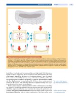
sion (
Fig. 5). Compared to multilevel ACDF, corpectomy offers the advantage of
reducing the host-graft interfaces.Swanketal.[263]haveshownthatthenon-
union rate of two-level ACDF was 36% while one-level corpectomy resulted in a
non-union rate of 10% (
Case Study 2). Similar results were obtained by Hilibrand
et al. [125], who reported a non-union rate of 34% for ACDF (one to four levels)
and 7% for corpectomy.
One-level corpectomies are best reconstructed using iliac crest autograft. The
angulation of the iliac crest limits its applicability for longer anterior reconstruc-
tions. Therefore, fibula strut allo grafts have been used with satisfactory results
[263]. However, the fusion rate of allograft fibula is somewhat lower than with
autograft [100, 263]. This limitation can be overcome with additional posterior
instrumented fusion [180]. Recently, cages constructs have been used for long
anterior column reconstructions [56, 187, 261, 268, 293]. The drawbacks of cage
buttressing for anterior cervical reconstructions include subsidence, limited
assessment of fusion status, and difficult revision surgery because of frequent
partial incorporation [180].
Three-level corpectomies
necessitate anterior-
posterior fixation
Anterior plating currently is recommended to increase fusion rate and
decrease the incidence of graft dislocation [153]. However, the ability of plate fix-
ation to stabilize a three-level corpectomy is limited [136, 242, 270] and addi-
tional posterior stabilization is recommended to circumvent implant failure and
non-union [73, 93, 162, 226].
Anterior Discectomy Without Fusion
A drawback of the classic Robinson-Smith technique is that the intervertebral
disc is removed to reach the location of the neural compromise. Attempts have
452 Section Degenerative Disorders
abc
de f
Figure 5. Technique of corpectomy and instrumented fusion
The cervical spine is exposed by an anteromedial approach. a The intervertebral discs are excised adjacent to the target
level.
b The medial three-thirds of the vertebral body are resected. The lateral wall is preserved to protect the vertebral
arteries.
c A high-speed diamond burr is used to remove the median part of the vertebral body. d The remaining part of
the posterior vertebral wall is elevated away from the spinal cord and resected with a Kerrison rongeur.
e Kerrison ron-
geur and curettes are used to remove posterior osteophytes and decompress spinal cord and exiting nerve roots.
f The
spine is reconstructed by insertion of a tricortical iliac bone block and anterior plating.
therefore been made to remove the disc herniation without completely resecting
the intervertebral disc. Indications of this technique are:
soft disc herniation
disc sequestration
young individual
no spondylosis
no segmental instability
Retrospective case series did not report a clinical outcome inferior to discectomy
and fusion [24, 25, 183, 192, 219, 220]. The disadvantages of this method, how-
ever, were:
recurrent herniation
motion segment degeneration
segmental instability
chronic neck pain
spontaneous fusion
Degenerative Disorders of the Cervical Spine Chapter 17 453
ab
c
d
ef
Case Study 2
A 56-year-old male had recurrent episodes of neck pain with occasional radiating pain to his right forearm for 18 months
before he developed acute onset excruciating arm pain followed by a progressive sensorimotor deficit of C6 on the right
side. Lateral radiograph (
a) showing cervical spondylosis at the level of C5/6 and C6/7. Sagittal T2W image (b) reveals cer-
vical spondylosis and disc protrusions at C5/6 and C6/7. Axial T2W image shows a sequestrated disc herniation at C5/6
(arrow) with compression of the exiting nerve root C6 (
c) and a disc protrusion at C6/7 with compromise of the C7 nerve
root (
d). The indication for surgery was prompted by the progression of the paresis. The patient underwent a corporec-
tomy of C6, decompression of the C6 and C7 nerve root, reconstruction with a tricortical iliac bone block and anterior
plating (
e, f). At 1 year follow-up, the sensorimotor deficit had completely recovered. The patient was fully functional but
occasionally had some episodes of benign neck pain.
Outcome of discectomy
without fusion is not inferior
to that of ACDF
In a prospective randomized study on 91 patients with single-level cervical root
compression, Savolainen et al. [244] analyzed three different treatment groups:
discectomy without fusion, fusion with autologous bone graft, and fusion with
autologous bone graft plus plating. Clinical outcomes were good for 76%, 82%,
and 73% of the patients, respectively. A slight kyphosis developed in 62.5% of
the patients who had undergone discectomy, 40% of the patients who had
undergone fusion, and 44% of the patients who had undergone fusion plus
454 Section Degenerative Disorders
plating [244]. This study indicates that discectomy without fusion is not inferior
to ACDF.
Techniques were developed to preser ve the intervertebral disc,whichoftenis
not substantially degenerated and can therefore be preserved. Verbiest [274] sug-
gested a lateral approach while Hakuba [112] described a trans-unco-discal
Disc preserving anterior
nerve root decompression
is feasible
approach. The latter approach is a combined anterior and lateral approach to the
cervical discs. Interbody fusion was not performed except for special cases with
significant kyphosis or instability [112]. Minimally invasive techniques were sug-
gested by Jho [140] and Saringer et al. [240], who reported on a microsurgical
anterior foraminotomy which provides direct anatomical decompression of the
compressed nerve root by removing the compressive spondylotic spur or disc
fragment. Saringer et al. [241] modified this technique by using an endoscopic
approach. Other authors removed the herniated disc under endoscopic view
using a transdiscal route [13, 84].
Total Disc Arthroplasty
Adjacent segment
degeneration is the main
argument for TDA
Adjacent segment degeneration (Fig. 6) has been mentioned as the main argu-
ment against spinal fusion and therefore favoring total disc arthroplasty (TDA).
However, the data on adjacent segment degeneration is sparse [14, 52, 124, 160].
Hilibrand et al. [124] followed 374 patients who had a total of 409 anterior cervi-
cal fusions for a maximum of 20 years. Symptomatic adjacent-segment disease
occurred at an incidence of 2.9% per year during the 10 years after operation.
About one-fourth of the patients who had an anterior cervical fusion were at risk
of developing symptomatic adjacent segment disease within 10 years. A single-
level arthrodesis involving C5/6 or C6/7 and preexisting radiographic evidence
of degeneration at adjacent levels appeared to be the greatest risk factors for new
abc
Figure 6. Adjacent segment degeneration
a Symptomatic cervical spondylosis at C5/6 with anterior and posterior osteophytes. b Postoperative lateral radiograph
after anterior cervical discectomy and fusion with a tricortical iliac bone graft (Robinson-Smith technique).
c Lateral
radiographs at 6 years follow-up demonstrate a perfect fusion at C5/6 with remodeling of the osseus structures (arrow-
heads). Note the adjacent segment degeneration at C4/5 (arrow).
Degenerative Disorders of the Cervical Spine Chapter 17 455
disease [124]. Importantly, no study so far was able to differentiate the effect of
natural history versus the effect of the arthrodesis on the development of adja-
cent segment degeneration [52, 101].
More than 15 different designs are now under pre-clinical and clinical evalua-
tion (e.g. Prestige II, Bryan, PCM, ProDisc-C, Cervicore, Discover) [199]. Current
TDA designs include one-piece implants and implants with single or double glid-
ing articulations with either metal-on-metal or metal-on-polymer bearing sur-
ab
c
de
Case Study 3
A 53-year-old female patient complained of persistent (4 months) right-sided shoulder/arm pain and was referred to our
shoulder specialists with suspected impingement syndrome. A thorough physical examination revealed a normal shoul-
der function but a decreased sensation at the lateral aspect of the radial forearm and thumb as well as weakness in dor-
siflexion of the hand. The biceps tendon reflex was diminished on the right. A lateral radiograph (
a) showed segmental
kyphosis at C4/5 and minimal cervical spondylosis at C5/6 and C6/7. Parasagittal T2W image (
b) revealed a lateral disc
protrusion at C5/6. Axial T2W image (
c) confirms the foraminal disc protrusion with compression of the exiting C6 nerve
root. Non-operative therapy (medication, physiotherapy) failed to provide persistent substantial pain relief. A nerve root
block (C6) completely alleviated the symptoms for 1 week. Discectomy, nerve root decompression and total disc arthro-
plasty at C5/6 was carried out (
d, e). Immediately after surgery, the patient had complete pain relief and was fully func-
tional 2 weeks after surgery. At the 2-year follow-up, the patient was still completely symptom-free.
456 Section Degenerative Disorders


