Spinal Disorders: Fundamentals of Diagnosis and Treatment Part 55 doc
Bạn đang xem bản rút gọn của tài liệu. Xem và tải ngay bản đầy đủ của tài liệu tại đây (300.09 KB, 10 trang )
ab
cd
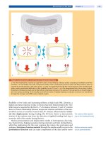
Figure 6. Surgical decompression of a spinal stenosis
a A midline approach exposes the interlaminar windows L3/4 and L4/5 as well as the facet joints to decompress a spinal
stenosis at these levels.
b The supra- and interspinous ligaments are resected under the preservation of the spinous pro-
cess. The interlaminar window is opened with a Kerrison rongeur and the compressing bone and hypertrophied flavum
are removed.
c It is important to realize that the narrowest part of the stenosis is always under the lamina. Therefore, the
lamina has to be resected (laminotomy) in the caudal third or half. The remaining part needs to be undercut from the
superior and inferior sides, respectively.
d In some cases, the undercutting of the lamina does not suffice for an adequate
decompression and the lamina needs to be resected.
riorate in longer follow-up [45, 49, 89]. Clinical results of decompression on open
(50–90%) [6, 80, 95] or microsurgical [53, 96] laminotomy are quite similar to
those achieved by laminectomy. Although it is generally assumed that laminec-
tomy may increase or cause vertebral instability [31, 35], no difference in clinical
outcomes or spondylolisthesis progression between the two treatment methods
was seen in two studies [95, 96], especially not when the motion segments were
528 Section Degenerative Disorders
fully stable preoperatively and were not made unstable by a total laminectomy
[29, 80].
Decompression and Spinal Fusion
The addition of fusion with or without instrumentation to surgical decompres-
sion is generally recommended when segmental instability is assumed. However,
the radiologic assessment of segmental instability remains a matter of debate.
Decompression and fusion are considered by many spine surgeons in case of:
segmental instability (degenerative spondylolisthesis and scoliosis)
concomitant moderate to severe back pain
necessity for a wide decompression
recurrent spinal stenosis
Instrumented fusion
provides higher fusion rates
and better long term
outcome
The best fusion technique (Case Introduction, Case Study 2)isstillcontroversial,
and the evidence in the literature favoring one technique over the other is still
sparse [27, 28, 63]. Most information relates to cases in which degenerative spon-
dylolisthesis is associated with spinal stenosis. Herkowitz et al. [31] prospectively
compared decompression alone versus decompression and non-instrumented
fusion in 50 patients who had spinal stenosis and degenerative spondylolisthesis.
The authors concluded that in the patients who had had a concomitant fusion,
the results were significantly better with respect to relief of pain in the back and
lower limbs. In a subsequent study, Fishgrund et al. [24] prospectively random-
a b c
de
Case Study 2
A 71-year-old female presented with
buttock and posterior thigh pain
only while walking. She was asymp-
tomatic while sitting, lying and
riding a bicycle. The painfree walking
distance was limited to about 200 m.
The standard lateral radiograph (
a)
exhibited a degenerative spondylo-
listhesis at the level of L4/5. A T2W
image (
b) confirmed the suspected
diagnosis of a concomitant spinal
stenosis at this level (arrow). Note
the hypertrophied flavum (arrow-
heads) and degenerative changes of the facet joints (arrows)(
c). Since the patient did not report any back pain, a lamina
preserving decompression was performed. The degenerative spondylolisthesis was addressed by a non-instrumented
fusion to improve long term outcome. At 2 years postoperatively the fusion was solid (arrows)(
d, e). The patient was pain
free and able to perform all her desired activities.
Lumbar Spinal Stenosis Chapter 19 529
ized 67 patients comparing instrumented (pedicle screw fixation) versus non-
instrumented fusion. Clinical outcome was excellent or good in 76% of the
instrumented and 85% of the non-instrumented cases. This difference was not
statistically significant. However,successful fusion was significantly higher in the
instrumented group (82 vs. 45%). The authors concluded that the use of pedicle
screws may lead to a higher fusion rate, but clinical outcome shows no improve-
ment in pain in the back and lower limbs. However, Kornblum et al. [57] demon-
strated the long term (5–14 years) benefits of a successful fusion over non-union
with respect to back and lower limb symptoms in patients with degenerative
spondylolisthesis and spinal stenosis.
The need for an additional
interbody fusion is not
supported by the literature
There is no evidence in the literature that an additional interbody fusion by an
anterior (ALIF) or posterior (PLIF, TLIF) approach improves outcome. Newer
techniques such as interspinous spacer stabilization are still evolving and conclu-
sions on clinical effectiveness are premature [105].
Operative Risks and Complications
Reoperation rates for decompressive laminectomy vary from 7% to 23% [32, 35,
40, 49]. In a cohort study [64], the cumulative incidence of reoperation among
patients who underwent surgery for spinal stenosis was slightly higher following
initial fusion (19.9%) than decompression alone (16.8%). Reoperation among
patients initially presenting with spondylolisthesis was lower with fusion
(17.1%) than with decompression alone (28%). These findings are supported by
controlled trials indicating better outcome for fusion than decompression alone
when spondylolisthesis is present [24, 31]. Interestingly, this data suggests that
over 60% of reoperations following fusion are associated with device complica-
tions or non-union, rather than new levels of disease or disease progression.
In a population based study of reoperation after back surgery [37], the sub-
group spinal stenosis showed a complication rate for laminectomy alone and
decompression with fusion of 4.6% and 7.7%, respectively. Reoperation after
laminectomy was seen in 10% of the cases, which was equal to the 10.2% after
decompression with fusion.
Patients with spinal stenosis
often present with
significant comorbidities
which influence the
surgical strategy
The morbidity associated with surgical treatment of lumbar stenosis in the
elderly is an important aspect as those patients often present with a number of
preexisting cardiovascular, pulmonary, or metabolic comorbidities [15, 18, 47,
49]. Advanced age does not increase the morbidity, nor does it decrease patient
satisfaction or lengthen the return to activity [25, 81]. An increased complication
rate has also been shown to be associated with spinal fusion performed for lum-
bar stenosis in elderly patients [15, 18, 94]. Therefore less invasive surgical
approaches may be of particular interest. Mortality rate has been found to be
approximately 0.6–0.8% [18, 92].
530 Section Degenerative Disorders
Recapitulation
Epidemiology.
Spinal stenosis can be found in up
to 80 % of individuals aged over 70 years. However,
about 20% of asymptomatic individuals demon-
strate signs of spinal stenosis on MRI indicating that
there is no strong correlation with the imaging find-
ings. The rate of spinal surgery for spinal stenosis is
about 10 per 100000 individuals per year.
Pathogenesis. The pathomechanism of central
spinal stenosis is predominantly related to a hyper-
trophy of the yellow ligament which is a result of a
compensatory mechanism to restabilize a segmen-
tal hypermobility. Furthermore, bony canal com-
promise is caused by the occurrence of facet joint
enlargement (osteoarthrosis), osteophyte forma-
tion,anddegenerative spondylolisthesis.Thisfi-
nally results in a progressive compression of the
cauda equina. A congenitally narrow spinal canal is
a rare cause of spinal stenosis. Claudication symp-
toms can be explained by the neurogenic compres-
sion and/or the vascular compression theory.Itis
assumed that both mechanisms play a role. Me-
chanical nerve root compression resultsinde-
creased nutrition, microvascular changes, edema
and fibrosis. The vascular compression theory sug-
gests that spinal stenosis has pathologic effects on
the blood supply of the cauda equina. It is assumed
that venous congestion within the nerve root(s)
between the levels of stenosis leads to a compro-
mised nutrition and results in clinical symptoms.
Clinical presentation. The prevailing symptom of
spinal stenosis is neurogenic claudication,which
can be described as numbness, weakness and dis-
comfort in the legs while walking or prolonged
standing. In contrast to vascular claudication,
symptoms improve by forward bending. Objective
neurological deficits are rarely present during rest.
These symptoms may or may not be associated
with back pain but usually patients suffer much
more from the claudication symptoms while they
can live with the back pain. Radicular claudication
is caused by a lateral recess or foraminal stenosis
and results in nerve root pain while walking and
prolonged standing.
Diagnostic work-up. The imaging modality of
choice is MRI, which allows a precise depiction of
the pathoanatomy in terms of the central and fo-
raminal stenosis. Standing radiographs are useful to
diagnose a concomitant degenerative spondylolis-
thesis or scoliosis. Radiographs may also indicate a
congenitally narrow spinal canal. Neurophysiologic
studies are indicated to confirm the significance of a
mild to moderate spinal stenosis with equivocal
symptoms. They are also helpful in confirming a radi-
culopathy in case of a lateral recess or foraminal ste-
nosis. In elderly patients, peripheral neuropathy is
frequent, which can be detected by electrophysiolo-
gy. The most important differential diagnosis is pe-
ripheral vascular disease, which has to be ruled out
by vascular status and in some cases angiography.
Non-operative treatment. Conservative measures
cannot influence the natural history of spinal steno-
sis, which is a progressive degenerative disease
leading to an increasing immobilization of the pa-
tient. However, non-operative treatment may be
considered in cases with only mild to moderate ste-
nosis and only minimal interference with lifestyle.
Treatment options consist of medication (analge-
sics, NSAIDs, muscle relaxants), administration of
calcitonin, postural education, physical therapy and
epidural injections. There is only sparse scientific
evidence in support of the clinical effectiveness of
any such measures compared to the natural history.
Operative treatment. The treatment of choice is
spinal decompression. In the early years, laminec-
tomy was considered the standard surgical treat-
ment and is still indicated in severe stenosis. How-
ever, reports on increasing segmental instability
have resulted in a shift to a more conservative ap-
proach preserving the posterior elements as much
as possible. Today, laminotomy is the preferred
treatment in cases presenting without additional
deformity or putative segmental instability. This ap-
proach can even be performed by minimal access
surgery under microscopic guidance. When degen-
erative spondylolisthesis or scoliosis or significant
concomitant back pain due to facet joint osteoar-
thritis is present, fusion is considered an important
adjunct to decompression. Instrumented fusion re-
sults in a higher fusion rate and a better long term
outcome than non-instrumented fusion. Many
spine surgeons therefore favor instrumented fusion
although the scientific evidence for this approach is
still weak.
Lumbar Spinal Stenosis Chapter 19 531
Key Articles
Ve rbiest H (1954) A radicular syndrome from developmental narrowing of the lumbar
vertebral canal. J Bone Joint Surg Br 36-B:230 – 7
Classic article on the clinical presentation of neurogenic claudication as a result of spinal
stenosis.
Amundsen T, Weber H, Nordal HJ, Magnaes B, Abdelnoor M, Lilleas F (2000)Lumbar
spinal stenosis: conservative or surgical management? A prospective 10-year study.
Spine 25(11):1424 – 35
A cohort of 100 patients with symptomatic lumbar spinal stenosis were given surgical or
conservative treatment and followed for 10 years. Nineteen patients with severe symp-
toms were selected for surgical treatment and 50 patients with moderate symptoms for
conservative treatment, whereas 31 patients were randomized between the conservative
(n=18) and surgical (n=13) treatment groups. After a period of 4 years, excellent or fair
results were found in half of the patients selected for conservative treatment, and in four-
fifths of the patients selected for surgery. Patients with an unsatisfactory result from con-
servative treatment were offered delayed surgery after 3–27 months. The treatment result
of delayed surgery was essentially similar to that of the initial group. The treatment result
for the patients randomized for surgical treatment was considerably better than for the
patients randomized for conservative treatment. Clinically significant deterioration of
symptoms during the final 6 years of the follow-up period was not observed. Patients with
multilevel afflictions, surgically treated or not, did not have a poorer outcome than those
with single-level afflictions. The authors concluded that the outcome was most favorable
forsurgicaltreatment.However,aninitialconservativeapproachseemsadvisablefor
many patients because those with an unsatisfactory result can be treated surgically later
withagoodoutcome.
Grob D, Humke T, Dv orak J (1995) Degenerative lumbar spinal stenosis. Decompression
with and without arthrodesis. J Bone Joint Surg Am 77:1036 – 41
The authors prospectively evaluated the results of decompression of the spine, with and
without spinal fusion, for the treatment of lumbar spinal stenosis without instability in 45
patients. The patients were randomly assigned to one of three treatment groups: Group I
was treated with decompression with laminotomy and medial facetectomy; Group II,
with decompression and arthrodesis of the most stenotic segment; and Group III, with
decompression and spinal fusion of all decompressed vertebral segments. After
24–32 months, all three groups had a significant improvement in walking distance. With
the numbers available, there were no significant differences in the results among the three
groups with regard to the relief of pain. The authors concluded that spinal fusion is not
necessary in patients presenting with spinal stenosis in the absence of segmental instabil-
ity.
Herkowitz HN, Kurz LT (1991) Degenerative lumbar spondylolisthesis with spinal ste-
nosis. A prospective study comparing decompression with decompr ession and inter-
transverse p rocess arthrodesis. J Bone Joint Surg Am 73:802 – 8
In a prospective study, 50 patients who had spinal stenosis associated with degenerative
lumbar spondylolisthesis were prospectively studied to determine if concomitant inter-
transverse-process arthrodesis provided better results than decompressive laminectomy
alone. After 2–4 years, patients with concomitant fusion had the significantly better
results with respect to relief of pain in the back and lower limbs.
FischgrundJS,MackayM,HerkowitzHN,BrowerR,MontgomeryDM,KurzLT(1997)
Degenerative lumbar spondylolisthesis with spinal stenosis: a prospective, randomized
study comparing decompressive laminectomy and arthrodesis with and without spinal
instrumentation. Spine 22(24):2807 – 12
In this prospective study patients with degenerative spondylolisthesis and spinal stenosis
were randomized into groups with and without pedicle screw instrumentation as an
adjunct to decompression and posterolateral fusion. After a 2-year follow-up, clinical
outcome was excellent or good in 76% of the patients with instrumentation and in 85%
without instrumentation. Successful fusion occurred in 82% of the instrumented cases
versus 45% of the non-instrumented cases (p<0.0015). However, successful fusion did
not influence patient outcome (p =0.435). The authors concluded that the use of pedicle
screws may lead to a higher fusion rate, but clinical outcome shows no improvement
regardingpaininthebackandlowerlimbs.
532 Section Degenerative Disorders
Key Articles
Kornblum MB, Fischgrund JS, Herkowitz HN, Abraham DA, Berkower DL, Ditkoff JS
(2004) Degenerative lumbar spondylolisthesis with spinal stenosis: a prospective long
term study comparing fusion and pseudarthrosis. Spine 29:726 – 33
A longer term follow-up (5–14 years) of the previous study indicated that clinical out-
come was excellent to good in 86% of patients with a solid fusion and in 56% of patients
with a non-union (p<0.01). The solid fusion group performed significantly better in the
symptom severity and physical function categories on the self-administered question-
naire. The authors concluded that in patients undergoing single-level decompression and
posterolateral arthrodesis for spinal stenosis and concurrent spondylolisthesis, a solid
fusion improves long-term clinical outcome.
Weinstein JN, Tosteson TD, Lurie JD, Tosteson AN, Blood E, Hanscom B, Herkowitz H,
Cammisa F, Albert T, Boden SD, Hilibrand A, Goldberg H, Berven S, An H (2008)Surgi-
cal versus nonsurgical therapy for lumbar spinal stenosis. N Engl J Med 358:794 – 810
In this very recent landmark study, study patients with a history of at least 12 weeks of
symptoms and spinal stenosis without spondylolisthesis were enrolled in either a ran-
domized cohort (n=289) or an observational cohort (n=365) at 13 U.S. spine clinics.
Treatment consisted either of decompressive surgery or usual non-surgical care. At
2 years, 67% of patients who were randomly assigned to surgery had undergone surgery,
whereas 43% of those who were randomly assigned to receive non-surgical care had also
undergone surgery. Despite the high level of non-adherence, the intention-to-treat analy-
sis of the randomized cohort showed a significant treatment effect favoring surgery on the
SF-36 scale for bodily pain. However, there was no significant difference in scores on phys-
ical function or on the Oswestry Disability Index. The as-treated analysis, which combined
both cohorts and was adjusted for potential confounders, showed a significant advantage
for surgery by 3 and 24 months postoperatively for all primary outcomes. In the combined
as-treated analysis, patients who underwent surgery showed significantly more improve-
ment in all primary outcomes than did patients who were treated non-surgically.
References
1. Airaksinen O, Herno A, Turunen V, Saari T, Suomlainen O (1997) Surgical outcome of 438
patients treated surgically for lumbar spinal stenosis. Spine 22:2278–82
2. Amonoo-Kuofi HS, Patel PJ, Fatani JA (1990) Transverse diameter of the lumbar spinal canal
in normal adult Saudis. Acta Anat (Basel) 137:124–8
3. Amundsen T, Weber H, Lilleas F, Nordal HJ, Abdelnoor M, Magnaes B (1995) Lumbar spinal
stenosis. Clinical and radiologic features. Spine 20:1178–86
4. Amundsen T, Weber H, Nordal HJ, Magnaes B, Abdelnoor M, Lilleas F (2000) Lumbar spinal
stenosis: conservative or surgical management?: A prospective 10-year study. Spine
25:1424–35; discussion 1435–6
5.ArnoldiCC,BrodskyAE,CauchoixJ,CrockHV,DommisseGF,EdgarMA,GarganoFP,
JacobsonRE,Kirkaldy-WillisWH,KuriharaA,LangenskioldA,MacnabI,McIvorGW,
Newman PH, Paine KW, Russin LA, Sheldon J, Tile M, Urist MR, Wilson WE, Wiltse LL
(1976) Lumbar spinal stenosis and nerve root entrapment syndromes. Definition and clas-
sification. Clin Orthop Relat Res:4–5
6. Aryanpur J, Ducker T (1990) Multilevel lumbar laminotomies: an alternative to laminec-
tomy in the treatment of lumbar stenosis. Neurosurgery 26:429–32; discussion 433
7. Atlas SJ, Deyo RA, Keller RB, Chapin AM, Patrick DL, Long JM, Singer DE (1996) The Maine
Lumbar Spine Study, Part III. 1-year outcomes of surgical and nonsurgical management of
lumbar spinal stenosis. Spine 21:1787–94; discussion 1794–5
8. Atlas SJ, Keller RB, Robson D, Deyo RA, Singer DE (2000) Surgical and nonsurgical manage-
ment of lumbar spinal stenosis: four-year outcomes from the Maine Lumbar Spine Study.
Spine 25:556–62
9.BakerAR,CollinsTA,PorterRW,KiddC(1995)LaserDopplerstudyofporcinecauda
equina blood flow. The effect of electrical stimulation of the rootlets during single and dou-
blesite,lowpressurecompressionofthecaudaequina.Spine20:660–4
10. Bennett GJ, Xie YK (1988) A peripheral mononeuropathy in rat that produces disorders of
pain sensation like those seen in man. Pain 33:87–107
11. Berney J (1994) [Epidemiology of narrow spinal canal]. Neurochirurgie 40:174–8
12. Boden SD (1996) The use of radiographic imaging studies in the evaluation of patients who
have degenerative disorders of the lumbar spine. J Bone Joint Surg Am 78:114–24
Lumbar Spinal Stenosis Chapter 19 533
13. Boden SD, Davis DO, Dina TS, Patronas NJ, Wiesel SW (1990) Abnormal magnetic-reso-
nance scans of the lumbar spine in asymptomatic subjects. A prospective investigation. J
Bone Joint Surg Am 72:403–8
14. Bolender NF, Schonstrom NS, Spengler DM (1985) Role of computed tomography and mye-
lography in the diagnosis of central spinal stenosis. J Bone Joint Surg Am 67:240– 6
15. Ciol MA, Deyo RA, Howell E, Kreif S (1996) An assessment of surgery for spinal stenosis:
time trends, geographic variations, complications, and reoperations. J Am Geriatr Soc
44:285–90
16. De Villiers PD, Booysen EL (1976) Fibrous spinal stenosis. A report on 850 myelograms with
a water-soluble contrast medium. Clin Orthop Relat Res:140–4
17. Delamarter RB, Bohlman HH, Dodge LD, Biro C (1990) Experimental lumbar spinal steno-
sis. Analysis of the cortical evoked potentials, microvasculature, and histopathology. J Bone
Joint Surg Am 72:110–20
18. Deyo RA, Cherkin DC, Loeser JD, Bigos SJ, Ciol MA (1992) Morbidity and mortality in asso-
ciation with operations on the lumbar spine. The influence of age, diagnosis, and procedure.
J Bone Joint Surg Am 74:536–43
19. Dyck P, Doyle JB, Jr. (1977) “Bicycle test” of van Gelderen in diagnosis of intermittent cauda
equina compression syndrome. Case report. J Neurosurg 46:667–70
20. Egli D, Hausmann O, Ramseier L, Schmid MR, Boos N, Curt A (2007) Confirmation of cauda
equina affection in severe lumbar spinal canal stenosis by electrophysiological recordings.
J Neurology (in press)
21. Epstein BS, Epstein JA, Jones MD (1978) Anatomicroradiological correlations in cervical
spine discal disease and stenosis. Clin Neurosurg 25:148–73
22. Eskola A, Pohjolainen T, Alaranta H, Soini J, Tallroth K, Slatis P (1992) Calcitonin treatment
in lumbar spinal stenosis: a randomized, placebo-controlled, double-blind, cross-over
study with one-year follow-up. Calcif Tissue Int 50:400–3
23. Fanuele JC, Birkmeyer NJ, Abdu WA, Tosteson TD, Weinstein JN (2000) The impact of spinal
problems on the health status of patients: have we underestimated the effect? Spine
25:1509–14
24. Fischgrund JS, Mackay M, Herkowitz HN, Brower R, Montgomery DM, Kurz LT (1997) 1997
Volvo Award winner in clinical studies. Degenerative lumbar spondylolisthesis with spinal
stenosis: a prospective, randomized study comparing decompressive laminectomy and
arthrodesis with and without spinal instrumentation. Spine 22:2807–12
25. Fredman B, Arinzon Z, Zohar E, Shabat S, Jedeikin R, Fidelman ZG, Gepstein R (2002)
Observations on the safety and efficacy of surgical decompression for lumbar spinal steno-
sis in geriatric patients. Eur Spine J 11:571–4
26. Fukusaki M, Kobayashi I, Hara T, Sumikawa K (1998) Symptoms of spinal stenosis do not
improve after epidural steroid injection. Clin J Pain 14:148–51
27. Gibson JN, Grant IC, Waddell G (1999) The Cochrane review of surgery for lumbar disc pro-
lapse and degenerative lumbar spondylosis. Spine 24:1820–32
28. Gibson JN, Waddell G (2005) Surgery for degenerative lumbar spondylosis: updated Coch-
rane Review. Spine 30:2312–20
29. Grob D, Humke T, Dvorak J (1995) Degenerative lumbar spinal stenosis. Decompression
with and without arthrodesis. J Bone Joint Surg Am 77:1036–41
30. Hart LG, Deyo RA, Cherkin DC (1995) Physician office visits for low back pain. Frequency,
clinical evaluation, and treatment patterns from a U.S. national survey. Spine 20:11–9
31. Herkowitz HN, Kurz LT (1991) Degenerative lumbar spondylolisthesis with spinal stenosis.
A prospective study comparing decompression with decompression and intertransverse
process arthrodesis. J Bone Joint Surg Am 73:802–8
32. Herno A, Airaksinen O, Saari T (1993) Long-term results of surgical treatment of lumbar
spinal stenosis. Spine 18:1471–4
33. Herno A, Airaksinen O, Saari T (1994) Computed tomography after laminectomy for lum-
bar spinal stenosis. Patients’ pain patterns, walking capacity, and subjective disability had
no correlation with computed tomography findings. Spine 19:1975–8
34. Herno A, Saari T, Suomalainen O, Airaksinen O (1999) The degree of decompressive relief
and its relation to clinical outcome in patients undergoing surgery for lumbar spinal steno-
sis. Spine 24:1010–4
35. Hopp E, Tsou PM (1988) Postdecompression lumbar instability. Clin Orthop Relat Res
227:143–51
36. Howe JF, Loeser JD, Calvin WH (1977) Mechanosensitivity of dorsal root ganglia and chron-
ically injured axons: a physiological basis for the radicular pain of nerve root compression.
Pain 3:25–41
37. Hu RW, Jaglal S, Axcell T, Anderson G (1997) A population-based study of reoperations after
back surgery. Spine 22:2265–70; discussion 2271
38. Iguchi T, Kurihara A, Nakayama J, Sato K, Kurosaka M, Yamasaki K (2000) Minimum 10-
year outcome of decompressive laminectomy for degenerative lumbar spinal stenosis. Spine
25:1754–9
534 Section Degenerative Disorders
39. Iguchi T, Wakami T, Kurihara A, Kasahara K, Yoshiya S, Nishida K (2002) Lumbar multilevel
degenerative spondylolisthesis: radiological evaluation and factors related to anterolisthesis
and retrolisthesis. J Spinal Disord Tech 15:93–9
40. Jansson KA, Blomqvist P, Granath F, Nemeth G (2003) Spinal stenosis surgery in Sweden
1987–1999. Eur Spine J 12:535–41
41. Javid MJ, Hadar EJ (1998) Long-term follow-up review of patients who underwent laminec-
tomy for lumbar stenosis: a prospective study. J Neurosurg 89:1–7
42. Johnsson KE (1995) Lumbar spinal stenosis. A retrospective study of 163 cases in southern
Sweden. Acta Orthop Scand 66:403 –5
43. Johnsson KE, Rosen I, Uden A (1992) The natural course of lumbar spinal stenosis. Clin
Orthop Relat Res:82–6
44.
Johnsson KE, Uden A, Rosen I (1991) The effect of decompression on the natural course
of spinal stenosis. A comparison of surgically treated and untreated patients. Spine 16:
615–9
45. Jonsson B, Annertz M, Sjoberg C, Stromqvist B (1997) A prospective and consecutive study
of surgically treated lumbar spinal stenosis. Part II: Five-year follow-up by an independent
observer. Spine 22:2938–44
46.KatzJN,DalgasM,StuckiG,KatzNP,BayleyJ,FosselAH,ChangLC,LipsonSJ(1995)
Degenerative lumbar spinal stenosis. Diagnostic value of the history and physical examina-
tion. Arthritis Rheum 38:1236–41
47. Katz JN, Lipson SJ, Brick GW, Grobler LJ, Weinstein JN, Fossel AH, Lew RA, Liang MH
(1995) Clinical correlates of patient satisfaction after laminectomy for degenerative lumbar
spinal stenosis. Spine 20:1155–60
48. Katz JN, Lipson SJ, Chang LC, Levine SA, Fossel AH, Liang MH (1996) Seven- to 10-year out-
come of decompressive surgery for degenerative lumbar spinal stenosis. Spine 21:92–8
49. Katz JN, Lipson SJ, Larson MG, McInnes JM, Fossel AH, Liang MH (1991) The outcome of
decompressive laminectomy for degenerative lumbar stenosis. J Bone Joint Surg Am 73:
809–16
50. Katz JN, Wright EA, Guadagnoli E, Liang MH, Karlson EW, Cleary PD (1994) Differences
between men and women undergoing major orthopedic surgery for degenerative arthritis.
Arthritis Rheum 37:687–94
51. Kelly DT (1997) 1996 Paul Dudley White International Lecture: Our Future Society: A
Global Challenge. Circulation 95:2459–2464
52. Kent DL, Haynor DR, Larson EB, Deyo RA (1992) Diagnosis of lumbar spinal stenosis in
adults: a metaanalysis of the accuracy of CT, MR, and myelography. AJR Am J Roentgenol
158:1135–44
53. Khoo LT, Fessler RG (2002) Microendoscopic decompressive laminotomy for the treatment
of lumbar stenosis. Neurosurgery 51:S146–54
54. Kirkaldy-Willis WH, Paine KW, Cauchoix J, McIvor G (1974) Lumbar spinal stenosis. Clin
Orthop 99:30–50
55. Kirkaldy-Willis WH, Wedge JH, Yong-Hing K, Reilly J (1978) Pathology and pathogenesis of
lumbar spondylosis and stenosis. Spine 3:319 –28
56. Kirkaldy-Willis WH, Wedge JH, Yong-Hing K, Tchang S, de Korompay V, Shannon R (1982)
Lumbar spinal nerve lateral entrapment. Clin Orthop Relat Res:171–8
57. Kornblum MB, Fischgrund JS, Herkowitz HN, Abraham DA, Berkower DL, Ditkoff JS (2004)
Degenerative lumbar spondylolisthesis with spinal stenosis: a prospective long-term study
comparing fusion and pseudarthrosis. Spine 29:726–33; discussion 733–4
58. Larsen JL, Smith D (1980) Size of the subarachnoid space in stenosis of the lumbar canal.
Acta Radiol Diagn (Stockh) 21:627–32
59. Lee HM, Kim NH, Kim HJ, Chung IH (1995) Morphometric study of the lumbar spinal canal
in the Korean population. Spine 20:1679 –84
60. Leonardi M, Pfirrmann CW, Boos N (2006) Injection studies in spinal disorders. Clin
Orthop Relat Res 443:168–82
61. Long DM, BenDebba M, Torgerson WS, Boyd RJ, Dawson EG, Hardy RW, Robertson JT,
Sypert GW, Watts C (1996) Persistent back pain and sciatica in the United States: patient
characteristics. J Spinal Disord 9:40–58
62. Lundborg G (1975) Structure and function of the intraneural microvessels as related to
trauma, edema formation, and nerve function. J Bone Joint Surg Am 57:938–48
63. Mardjetko SM, Connolly PJ, Shott S (1994) Degenerative lumbar spondylolisthesis. A meta-
analysis of literature 1970–1993. Spine 19:2256S–2265S
64.
Martin BI, Mirza SK, Comstock BA, Gray DT, Kreuter W, Deyo RA (2007) Reoperation rates fol-
lowing lumbar spine surgery and the influence of spinal fusion procedures. Spine 32:382–7
65. Miller JA, Schmatz C, Schultz AB (1988) Lumbar disc degeneration: correlation with age,
sex, and spine level in 600 autopsy specimens. Spine 13:173–8
66. Niggemeyer O, Strauss JM, Schulitz KP (1997) Comparison of surgical procedures for
degenerative lumbar spinal stenosis: a meta-analysis of the literature from 1975 to 1995. Eur
Spine J 6:423–9
Lumbar Spinal Stenosis Chapter 19 535
67. Olmarker K, Rydevik B (1992) Single- versus double-level nerve root compression. An
experimental study on the porcine cauda equina with analyses of nerve impulse conduction
properties. Clin Orthop Relat Res:35– 9
68. Olmarker K, Rydevik B, Hansson T, Holm S (1990) Compression-induced changes of the
nutritional supply to the porcine cauda equina. J Spinal Disord 3:25–9
69. Olmarker K, Rydevik B, Holm S (1989) Edema formation in spinal nerve roots induced by
experimental, graded compression. An experimental study on the pig cauda equina with
special reference to differences in effects between rapid and slow onset of compression.
Spine 14:569–73
70. Ooi Y, Mita F, Satoh Y (1990) Myeloscopic study on lumbar spinal canal stenosis with special
reference to intermittent claudication. Spine 15:544–9
71. Panjabi MM, Goel V, Oxland T, Takata K, Duranceau J, Krag M, Price M (1992) Human lum-
bar vertebrae. Quantitative three-dimensional anatomy. Spine 17:299–306
72. Piera V, Rodriguez A, Cobos A, Hernandez R, Cobos P (1988) Morphology of the lumbar
vertebral canal. Acta Anat (Basel) 131:35–40
73. Podichetty VK, Segal AM, Lieber M, Mazanec DJ (2004) Effectiveness of salmon calcitonin
nasal spray in the treatment of lumbar canal stenosis: a double-blind, randomized, placebo-
controlled, parallel group trial. Spine 29:2343–9
74. Portal A (1802) Cours d’anatomie medicale ou elements de l’anatomie de l’homme, vol 1.
Badoin, Paris, pp 299
75. Porter RW, Hibbert C (1983) Calcitonin treatment for neurogenic claudication. Spine
8:585–92
76. Porter RW, Ward D (1992) Cauda equina dysfunction. The significance of two-level pathol-
ogy. Spine 17:9–15
77. Postacchini F (1996) Management of lumbar spinal stenosis. J Bone Joint Surg Br 78: 154–64
78. Postacchini F (1999) Surgical management of lumbar spinal stenosis. Spine 24:1043–7
79. Postacchini F, Cinotti G, Gumina S, Perugia D (1993) Long-term results of surgery in lumbar
stenosis. 8-year review of 64 patients. Acta Orthop Scand Suppl 251:78–80
80. Postacchini F, Cinotti G, Perugia D, Gumina S (1993) The surgical treatment of central lum-
bar stenosis. Multiple laminotomy compared with total laminectomy. J Bone Joint Surg Br
75:386–92
81. Ragab AA, Fye MA, Bohlman HH (2003) Surgery of the lumbar spine for spinal stenosis in
118 patients 70 years of age or older. Spine 28:348–53
82. Rauschning W (1987) Normal and pathologic anatomy of the lumbar root canals. Spine
12:1008–19
83. Richter M, Kluger P, Puhl W (1999) [Diagnosis and therapy of spinal stenosis in the elderly].
Z Orthop Ihre Grenzgeb 137:474–81
84. Rivest C, Katz JN, Ferrante FM, Jamison RN (1998) Effects of epidural steroid injection on
pain due to lumbar spinal stenosis or herniated disks: a prospective study. Arthritis Care
Res 11:291–7
85. Rydevik B, Holm S, Brown MD, Lundborg G (1990) Diffusion from the cerebrospinal fluid as
a nutritional pathway for spinal nerve roots. Acta Physiol Scand 138:247–8
86. Rydevik B, Lundborg G, Skalak R (1989) Biomechanics of peripheral nerves. In: Nordin M,
Frankel VH (eds) Basic biomechanics of the musculoskeletal system. Lea & Febiger, Phila-
delphia, pp 75 –87
87. Sasaki K (1995) Magnetic resonance imaging findings of the lumbar root pathway in
patients over 50 years old. Eur Spine J 4:71–6
88. SchmidMR,StuckiG,DuewellS,WildermuthS,RomanowskiB,HodlerJ(1999)Changesin
cross-sectional measurements of the spinal canal and intervertebral foramina as a function
of body position: in vivo studies on an open-configuration MR system. AJR Am J Roentge-
nol 172:1095–102
89. Scholz M, Firsching R, Lanksch WR (1998) Long-term follow up in lumbar spinal stenosis.
Spinal Cord 36:200–4
90. Schonstrom NS, Bolender NF, Spengler DM (1985) The pathomorphology of spinal stenosis
as seen on CT scans of the lumbar spine. Spine 10:806–11
91. Senegas J, Etchevers JP, Vital JM, Baulny D, Grenier F (1988) Recalibration of the lumbar
canal, an alternative to laminectomy in the treatment of lumbar canal stenosis. Rev Chir
Orthop Reparatrice Appar Mot 74:15–22
92. Silvers HR, Lewis PJ, Asch HL (1993) Decompressive lumbar laminectomy for spinal steno-
sis. J Neurosurg 78:695–701
93. Spratt KF, Keller TS, Szpalski M, Vandeputte K, Gunzburg R (2004) A predictive model for
outcome after conservative decompression surgery for lumbar spinal stenosis. Eur Spine J
13:14–21
94. Stromqvist B, Jonsson B, Fritzell P, Hagg O, Larsson BE, Lind B (2001) The Swedish National
Registerforlumbarspinesurgery:SwedishSocietyforSpinalSurgery.ActaOrthopScand
72:99–106
95. Thomas NW, Rea GL, Pikul BK, Mervis LJ, Irsik R, McGregor JM (1997) Quantitative out-
536 Section Degenerative Disorders
come and radiographic comparisons between laminectomy and laminotomy in the treat-
ment of acquired lumbar stenosis. Neurosurgery 41:567–74; discussion 574–5
96. Tsai RY, Yang RS, Bray RS, Jr. (1998) Microscopic laminotomies for degenerative lumbar
spinal stenosis. J Spinal Disord 11:389–94
97. Verbiest H (1954) A radicular syndrome from developmental narrowing of the lumbar ver-
tebral canal. J Bone Joint Surg Br 36-B:230 –7
98. Verbiest H (1979) The significance and principles of computerized axial tomography in
idiopathic developmental stenosis of the bony lumbar vertebral canal. Spine 4:369–78
99. Videman T, Nurminen M, Troup JD (1990) 1990 Volvo Award in clinical sciences. Lumbar
spinal pathology in cadaveric material in relation to history of back pain, occupation, and
physical loading. Spine 15:728–40
100. Wang TM, Shih C (1992) Morphometric variations of the lumbar vertebrae between Chi-
nese and Indian adults. Acta Anat (Basel) 144:23–9
101. Weishaupt D, Schmid MR, Zanetti M, Boos N, Romanowski B, Kissling RO, Dvorak J, Hod-
ler J (2000) Positional MR imaging of the lumbar spine: does it demonstrate nerve root
compromise not visible at conventional MR imaging? Radiology 215:247–53
102. Wildermuth S, Zanetti M, Duewell S, Schmid MR, Romanowski B, Benini A, Boni T, Hodler
J (1998) Lumbar spine: quantitative and qualitative assessment of positional (upright flex-
ion and extension) MR imaging and myelography. Radiology 207:391–8
103. Yoshida M, Shima K, Taniguchi Y, Tamaki T, Tanaka T (1992) Hypertrophied ligamentum
flavum in lumbar spinal canal stenosis. Pathogenesis and morphologic and immunohisto-
chemical observation. Spine 17:1353–60
104. Yoshizawa H, Kobayashi S, Morita T (1995) Chronic nerve root compression. Pathophysio-
logic mechanism of nerve root dysfunction. Spine 20:397–407
105. Zucherman JF, Hsu KY, Hartjen CA, Mehalic TF, Implicito DA, Martin MJ, Johnson DR,
2nd,SkidmoreGA,VessaPP,DwyerJW,PuccioS,CauthenJC,OzunaRM(2004)Apro-
spective randomized multi-center study for the treatment of lumbar spinal stenosis with
the X STOP interspinous implant: 1-year results. Eur Spine J 13:22–31
Lumbar Spinal Stenosis Chapter 19 537


