Spinal Disorders: Fundamentals of Diagnosis and Treatment Part 57 pps
Bạn đang xem bản rút gọn của tài liệu. Xem và tải ngay bản đầy đủ của tài liệu tại đây (208.51 KB, 10 trang )
Imaging Studies
Debate continues about the need for standard radiographs for the initial evalua-
tion of patients with predominant back pain. MRI has become the imaging
modality of choice in evaluating LBP patients. However, lumbosacral transi-
tional anomalies can be missed when only sagittal and axial views are obtained.
In our center, we only omit standard radiographs in the presence of recent ante-
roposterior and lateral radiographs. A detailed description of the imaging
modalities for the lumbar spine is included in Chapter
9 .
Standard Radiographs
Standard radiographs
are rarely diagnostic
Standard radiographs are helpful in diagnosing lumbosacral transitional anoma-
lies which may be overlooked on MRI in cases without coronal sequences. Stan-
dard radiographs are rarely helpful in reliably identifying the pain source. How-
ever, non-specific findings indicating a painful disc degeneration or facet joint
osteoarthritis are:
disc space narrowing with endplate sclerosis
severe facet joint osteoarthritis
Flexion/Extension Films
Flexion/extension views
cannot reliably distinguish
between normal and
symptomatic lumbar motion
Functional views are generally regarded as unreliable for the diagnosis of a seg-
mental instability because of the wide range of normal motion [248]. However,
excessive segmental motion (>4 mm) or subluxation of the facet joint that is rare
in asymptomatic individuals, and is not even observed in patients who exhibit
extreme ranges of motion (e.g. contortionists) [120]. However, the inability to
reliably diagnose or attribute segmental instability to a specific level by imaging
studies prompts the taking of great care with this diagnostic label (
Case Study 2).
Magnetic Resonance Imaging
MRI has surpassed computed tomography (CT) because of its tissue contrast and
multiplanar capabilities. MRI is a very sensitive but less specific imaging modal-
Severe Modic changes
and facet joint OA
are uncommon in
asymptomatic individuals
ity because of the vast majority of alterations which can be observed in asymp-
tomatic individuals [22]. There are only very few alterations which are uncom-
mon in asymptomatic individuals younger than 50 years [272], i.e.:
severe facet joint osteoarthritis
endplate changes (so-called Modic changes) [195]
On the contrary, annular tears can be found in up to 30% of asymptomatic indi-
viduals and are therefore not a good predictor.
In the context of lumbar spondylosis with predominant back pain, MR scans
should be graded specifically with regard to:
disc degeneration [215]
vertebral endplate changes [195]
facet joint osteoarthritis [273]
In particular, Type I Modic changes are considered to be related to discogenic
LBP [195]. However, Weishaupt et al. [275] have demonstrated that moderate to
Moderate to severe
Modic changes correlate
with positive provocative
discography
severe Type I and II Modic changes are correlated with discogenic LBP based on
provocative discography (
Case Introduction). Although CT provides better imag-
ing of bone, MRI does not provide less information regarding facet joint osteoar-
thritis than CT [273].
Degenerative Lumbar Spondylosis Chapter 20 549
ab c
d
Case Study 2
A 28-year-old female presented with severe LBP which had
been persistent for 4 months. The pain became worse dur-
ing the day while moving and was better during rest and
at night. In the morning, the patient was symptom-free.
The patient reported frequent sensations of sharp pain in
her lumbar spine during motion but no pain radiation into
the legs. Lateral radiograph showing a normal spine (
a).
Functional views (
b, c) demonstrated increased motion
(compared to adjacent levels) at L4/5 with increased seg-
mental kyphosis, slight anterior displacement of L4, and
subluxation of the facet joints (arrow). The MRI was unre-
markable (not shown). A facet joint block (
d)atL4/5
resulted in a symptom-free period for several weeks. The
patient was diagnosed with mechanical LBP (instability syndrome). Although very suggestive, the increased motion at
L4/5 should only tentatively be attributed to the increased mobility at L4/5 because of the large variation in segmental
motion in asymptomatic individuals. She was admitted to an intensive rehab program with emphasis on stabilizing exer-
cises which resolved her symptoms. At 1 year follow-up, the patient was completely painfree and unrestricted for all
activities.
Computed Tomography
The current role of CT in the evaluation of patients suffering from lumbar spon-
dylosis is the assessment of fusion status and for patients with contraindications
for MRI (e.g. pacemaker). In the latter case, MRI is often combined with myelo-
graphy (myelo-CT) to provide conclusions on potential neural compression.
CT is the method of choice
for the assessment
of spinal fusion
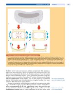
Computed tomography (Fig. 2) is the method of choice for the assessment of
thefusionstatus[228].However,CTinconjunctionwith2Dcoronalandsagittal
image reformation is more sensitive in diagnosing lumbar fusions than non-
union (Fucentese and Boos, unpublished data).
550 Section Degenerative Disorders
a
b
c
Figure 2. Computed tomography
Computed tomography is the imaging mo-
dality of choice for the assessment of spinal
fusion. Even in the presence of implants, the
bony bridges are well visualized. Bony
bridges outside a fusion cage are a more
reliable sign of solid fusion than when they
appear inside.
a Axial view; b sagittal refor-
mation;
c coronal reformation
Injection Studies
Injection studies
are helpful in identifying
the pain source
Thehighprevalenceofasymptomaticdiscalterationspromptstheneedforfur-
ther diagnostic tests to confirm that a specific structural abnormality is the
source of the pain. Spinal injections play an important role, although the scien-
tific evidence in the literature for their diagnostic efficacy is poor. Furthermore,
the predictive power of an injection study to improve patient selection for sur-
gery is poorly explored and documented [169]. A detailed description of the
strength and weaknesses of these diagnostic studies is included in Chapter
10 .
Provocative Discography
Discography remains
the only method to verify
discogenic LBP
Discography was introduced to image intervertebral disc derangement [172].
Currently, discography predominantly serves as a pain provocation test to differ-
entiate symptomatic and asymptomatic disc degeneration. The diagnostic effi-
cacy of this test remains a matter of debate [43, 202, 269] (see Chapter
10 ). The
assessment of the diagnostic accuracy of provocative discography for discogenic
LBP is problematic since no gold standard is available [43].
Always include an MR
normal level as internal
control
A reasonable practical approach is to include an adjacent MR normal disc level
as internal control [169, 275]. Accordingly, a positive pain response would
include an exact pain reproduction at the target level and no pain provocation or
only pressure at the normal disc level (
Case Introduction). In our center, patients
are only selected for provocative discography if they are potential candidates for
surgery, i.e. when the diagnostic test will influence treatment strategy. However,
careful interpretation of the findings is still mandatory with reference to the clin-
ical presentation [43]. Furthermore, provocative discography has failed to
improve patient selection to obtain better clinical outcome after surgery [177].
Degenerative Lumbar Spondylosis Chapter 20 551
Facet Joint Injections
Diagnosis of painful facet
joints by injections
must be made cautiously
The differentiation between symptomatic and asymptomatic facet joint osteoar-
thritis based on imaging studies alone is impossible [169]. So far, facet joint injec-
tions have been used for this purpose but are not without shortcomings (see
Chapter
10 ). Some authors suggest that a facet joint syndrome can be diagnosed
based on pain relief by an intra-articular anesthetic injection or provocation of
the pain by hypertonic saline injection followed by subsequent pain relief after
injection of local anesthetics [44, 173, 185, 199]. Interpretation of the pain
response is difficult because the facet joints are innervated by two to three seg-
mental posterior branches and the local anesthetic may diffuse to adjacent levels
if the injection is done non-selectively (i.e. without prior contrast medium injec-
tion) [169]. We recommend using contrast injection to document the correct
needle position and filling of the joint capsule (
Case Study 1). Uncontrolled diag-
nostic facet joint blocks exhibit a false-positive rate of 38% and a positive predic-
tive value of only 31% [239]. It is therefore mandatory to perform repetitive in-
filtrations to improve the diagnostic accuracy [239]. However, there are no
convincing pathognomonic, non-invasive radiographic, historical, or physical
examination findings that allow the reliable identification of lumbar facet joints
as a source of low-back pain and referred lower extremity pain [69, 70].
Temporary Stabilization
Temporary stabilization
does not predict fusion
outcome
The diagnosis of segmental instability remains a matter of intensive debate. How-
ever, it would be unreasonable to assume that abnormal segmental mobility is
non-existent or cannot be painful. Imaging studies, particularly functional
views, have failed to reliably predict segmental instability because of the wide
normal range of motion. The correct identification of the unstable level(s) is
challenging. The temporary stabilization with a pantaloon cast [223] has the
drawback of being unselective and requires further diagnostic testing, e.g. by
facet joint blocks. Stabilization of the putative abnormal segments by an external
transpedicular fixator has been suggested by several authors [74, 237, 254] with
mixed results in terms of outcome prediction. Based on an analysis of 103 cases,
Bednar [10] could not support using the external spinal skeletal fixation as a pre-
dictor of pain relief after lumbar arthrodesis.
Patient Selection for Treatment
The important role of non-biological factors for the outcome of surgical proce-
dures particularly for patients with predominant LBP is well documented. We
have therefore dedicated Chapter
7 to this topic. Various domains must be con-
sidered, i.e.:
medical factors
psychological factors
sociological factors
work-related factors
Non-biological factors
are important outcome
predictors
In clinical practice, however, it is extremely difficult to identify and systemati-
cally assess risk factors that can be used to accurately predict the outcome of sur-
gery. So far, there is insufficient evidence to exclude patients from surgery on the
grounds of specific risk factors [183]. Nonetheless, in the presence of selected fac-
tors (see Chapter
7 ), surgery should at least be delayed until attempts have been
made to modify risk factors that are amenable to change and all possible conser-
vative means of treatment are exhausted.
552 Section Degenerative Disorders
Non-operative Treatment
Most patients with predominant low-back pain without radiculopathy or claudi-
cation symptoms can be managed successfully by non-operative treatment
modalities (
Case St udy 2). The general objectives of treatment are (Table 3):
Table 3. General objectives of treatment
pain relief improvement of social activities
improvement of health-related quality of life improvement of recreational activities
improvement of activities of daily living improvement of work capacity
When the diagnostic assessment has identified a specific source of back pain
(
Table 1), the conservative treatment option does not differ from those applied to
non-specific disorders, which are extensively covered in Chapter
21 .Themain-
stay of non-operative management rests on three pillars:
pain management (medication)
functional restoration (physical exercises)
cognitive-behavioral therapy (psychological intervention)
Cognitive behavioral
interventions are necessary
to address fears and
misbeliefs
Pharmacologic pain management is outlined in Chapter 5 . Spinal injections
(e.g. facet joint blocks) may be a reasonable adjunct in controlling the pain for a
short term period [109, 169]. The first important aspect is a multidisciplinary
functional restoration program and psychological interventions to influence
patient behavior (see Chapter
21 ). The second important aspect is the timeli-
ness of the treatment intervention. The longer pain and functional limitations
persist, the less likely is pain relief, functional recovery and return to work (see
Chapter
6 ). Patients presenting with specific degenerative back pain usually
experience their pain and functional limitations for more than 3 months. These
patients should promptly be included in a multidisciplinary functional work
conditioning program. There is increasing evidence that patients with chronic
LBP benefit from a multidisciplinary treatment with a functional restoration
approach when compared with inpatients or outpatient non-multidisciplinary
treatments [263]. Two recent high quality randomized controlled trials (RCTs)
demonstrated that such a program is equally effective as surgery in treating
patients with lumbar spondylosis [31, 77].
It is as simple as it is obvious that the outcome of any treatment is critically
dependent on patient selection and this is also valid for non-operative treatment
(see Chapter
7 ). Favorable indications for non-operative treatment include
(
Table 4):
Table 4. Favorable indications for non-operative treatment
minor to moderate structural alterations short duration of persistent symptoms
(<6 months)
LBP of variable intensity and location absence of risk factor flags
intermittent symptoms highly motivated patient
Degenerative Lumbar Spondylosis Chapter 20 553
Operative Treatment
General Principles
Spinal fusion is thought
to eliminate painful motion
Spinal fusion is the most commonly performed surgical treatment for lumbar
spondylosis [66]. The paradigm of spinal fusion is based on the experience that
painful diarthrodial joints or joint deformities can be successfully treated by
arthrodesis [66, 121]. Since its introduction in 1911 by Albee [3] and Hibbs [127],
spinal fusion was initially only used to treat spinal infections and high-grade
spondylolisthesis. Later this method was applied to treat fractures and deformity.
Today approximately 75% of the interventions are done for painful degenerative
disorders [66]. Despite its frequent use, spinal fusion for lumbar spondylosis is
still not solidly based on scientific evidence in terms of its clinical effectiveness
[66, 102, 103, 264]. For a long time it was hoped that outcome of spinal fusions
could be significantly improved when the fusion rates come close to 100%. How-
ever, it is now apparently clear that outcome is not closely linked to the fusion sta-
tus [24, 90, 91, 102, 103, 256].
The standard concept advocated in the literature is that surgical treatment is
indicated when an adequate trial of non-operative treatment has failed to
improve the patient’s pain or functional limitations [122, 264]. However, there is
no general consensus in the literature on what actually comprises an adequate
trial of non-operative care. Based on a meta-analysis, van Tulder et al. [264] con-
cluded that fusion surgery may be considered only in carefully selected patients
after active rehabilitation programs for a period of 2 years have failed. The gen-
eral philosophy that surgery is only indicated if long-term non-operative care has
failed is challenged by the finding that the longer pain persists the less likely it is
that it will disappear. This notion is supported by recent advances in our under-
standing of the pathways and molecular biology of persistent (chronic) pain (see
Chapter
5 ). It has also been known for many years that returning to work
becomes very unlikely after 2 years [268].
Surgery if needed should be
done in a timely manner
Wethereforeadvocateamoreactive approach in patient selection for surgery,
i.e. not only offering surgery as the last resort after everything else has failed
because of the adverse effects of pain chronification. Patients should be evaluated
early (i.e. within 3 months), searching for a pathomorphological abnormality which
is likely to cause the symptoms. This evaluation must be based on a thorough clini-
cal assessment, imaging studies and diagnostic tests. If a pathomorphological alter-
ation in concordance with the clinical symptoms can be found, the patient should
be selected for potential surgery. Prior to surgery, the patient should then be inte-
grated in a fast track aggressive functional rehabilitation program (not longer than
3 months). If this program fails, the structural correlate should be treated surgically
if multilevel (>2 levels) fusion can be avoided. In multilevel degeneration of the
lumbar spine requiring more than two-level fusion, the clinical outcome is less sat-
isfactory in our hands and we are more conservative. We acknowledge that this
approach is anecdotal and not yet based on scientific evidence, but it seems to be
reasonable and works satisfactorily in a large spine referral center.
Favorable indications for surgery include (Table 5):
Table 5. Favorable indications for operative treatment
severe structural alterations short duration of persistent symptoms (<6 months)
one or two-level disease absence of risk factor flags
clinical symptoms concordant with the structural correlate highly motivated patient
positive pain provocation and/or pain relief tests initial response to a rehab program but frequent
recurrent episodes
554 Section Degenerative Disorders
Only a few morphological imaging abnormalities have been identified which
rarely occur in a group of asymptomatic individuals below the age of 50 years
[274] and may therefore predict the pain source when occurring in symptomatic
patients. Severe structural alterations which may predict a fa vorable outcome of
surgery include:
severe facet joint osteoarthritis
disc degeneration with severe endplate abnormalities (Modic Types I and II)
These abnormalities represent favorable predictors for surgery, particularly
when present at only one or two levels with the rest of the thoracolumbar spine
unremarkable, cause concordant symptoms and consistently respond to pain
provocation and relief test. As outlined above, the duration of symptoms should
be short to avoid the adverse effects of a chronic pain syndrome. It has been our
anecdotal observation that patients have a favorable outcome if they had
responded successfully to a m ultidisciplinary restoration program but have fre-
quent recurrent episodes.
Biology of Spinal Fusion
A basic understanding of the general principles of bone development and bone
healing as well as the biologic requirements for spinal fusion in the lumbar spine
are a prerequisite to choosing the optimal fusion technique [13]. A comprehen-
sive review of this topic is far beyond the scope of this chapter and the reader is
referred to some excellent reviews [13, 92, 93, 209, 232, 240].
In contrast to fracture healing, the challenge in spinal fusion is to bridge an
anatomic region with bone that is not normally supported by a viable bone [34].
Spinal arthrodesis can be generated by a fusion of:
adjacent laminae and spinous processes
facet joints
transverse processes
intervertebral disc space
Vascular supply to the
fusion area is important
An osseous fusion of the transverse processes is the most common type of fusion
performed in the lumbar spine [16]. MacNab was one of the first to realize that
the success of intertransverse fusion over posterior fusion (i.e. bone apposition
on the laminae and spinous processes) was based on the blood supply to the
fusionbedwhichallowedforarevascularizationandreossificationofthegraft
[176]. The early interbody fusion technique (inserting bone into the interverte-
bral disc space after discectomy) was hampered by graft subsidence or graft fail-
ure because of the heavy loads in the lumbar spine and did not provide favorable
results without instrumentation (see below).
The prerequisite of successful spine fusion is three distinct properties of the
applied graft material, i.e. [164, 259]:
osteogenesis
osteoconduction
osteoinduction
The optimal graft material
should be osteogenic,
osteoconductive
and osteoinductive
Osteogenicity is the capacity of the graft material to directly form bone and is
dependent on the presence of viable osteogenetic cells. This property is only
exhibited by fresh autologous bone and bone marrow. Osteoconduction is the
process of living tissue to grow onto a surface or into a scaffold, which results in
new bone formation and incorporation of that structure [59]. Particularly can-
cellous bone with its porous and highly interconnected trabecular architecture
allows easy ingrowth of surrounding tissues. Osteoconduction is also observed
Degenerative Lumbar Spondylosis Chapter 20 555
in fabricated materials that have porosity similar to that of bone structure, e.g.
coralline ceramics, hydroxyapatite beads, combinations of hydroxyapatite and
collagen, porous metals and biodegradable polymers [59]. Osteoinduction indi-
cates that primitive, undifferentiated and pluripotent cells are stimulated to
develop into bone-forming cells [4]. Urist [257, 258] coined the term “bone mor-
phogenetic proteins” (BMPs) for those factors that stimulate cells to differentiate
into osteogenic cells.
Bone Grafts
Autologous bone
is still the gold standard
Autologous bone is generally considered the “gold standard” as a graft material
for spinal fusion and exhibits osteogenetic, osteoconductive and osteoinductive
properties [115]. Autologous bone for spinal fusion is harvested from the ante-
rior or posterior iliac crest as cancellous bone, corticocancellous bone chips or
tricortical bone blocks. The drawback of autologous bone is related to the limited
quantity and potential donor site pain [63, 80, 125].
Allografts potentially
transmit infectious disease
These drawbacks have led to the use of allograft bone early in the evolution of
spinal fusion. Allografts are used in different forms for spinal fusion. They are
predominately used as structural allografts (e.g. femoral ring allografts) but are
available in other forms (e.g. corticocancellous bone chips). Bone allografts
exhibit strong osteoconductive, weak osteoinductive but no osteogenetic proper-
ties [152, 232]. Fresh allografts elicit both local and systemic immune responses
diminishing or destroying the osteoinductive and conductive properties. Freez-
ing or freeze-drying of allografts is therefore used clinically to improve incorpo-
ration [107], but mechanical stability of the graft is reduced by freeze drying
(about 50%) [232]. However, the major drawback of those allografts is the poten-
tial transmission of infections (particularly hepatitis C, HIV) [64]. Gamma irr a-
diation of at least 34 kGy is recommended to substantially reduce the infectivity
titer of enveloped and non-enveloped viruses [220]. However, screening proce-
dures remain mandatory. Autologous or allogenic cortical grafts are at least ini-
tially weight-bearing but all bone grafts are finally resorbed.
Cancellous allografts
are completely replaced
by autologous bone
or resorbed
Cancellous grafts are completely replaced in time by creeping substitution,
whereas cortical grafts remain as an admixture of necrotic and viable bone for a
prolonged period of time [107]. Bone graft incorporation within the host,
whether autogenous or allogeneic, depends on various factors [152]:
type of graft
site of transplant
quality of transplanted bone and host bone
host bed preparation
preservation techniques
systemic and local disease
mechanical properties of the graft
Although the role of cancellous allograft as a delivery vehicle for other osteoin-
ductive factors is conceptually reasonable, data is lacking to support this applica-
tion at this time [162]. Femoral ring allografts for anterior interbody fusions have
gained increasing popularity because of their capability for an initial structural
support [191]. The decreased fusion rate associated with allografts becomes
more significant in multilevel surgery and in patients who smoke [65].
Bone Graft Substitutes
Bone graft substitutes are increasingly being used for spinal fusion because of the
minimal but inherent risk of a transmission of infectious disease with allografts
556 Section Degenerative Disorders
[115]. Among the characteristics of an optimal bone graft substitute are:
high degree of biocompatibility
lack of immunogenicity and toxicity
ability for biodegradation
ability to withstand sterilization
availability in different sizes, shapes and amounts
reasonable cost
The most commonly used bone graft substitutes in spinal fusion are:
calcium phosphates
demineralized bone matrix (DBM)
Calcium Phosphates
Calcium phosphate materials can be classified by chemical composition and ori-
gin [i.e. natural or synthetic (ceramic) forms] and include:
hydroxyapatite (HA)
tricalcium phosphate (TCP)
natural coralline
This group of materials closely resembles the mineral composition, properties
and microarchitecture of human cancellous bone and has a high affinity for
binding proteins [162]. HA is relatively inert and biodegrades poorly. Due to its
brittleness and slow resorption, remodeling may be hindered and the material
can become a focus of mechanical stress [232]. In contrast, TCP composites
exhibit greater solubility than HA and typically undergo biodegradation within
approximately6weeks,whichmaybetooearlyforamaturationofthefusion
mass [162, 232]. Coralline HA (CHA) was developed in 1971 with the aim of pro-
viding a more consistent pore size and improved interconnectivity [198]. These
natural ceramics are derived from sea corals and are structurally similar to can-
cellous bone. The coral calcium carbonate undergoes a hydrothermal reaction
where calcium carbonates are transformed into HA [162].
Calcium phosphates
are of limited effectiveness
These materials are available in various preparations including putty, granular
material, powder, pellets or injectable calcium phosphate cement [20]. In con-
trast to early reports suggesting the capability for osteogenic stimulation, it is
now believed that calcium phosphates have only osteoconductive properties
[232]. Purely osteoconductive substitutes are less effective in posterolateral spine
fusion, but may be suitable for interbody fusion when it is rigidly immobilized
[13]. Although selective data both from animal and clinical studies appears
promising, there is still only limited evidence for the clinical effectiveness of
these materials to generate or at least enhance spinal fusion [232].
Demineralized Bone Matrix
A group of low-molecular-weight glycoproteins contained in the organic phase
(particularly BMPs) are responsible for the bone inductive activity [166]. DBM is
produced through a mild acid extraction of cortical bone and is processed to
reduce risk of infection and immunogenic host response. The mild demineraliza-
tion removes the mineral content of the bone, leaving behind collagen and non-
collagenous proteins including the BMP, which becomes locally available to the
cellular environment [166]. DMB is supplied in a variety of forms such as gel,
malleable putty, flexible strips or injectable bone paste. Lee et al. [166] have
pointed out that the amount of osteoinductive ability may rely on its preparation
and the type of carrier with which it is combined.
Degenerative Lumbar Spondylosis Chapter 20 557
DBM predominantly
is a bone graft extender
Even though DBM is considered osteoinductive, this effect is much weaker as
compared with commercially available recombinant BMPs. The use of current
available DBMs is primarily as a bone graft extender or enhancers but caution is
necessary as bone graft substitutes [5, 13].
Bone Promoters
Since their discovery by Urist in 1965 [257], BMPs have been the focus of inten-
sive research and clinical testing aiming to develop treatment strategies to
enhance bone healing and generate arthrodesis. The role of BMPs in bone forma-
tion during development and in fracture healing is now well established [225].
BMPsaremembersofthetransforminggrowthfactor- supergene family [40]
and so far more than 15 BMPs have been identified [225]. BMPs function as a dif-
ferentiation factor and act on mesenchymal stem cells to induce bone formation
[34].
The majority of preclinical and clinical studies for spinal fusion (interbody
and posterolateral) have been done using [15, 68, 106, 139, 142, 145, 260, 261]:
BMP 2
BMP 7 (osteogenic protein-1, OP-1)
BMPs promote fusion but
cost-effectiveness is unclear
The BMPs are delivered to the fusion site on carriers, e.g. HTA/TCP [15] or colla-
gen matrix [145]. When used at an optimized concentration and with an appro-
priate carrier, BMPs can be successfully used as bone graft replacement [34].
However, only increasing experience and longer term follow-up will show
whether these new fusion techniques will surpass the level of safety and clinical
feasibility and can be established as a cost-effective treatment.
Surgical Techniques
For a long time, spinal fusion has been the treatment of choice when addressing
symptomatic lumbar spondylosis. Motion preserving implant technologies have
emerged which offer theoretical advantages over fusion. The early motion pre-
serving technologies such as Graf li gamentoplasty [96, 144, 226] and Dynesys
stabilization [237, 238] have demonstrated favorable outcomes for selected
patients. Similarly, the early outcome was promising for total disc arthroplasty
[62, 116, 190, 284] and posterior interspinous spacers [49, 153, 286]. However, the
new technologies must pass the test of time, i.e. long-term follow-up in RCTs,
before they can be broadly accepted as alternative fusion techniques. So far, no
evidence has been reported to demonstrate that these new techniques are supe-
rior to spinal fusion.
The scientific literature exhibits a plethora of articles covering the outcome of
surgical treatment. The vast majority of these papers cover technical aspects,
safety and early clinical results without adequate control groups. Many of the
studies incorporated a whole variety of indications, which limits conclusions on
degenerative lumbar spondylosis without neurological compromise. However,
The scientific evidence for
spinal fusion in lumbar
spondylosis is poor
when the scientific literature is reduced to Level A evidence (i.e. consistent evi-
dence in multiple high-quality RCTs), only 31 RCTs can be identified through
March 2005 [102, 103]. These facts greatly limit treatment recommendations on
degenerative lumbar spondylosis. In this chapter, we therefore attempt to base
treatment recommendations on the best available evidence.
558 Section Degenerative Disorders


