Spinal Disorders: Fundamentals of Diagnosis and Treatment Part 58 potx
Bạn đang xem bản rút gọn của tài liệu. Xem và tải ngay bản đầy đủ của tài liệu tại đây (427.9 KB, 10 trang )
Non-instrumented Spinal Fusion
Lumbar arthrodesis can be achieved by three approaches. The most commonly
used technique is posterolateral fusion (PLF), which comprises a bone grafting
of the posterior elements. As an alternative, the bone grafting can be performed
after disc excision and endplate decancellation (interbody fusion)byaposterior
approach (posterior lumbar interbody fusion, PLIF) or the anterior approach
(anterior lumbar interbody fusion, ALIF). The so-called combined or 360 degree
fusion is the combination of both techniques.
Posterolateral Fusion
Posterolateral fusion
remains the fusion gold
standard
Posterolateral fusion was first described by Watkins in 1953 [270] and remains
the gold standard for spinal fusion. The technique consisted of a decortication of
the transverse spinous processes, pars interarticularis and facet joints with appli-
cation of a large corticocancellous iliac bone block. This method has been modi-
fied by Truchly and Thompson [255], who used multiple thin iliac bone strips as
graft material instead of a single corticocancellous bone block because of fre-
quent graft dislocation [255]. In 1972, Stauffer and Coventry [245] presented the
technique still used today by most surgeons, which consisted of a single midline
approach (
Fig. 3). However, the bilateral approach had a revival some years later
when Wiltse et al. [278] suggested an anatomic muscle splitting approach which
was modified by Fraser [118].
ab
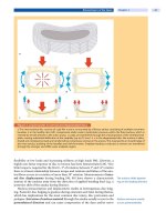
Figure 3. Surgical technique of posterolateral fusion
Careful preparation of the fusion bed is important and consists of: a decortication of the transverse process and facet
joints and isthmus;
b placement of autologous corticocancellous bone chips over the facet joints and transverse pro-
cesses.
Degenerative Lumbar Spondylosis Chapter 20 559
Non-instrumented
posterolateral fusion
remains the benchmark
for comparison of fusion
techniques
Boos and Webb [24] reviewed 16 earlier non-randomized studies (1966–1995)
with a total of 1264 cases and found a mean fusion rate of 87% (range, 40–96%)
and an average rate of satisfactory outcome of 70% (range, 52–89%). The results
reportedinthearticlebyStaufferandCoventry[245]remainabenchmarkfor
non-instrumented posterolateral fusion. Eighty-nine percent of those whose
fusion was done as a primary procedure for degenerative disc disease achieved
good clinical results and 95% were judged to have a solid fusion. These favorable
results were not surpassed by many studies which followed.
Posterior Lumbar Interbody Fusion
Posterior disc excision and insertion of bone grafts was first described by Jaslow
in 1946 [138] and popularized by Cloward [52, 54] and others as posterior lumbar
interbodyfusion(PLIF)(
Fig. 4). The disadvantage of PLIF was the need for an
extensive posterior decompression to allow for a graft insertion which destabi-
lized the spine. Furthermore, graft insertion necessitates a substantial retraction
of the nerve roots which carries the risk of nerve root injuries and significant
postoperative scarring.
PLIF increases fusion rate PLIF resulted in a somewhat higher fusion rate and better clinical outcome
than posterolateral fusion. Based on an analysis of 1372 cases reported in 8 stud-
ies [53, 56, 130, 131, 165, 171, 194, 219], mean fusion rate was 89% (range,
82–94%) and the average rate of satisfactory outcome was 82% (range,
78–98%) [24].
Anterior Lumbar Interbody Fusion
Anterior spinal fusion was first described by Capener in 1932 for the treatment
of spondylolisthesis [39]. However, Lane and Moore [163] were the first to per-
form anterior lumbar interbody fusion (ALIF) on a larger scale [163]. Iliac tri-
cortical bone autograft as well as femoral, tibia, or fibula diaphyseal allografts
were used for this technique. Particular femoral ring allografts have been
recently used as cost-effective alternatives to cages and offer some advantages
regarding the biology of the fusion compared to cages [167, 191]. The advantage
of ALIF was that the paravertebral muscles and neural structures remained
intact. A further technical advantage is that disc excision and graft bed prepara-
tioncanbedonebetterthanwithPLIF.Ontheotherhand,theabdominalaccess
is associated with specific approach related problems such as retrograde ejacu-
lation in male patients (range, 0.1–17%) [29, 76, 254] and vascular injuries
(range, 0.8–3.4%) [29, 210].
Stand-alone ALIF
has not been successful
The results in the literature were largely variable. An analysis of 1072 cases
reported in 10 studies revealed a mean fusion rate of 76% (range, 56–94%) and
an average satisfactory outcome rate of 76% (range, 36–92%) [24]. Compared to
the favorable results Stauffer and Coventry achieved with a posterolateral fusion
[245], the ALIF results of the same authors [244] were disappointing (fusion rate
56%, satisfactory outcome 36%). Stauffer and Coventry [244] concluded that
ALIF should be utilized as a salvage procedure in those infrequent cases in which
posterolateral fusion is inadvisable because of infection or unusual extensive
scarring [244]. Graft dislocation and subsidence as well as moderate fusion rate
caused the “stand-alone” ALIF to fall out of favor for some years.
Instrumented Spinal Fusion
With the advent of pedicle screw fixation devices in the 1980s and the introduc-
tion of fusion cages in the 1990s, spinal instrumentation was widely used with the
560 Section Degenerative Disorders
ab
c
d
Figure 4. Surgical technique of posterior lumbar interbody fusion
a Pedicle screws are inserted at the target levels. A wide decompression is necessary to insert the cages safely through
the spinal canal. The intervertebral disc is removed as completely as possible but without jeopardizing the anterior outer
anulus (vascular injuries). The cartilage endplates are removed with curettes. Cages are inserted by retracting the nerve
root and thecal sac medially.
b, c Prior to insertion, the disc space is filled with cancellous bone graft particularly anteri-
orly.
d The rod is inserted and fixed to the screws. A posterolateral fusion is added.
rationale that the improved segmental stability may enhance the fusion rate and
simultaneously improve clinical outcome. The biomechanical background of spi-
nal instrumentations is reviewed in Chapter
3 .
Pedicle Screw Fixation
Pedicle screw fixation
is the gold standard
for lumbar stabilization
The pedicle is the strongest part of the vertebra, which predestines it as an
anchorage for screw fixation of the vertebral segments. Pedicle screw fixation had
Degenerative Lumbar Spondylosis Chapter 20 561
Roy-Camille first used
pedicle screws
its origins in France. From 1963, Raymond Roy-Camille first used pedicle screws
with plates to stabilize the lumbar spine for various disorders [230]. Some years
later, Louis and Maresca modified Roy-Camille’s plate and technique to better
adapt to the lumbosacral junction [174, 175]. Based on the pioneering work of
Fritz Magerl [179], the concept of angle-stable pedicular fixation was introduced,
which led to the development of the AO Internal Fixator [1, 67]. Around the same
time, Steffee [246] developed the variable screw system (VSP), a plate pedicle
screw construct. A further milestone in the development was the introduction of
a new screw-rod system by Cotrel and Dubousset in 1984 [60]. The versatile
Pedicle screw fixation
is most commonly used
in conjunction with
posterolateral fusion
Cotrel-Dubousset instrumentation system became widely used for the treatment
of degenerative disorders. The current system offers the advantage of polyaxial
screw heads which facilitate the rod screw connection. The most frequently used
fusion technique today is to combine pedicle screw fixation with posterolateral
fusion (
Case Study 1).
The fusion rates with the pedicle screw system average 91% (range 67–100%)
with satisfactory clinical outcome ranging between 43% and 95% (mean 68%)
[24]. Many surgeons applied the pedicle screw stabilization system
Pedicle screw fixation
enhances fusion rate
but not clinical outcome
with the rationale that the enhanced fusion rate would also improve outcome.
However, at the end of the 1990s it became obvious that pedicle screw fixation
may increase the fusion rate but not necessarily clinical outcome [24, 102].
Translaminar Screw Fixation
Translaminar screws are an
alternative to pedicle screws
An alternative method of screw fixation in the lumbar spine was first described
in 1959 by Boucher [26]. These oblique facet screws were used to block the
zygapophyseal joints. However, the stability of these screws crossing the facet
joints obliquely was unsatisfactory. Magerl [180] developed the so-called trans-
laminar screw fixation which crossed the facet more perpendicularly, increas-
ing stability [126]. The initial clinical results were promising [113, 129, 136,
184]. The advantage is that the screws can be used as a minimally invasive pos-
teriorstabilizationtechniqueandcanoftenbecombinedwithananteriorinter-
body fusion [191], which can also be done minimally invasively (see below,
Case Introduction
)[21].
Cage Augmented Interbody Fusion
Cages stabilize
the anterior column
and increase fusion rate
The application of interbody fusion cages for fusion enhancement is based on the
rationale that a strong structural support is needed for the anterior column
which does not migrate or collapse [122]. Interbody cages were designed and
firstusedbyBagby and Kuslich (BAK cage) in the 1990s and consisted of threa-
ded hollow cylinders filled with bone graft [160, 161]. Today, different designs
and materials are available for anterior and posterior use (
Table 6):
Table 6. Cage materials and design
Designs Materials
threaded, cylindrical cages titanium
ring-shaped cages with and without mesh structure carbon
box-shaped cages polyetheretherketone (PEEK)
The cages were originally designed as stand-alone anterior or posterior fusion
devices. The initial studies in the literature reported promising results [161, 224,
233] and some authors reported satisfactory long term outcome [27]. However,
the biomechanical (stability, no cage subsidence) and biologic (load sharing with
562 Section Degenerative Disorders
abc
Figure 5. Circumferential fusion
a Young (28 years) female patient with endplate changes (Modic Type II) undergoing pedicle screw fixation L5/S1 and
posterolateral fusion in combination with a cage augmented anterior lumbar interbody fusion. Postoperative
b antero-
posterior view and
c lateral view.
The outcome of stand-alone
cages is not favorable
the graft) requirements for spinal fusion were challenging (see Chapter 3 )and
resulted ina high failure rate [73, 189]. The problems associated with stand-alone
cages led to the recommendation of the use of cages only in conjunction with spi-
nal instrumentation (
Fig. 5) [37, 45].
Although a bilateral cage insertion is generally recommended for biomechan-
ical reasons, it is not always possible to insert two cages when the disc space is
still high and the spinal canal rather narrow. Recently, it has been shown that uni-
Unilateral cage insertion
may suffice in selected cases
lateral cage insertion leads to comparable results to bilateral cage placements
[82, 196]. The shortcomings of the PLIF technique (i.e. retraction of nerve roots
and potential epidural fibrosis) led to a modified technique by a transforaminal
route (transforaminal lumbar interbody fusion, TLIF). After unilateral resec-
tion of the facet joints, the disc is exposed and excised without retraction of the
thecal sac and nerve roots before a cage is implanted. TLIF should only be used
in conjunction with spinal instrumentations. The reported results with this tech-
nique are promising [105, 117, 123, 231, 235].
Circumferential Fusion
Circumferential fusion (i.e. interbody and posterolateral fusion) was first used
for the treatment of spinal trauma and deformity, then expanded to failed previ-
ous spinal fusion operations and is now used also as a primary procedure for
chronic low-back pain [122]. Theoretically, this technique should increase the
fusion rate by maximizing the stability within the motion segment and enhance
outcome because of an elimination of potential pain sources in anterior and pos-
terior spinal structures. Today, circumferential fusion is almost always done in
conjunction with instrumentation. Interbody fusion can be done by a posterior
(PLIF) (
Fig. 4)oranteriorapproach(ALIF)(Figs. 5, 6) depending on the individ-
Outcome of PLIF and ALIF
appears to be comparable
ual pathology and surgeons’ preferences. There seems to be no difference
between both approaches in terms of clinical outcome [178].
Degenerative Lumbar Spondylosis Chapter 20 563
ab
cd
Figure 6. Surgical technique of anterior lumbar interbody fusion
The lumbosacral junction is exposed by a minimally invasive retroperitoneal approach. a The intervertebral disc is
excised;
b the endplates can be distracted with a spreader and the endplate cartilage is removed with curettes; c the disc
space is filled with cancellous bone and supported with two cages. Ring-shaped cage design allows sufficient bone graft
to be placed around the cages.
d Pedicle screw fixation is added in conjunction with posterolateral fusion.
Combined interbody
and posterolateral fusion
has the highest fusion rate
Several studies have consistently demonstrated that circumferential fusion
increases therate of solid fusion [48,91], with fusion rates ranging from91% to 99%
[48, 91, 242, 252]. However, it remains controversial whether circumferential fusion
improves clinical outcome [91, 267]. Fritzell et al. [91] did not find a significant dif-
ference in outcome when comparing non-instrumented, instrumented posterolat-
eral or circumferential fusion. On the contrary, Videbaek et al. [267] have demon-
strated that patients undergoing circumferential fusion have a significantly better
long term outcome compared to posterolateral fusion in terms of disability (Oswe-
stry Disability Index) and physical health (SF-36). Some patients continue to have
pain after posterolateral spinal fusion despite apparently solid arthrodesis. One
potential etiology is pain that arises from adisc within the fused levels and has posi-
tive pain provocation on discography. These patients benefit from an ALIF [8].
564 Section Degenerative Disorders
Minimally Invasive Approaches for Spinal Fusion
Access technology should
decrease collateral muscle
damage during fusion
surgery
In the last two decades, attempts have been made to minimize approach-related
morbidity [98, 154, 247]. Particularly, the posterior approach to the lumbosacral
spine necessitates dissection and retraction of the paraspinal muscles. The mus-
cle retraction was shown to cause a significant muscle injury dependent on the
traction time [147–150]. The use of translaminar screw fixation in conjunction
with an ALIF has been suggested to minimize posterior exposure of the lumbar
spine [9, 137, 159, 191, 241] (
Case Introduction). Newer posterior techniques use
a tubular retractor system for pedicle screw insertion and percutaneous rod
insertion that avoids the muscle stripping associated with open procedures [71,
83, 98].
Laparoscopic techniques foranteriorinterbodyfusionweredevelopedinthe
1990s to minimize surgical injury related to the anterior approach [38, 170, 252,
281]. This technique was favored in conjunction with the use of cylindrical cages
and may exhibit some immediate postoperative advantages (e.g. less blood loss,
shorter postoperative ileus, earlier mobilization) [61, 78]. However, this tech-
nique did not prevail because of the tedious steep learning curve, longer opera-
tion time, expensive laparoscopic instruments and tools and need for a general
surgeon familiar with laparoscopy without providing superior clinical results
[50, 200, 281]. Many surgeons today prefer a mini-open anterior approach to the
lumbar spine using a retraction frame (
Case Introduction), which allows a one or
two level anterior fusion to be performed through a short incision [2, 186]. It also
allows for a rapid extension of the exposure in case of complications such as an
injury to a large vessel.
Minimally invasive
approaches have not yet
demonstrated superior
outcomes
Many initial reports have shown similar clinical results in terms of spinal
fusion rates for both traditional open and minimally invasive posterior
approaches [71, 84]. However, the anterior minimally invasive procedures are
often associated with a significantly greater incidence of complications and tech-
nical difficulty than their associated open approaches [71].
F usion Related Problems
Revision Surgery for Non-union
Revision surgery for non-union remains costly and difficult. Diagnosis of non-
union by radiological assessment is not easy and solid fusion determined from
radiographs ranged from 52% to 92% depending on the choice of surgical proce-
dure [47].
Functional and clinical
results of lumbar fusion
are often not in correlation
Similarly to a primary intervention, the single most important factor in
achieving a successful clinical outcome is patient selection [75]. It is well antici-
pated that functional and clinical results of lumbar fusion are often not in corre-
lation and the rate of non-union has no significant association with clinical
results in the first place [81, 277], which challenges the clinical success of revision
surgery for non-union.
The best lumbar fusion rates
are achieved
by a circumferential fusion
Interbody fusion is advocated to repair non-union because revision surgery
by posterolateral fusion has not been overly successful [55, 75]. Circumferential
fusion provides the highest fusion rate. It is therefore recommended to perform
a 360-degree fusion during a revision operation [47]. However, patients with a
non-union after stand-alone cage augmented fusion (PLIF or ALIF) may well
benefit from a revision posterolateral fusion and pedicle screw fixation [45].
Despite successful fusion
repair, clinical outcome
is often disappointing
Although solid fusion after non-union can be achieved in 94–100% of
patients with appropriate techniques [36, 42, 99], there is only a poor correlation
of the radiographic and clinical results [42]. After repair of pseudoarthrosis, Car-
Degenerative Lumbar Spondylosis Chapter 20 565
penter et al. reported a solid fusion rate of 94% without significant association
with clinical outcome, patient’s age, obesity and gender [42]. Similar findings
were made by Gertzbein et al. [99]. These authors reported a fusion rate of 100%
even in the face of factors often placing patients at high risk for developing a
pseudarthrosis, i.e. multiple levels of previous spinal surgery, including previous
pseudarthrosis, and a habit of heavy smoking. However, the satisfactory outcome
rate was only somewhat better than 50%, based on a lack of substantial pain
improvement and return to work [99]. It is therefore mandatory to inform surgi-
cal candidates that the risk of an unsatisfactory outcome is high despite solid
fusion.
Adjacent Segment Degeneration
Adjacent segment degeneration following lumbar spine fusion remains a well
known problem, but there is insufficient knowledge regarding the risk factors
that contribute to its occurrence [158]. Biomechanical and radiological investi-
gations have demonstrated increased forces, mobility, and intradiscal pressure in
adjacent segments after fusion [72]. Although it is hypothesized that these
changes lead to an acceleration of degeneration, the natural history of the adja-
cent segment remains unaddressed [72]. When discussing the problem of adja-
cent segment degeneration it is important to:
take the preoperative degeneration grade into account
differentiate asymptomatic and symptomatic degeneration
consider the natural history of the adjacent motion segment
Adjacent segment
degeneration is a
frequent problem
There is no significant correlation between the preoperative arthritic grade and
the need for additional surgery [100]. Radiographic segmental degeneration
weakly correlates with clinical symptoms [208] and the age of the individual [46,
104, 213]. There are conflicting results on the influence of the length of spinal
fusion [46]. Pellise et al. [213] found that radiographic changes suggesting disc
degeneration appear homogeneously at several levels cephalad to fusion and
seem to be determined by individual characteristics. Ghiselli et al. [100] reported
a rate of symptomatic degeneration at an adjacent segment warranting either
decompression or arthrodesis to be 16.5% at 5 years and 36.1% at 10 years. It
remains to be seen whether disc arthroplasty will alter the rate of adjacent seg-
ment degeneration [128].
Motion Pr eserving Surgery
Motion preservation
surgery is still emerging
With the advent of motion preserving surgical techniques, there is a great excite-
ment among surgeons and patients that the drawbacks of spinal fusion can be
overcome. So far, the initial results are equivalent to those obtained with spinal
fusion and it is hoped that there is a decrease in the rate of adjacent segment degen-
eration. The success of the paradigm shift toward motion preservation is still
unproven but it makes intuitive and biomechanical sense [6]. A review of the bio-
mechanical background of motion preserving surgery is included in Chapter
3 .
Total Disc Arthroplasty
Attempts to artificially replace the intervertebral discs were already made in the
1950s by Fernstrom [79]. However, the ball like intercorporal endoprosthesis was
prone to failures (i.e. loosening and migration). The disc prosthesis with the lon-
gest history is the SB-Charit´e prosthesis, which dates back to 1982. The prosthe-
sis was developed by Kurt Schellnack and Karin Büttner-Janz at the Charit´eHos-
566 Section Degenerative Disorders
pital in Berlin. The prosthesis has meanwhile undergone several redesigns. The
SB-Charit´e III disc prosthesis (Depuy Spine) was the first to receive FDA approval
in 2004. In recent decades various alternative designs have been developed such
as the ProDis-L (Synthes, FDA approval 2006), Maverick (MedtronicSofamorDa-
nek), Flexicore (Stryker), Kineflex (SpinalMotion) and ActivL (B. Braun/Aesku-
lap) total disc replacement systems.
Indications and contraindications for total disc arthroplasty (TDA) are
(
Table 7):
Table 7. Total disc arthroplasty
Indications Contraindications
age 18–60 years osteoporosis
severe back pain multilevel disc degeneration
severe disability (ODI >30–40) facet joint osteoarthritis
failed non-operative treatment for >6 months spinal deformity or instability
single or two-level disc degeneration prior lumbar fusion
obesity
consuming illness (tumor, infection, inflammatory disorders)
metabolic disorders
known allergies
Modified from Zigler et al. [283] and Guyer et al. [116]
ODI Oswestry Disability Index
German and Foley [97] have highlighted that particular attention should be paid
to the presence of facet joint osteoarthritis, as this has been associated with poor
clinical outcomes after arthroplasty [187, 262]. Total disc arthroplasty (
Fig. 7)has
meanwhile passed the level of technical feasibility and safety [11, 51, 168, 187].
However, major concerns remain regarding revision arthroplasty, which can
cause life-threatening complications (e.g. in case of a major vessel injury during
reoperation).
abc
Figure 7. Total disc arthoplasty
Female patient (48 years) with endplate (Modic) changes at L5/S1 treated by total disc replacement with Prodisc (Syn-
thes).
a Sagittal T2 weighted MRI scan demonstrating Modic Type II changes at L5/S1. Postoperative b anteroposterior
view; and
c lateral view showing correct positioning of the TDA.
Degenerative Lumbar Spondylosis Chapter 20 567
Two randomized controlled FDA IDE trials compared TDA with spinal fusion.
In the first trial, the SB-Charit´e disc prosthesis was compared with stand-alone
BAK cages with autograft from the iliac crest for one-level disc disease L4–S1
[12, 188]. The second trial compared the ProDisc-L total disc arthroplasty with
circumferential spinal fusion for the treatment of discogenic pain at one verte-
bral level between L3 and S1 [282]. Both prospective, randomized, multicenter
Short-term clinical outcome
of TDA is comparable
to spinal fusion
studies demonstrated that quantitative clinical outcome measures following
TDAareatleastequivalenttoclinicaloutcomeswithconventionalfusiontech-
niques.
Although these results are promising, only longer term follow-up will show
whether TDA is superior to spinal fusion and reduce the rate of adjacent segment
degeneration [97].
Dynamic Stabilization
Abnormal loading patterns
are a cause of pain
Mulholland [201] has hypothesized that abnormal patterns of loading rather
than abnormal movement are the reason that disc degeneration causes back pain
in some patients. Abnormal load transmission is the principal cause of pain in
osteoarthritic joints. Both osteotomy and total joint replacement succeed
because they alter the load transmission across the joint [201]. In this context, the
spine is painful in positions and postures rather than on movement [201]. The
The dynamic stabilization
system may alter abnormal
loading and thus
be effective
rationale for dynamic or “soft” stabilization of a painful motion segment is to
alter mechanical loading by unloading the disc but preserving lumbar motion in
contrast to spinal fusion [205]. The Graf ligamentoplasty was the first dynamic
stabilization system widely used in Europe [30, 96, 111]. The principle of the Graf
system was to stabilize the spine in extension (locking the facet joints) using ped-
icle screws connected by a non-elastic band. This system increased the load over
the posterior anulus, caused lateral recess and foraminal stenosis and was only
modestly successful [201].
Best indications
for dynamic stabilization
are not well established
The Dynesys system is based on pedicle screws connected with a polyethylene
cord and a polyurethane tube reducing movement both in flexion and extension
[238, 249]. However, often it also unloads the disc to a degree that is unpredict-
able [201]. Non-randomized studies reported promising results [221, 249, 276].
However, Grob et al. [112] reported that only half of the patients declared that the
operation had helped and had improved their overall quality of life, and less than
half reported improvements in functional capacity. The reoperation rate after
Dynesys was relatively high. Only long-term follow-up data and controlled pro-
spective randomized studies will reveal whether dynamic stabilization is supe-
rior to spinal fusion for selected patients [238].
The clinical effectiveness of
interspinous stabilization
remains to be proven
Recently, interspinous implants have been introduced as minimally invasive
dynamic spine stabilization systems, e.g. X-Stop (St. Francis Medical Technolo-
gies), Diam (Medtronic), and Wallis (SpineNext). The interspinous implants act
to distract the spinous processes and restrict extension. This effect will reduce
posterior anulus pressures and theoretically enlarge the neural foramen [49].
These implants are therefore predominantly used for degenerative disc disor-
ders in conjunction with spinal stenosis [157, 251, 285]. Further case-control
studies and RCTs still have to identify the appropriate indications and clinical
efficacy.
Comparison of Treatment Modalities
During the last decade, several high quality prospective randomized trials have
elucidated the effect of conservative versus operative treatment on clinical out-
come for lumbar degenerative disorders.
568 Section Degenerative Disorders


