Spinal Disorders: Fundamentals of Diagnosis and Treatment Part 69 ppt
Bạn đang xem bản rút gọn của tài liệu. Xem và tải ngay bản đầy đủ của tài liệu tại đây (433.68 KB, 10 trang )
Neurological Examination
Always check abdominal
reflexes
The treating surgeon must complete a thorough physical examination not limited
to the musculoskeletal examination. Literally, a head to toe examination is
required to search for NMD. Missing abdominal reflexes can be a subtle sign of
neurogenic scoliosis. Flaccid faces can be suggestive of subtle myopathies while
asymmetrical shoe size can be a subtle sign of syringomyelia. Having the patient
walk and run while looking for gait pattern and upper extremity posturing can
elucidate a subtle spastic diplegia. Lower extremity morphological asymmetry
such as a unilateral cavus must alert the surgeon that there may be underlying
spinal cord pathology warranting further investigation. A detailed neurological
examination must be carried out to assess for both sensory and motor deficits.
Testing reflexes and looking for long tract signs such as Babinski’sand Hoffman’s
signs, clonus, and spasticity are all part of a first visit examination of a newly
diagnosed scoliosis. If weakness is present, differentiating proximal from distal
distribution may help in differentiating neuropathies from myopathies. Looking
for proximal girdle strength should also be tested by asking the child to stand
unassisted from a sitting position. If the child is unable to do so or uses their
hands to push themselves up by adapting a wide base gait and locks the knees in
extension with the hands and uses the hands to push themselves along on their
legs, then this is considered a positive Gower test. Romberg’s test should also be
performed to test cerebellar function (testing balance with eyes closed, feet side
bysideandarmforwardflexed).Signsofcalfhypotrophyarealsodocumentedas
a diagnosis of Charcot-Marie-Tooth disease can be made.
Diagnostic Work-up
Medical Assessment
Pulmonary function less
than35%ofpredicted
is associated with increased
risk of ventilation
dependency
Confirming the diagnosis of neuromuscular scoliosis is best done in a multidisci-
plinary fashion by including the neurologist and geneticist. To achieve a final
diagnosis, nerve and muscle biopsy may be warranted. Managing spinopelvic
deformity in the neuromuscular patient remains a challenging task. These
patients tend not only to have severe deformities, but they also have associated
pathologies that are directly or indirectly related to their spinal deformity that
puts them at higher risk of morbidity and mortality (
Case Study 2). This multi-
disciplinary team should include a pulmonologist, a cardiologist, dieticians, a
physiotherapist, and an occupational therapist. Particular attention must be paid
to pulmonary functions as many patients have severe restrictive pulmonary dis-
ease. Pulmonary function of less than 35% predicted is associated with a pro-
tracted postoperative course with an increased risk of ventilation dependency.
Cardiac arrhythmias secondary to conduction abnormalities and even possible
ventricular hypokinesis can be seen in dystrophy patients, in particular those
with Duchenne muscular dystrophy. A large proportion of patients with neuro-
muscular scoliosis have concomitant dietary problems leading to malnutrition
Check for the nutritional
status
(low total protein and a low leukocyte count). As nutritional status [51] has a
direct impact on the risk of deep wound infections, perioperative nutritional
optimization in the form of continuous feeds via a nasogastric tube or total par-
enteral nutrition (intravenous caloric and protein supplements) during hospital-
ization is recommended.
Neuromuscular Scoliosis Chapter 24 673
ab
c
d
e
f
Case Study 2
An 11-year-old girl with a mid-tho-
racic functional myelomeningocele
presented with progressive neuro-
genic kyphosis (
a, b). The patient had
had a tracheotomy for central apnea
since the age of 6 years. Sitting and
wheelchair adaptation had become
progressively more difficult. The tho-
racolumbar kyphosis was compound-
ing her already compromised respira-
tory status due to loss of spinal
height. The pathophysiology of mye-
lomeningocele kyphotic progressive
674 Section Spinal Deformities and Malformations
g h
Case Study 2 (Cont.)
deformities is secondary to the fol-
lowing “mechanical” considerations:
loss of posterior tension band, erec-
tor spinal musculature becoming a
“flexion” vector as it subluxes ante-
rior laterally, and anterior column
deficiency. A hyperextension X-ray
shows the kyphosis to have cor-
rected but only partially (
c). Surgical
treatment included first stage poste-
rior spinal instrumentation and cor-
rection with a pedicle subtraction
osteotomy at L2. Distal fixation was
achieved by using a Donn McCarthy
presacral ala rod supplemented with
a far lateral pedicle screw preventing
distal fixation pull-out. Proximal ped-
icle screws were used flanking the
osteotomy while proximally the
fusion and instrumentation was
extended to T2 to avoid junctional
kyphosis (
d, e). The patient had 2nd
stage anterior interbody fusion
across the kyphotic segment as pos-
terior bone mass was inadequate for
solid fusion. In the span of 5 months,
the patient developed severe junc-
tional kyphosis (
f)withrequired
extension of the instrumentation to
the first lordotic cervical segment.
Junctional kyphosis was assessed
and noted to be relatively flexible on
extension film; thus no anterior
release was done prior to final sur-
gery (
g, h). Inferior facettes were
resected, providing adequate correc-
tion and sagittal balance.
Imaging Studies
Plain Radiographs
Standard radiographs
(standing or sitting) remain
the imaging modality
of choice
Obtaining reliable spine X-rays is a challenge in this patient population as some
are unable to stand, to sit or even to lie still for the X-rays. Taking this into consid-
eration, standard unassisted upright standing or sitting AP and lateral X-rays
have an added variability, thus making curve monitoring more difficult. In some
cases supine X-rays are the only X-rays feasible. As part of the preoperative imag-
ing, supine bending films and/or traction films should be obtained to guide sur-
gical planning. The bending films and even the traction films will provide some
insight into the spinal muscular atrophy patient; however, in the spastic quad lit-
tlewillbegainedasthepatientwillnotrelaxforthesurgeontoseetheresidual
rigid deformity. Obtaining an intraoperative X-ray with the patient under gen-
eral anesthesia can provide added information to decide whether the patient
needs an anterior release. More important is an intraoperative physical examina-
tion to assess curve and pelvis flexibility. An absolute Cobb measurement must
NMD curve typically
presents with a long
collapsing C-shaped curve
not be taken without clinical correlation.
Alongcollapsing C -shaped curve pattern is the classic spinal deformity found
in the neuromuscular patient (
Case Study 1). Granted that this is the classic curve
Neuromuscular Scoliosis Chapter 24 675
But NMD can present
with any other curve pattern
pattern, any curve pattern can be found. Left-sided curves, particularly in males,
have been associated with syringomyelia. The absence of Dickson’s apical lordo-
sis [9] on the lateral X-ray should raise the suspicion of neuromuscular scoliosis
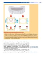
[39]. Stagnara described that as the spine rotates 90° the lateral deviation (scolio-
sis) of the spine is then oriented in the sagittal plan, resulting in apparent kypho-
sis [46] (
Fig. 3). The other type of ky photic deformity in neuromuscular scoliosis
is secondary to loss of the posterior tension band such as in myelomeningocele
[20] (
Case Study 2) or in myopathy scoliosis. This kyphosis can result in signifi-
cant loss of spinal height, resulting in internal organ crowding and skin break-
down over the gibbus.
a
b
c
d
Figure 3. Neurogenic kyphoscoliosis
The rotational deformity of scoliosis causes an apparent kyphosis. a, b The clinical coronal deformity appears moderate.
However, due to the severe rotational deformity compounded by severe pelvic obliquity, the PA X-ray is actually more of
a lateral of the spine.
c, d The apparent severe sagittal kyphotic deformity is in fact the coronal scoliotic deformity. This
is apparent as one notes the lumbar vertebrae are oriented in a PA orientation. This case illustrates the true three-dimen-
sional nature of spinal deformities.
676 Section Spinal Deformities and Malformations
Magnetic Resonance Imaging
Rule out intradural
pathology by MRI
Any scoliotic patients with a hint of neurological signs or symptoms [8, 49] or
with neuroectodermal skin lesions must have MRI performed of the entire spine
(occiput to sacrum) to assess the presence of any intradural lesions: syringomye-
lia, tethered cord, and spinal tumor. Malignant curve progression warrants MRI
as it may also be a sign of intradural pathology.
Non-operative Treatment
When consulting patients for the type of treatment, a thorough knowledge of the
natural history is mandatory. The natural history in neuromuscular scoliosis is
closely linked to the underlying disease.
Natural History
The life expectancy of
NMD patients is diminished
In general, patients with neuromuscular scoliosis have a diminished life expec-
tancy compared with the general population which is mainly secondary to their
underlying neuromuscular diagnosis. Spinal deformity if severe can negatively
impact their life expectancy, particularly scoliotic deformities leading to cardio-
pulmonary compromise [18] (
Table 1).
The natural history of neuromuscular spinal deformity is one of curve pro-
gression irrespective of etiology. Granted that there are many different factors
influencing curve progression, there are some neuromuscular curves which do
not progress; however, the majority will.
Factors influencing curve progression are as follows:
age of onset of NMD
severity and rapidity of weakness
evolving or static neuromuscular disease
skeletal maturity
ambulation status
severity of curves
Few papers have specifically looked at the natural history and curve progression
of patients with neuromuscular scoliosis [15, 20, 25]. Their curve progression has
Severe curve progression
occurs mainly during
peak growth
been reported to be from 7° to 40° per year. The severe progression occurs mainly
during patients’ peak growth compounded with loss of an autoregulatory spinal
alignment process which their underlying neuromuscular condition impedes.
For example, in Duchenne muscular dystrophy, the rate of curve progression
in untreated boys overall averages 7° per year. Oda et al. [36], after reviewing the
natural history of scoliosis in DMD, found that there were three courses of curve
progression:
Type I curves comprise progressive collapsing kyphoscoliosis with signifi-
cant rotatory deformity extending into the pelvis which always reach 30°
before the age of 15 years, with a rapid progression of 15°–20° per year
thereafter.
Type II curves are characterized by hyperlordosis with a progressive scoli-
otic deformity. The patients with double major curves tend not to have pel-
vicobliquityandhavestablecurves,whilepatientswithlumbarorthoraco-
lumbar curves tend to have pelvic obliquity and progress as type I curves.
Type III curves have straight sagittal spines and have non-progressive scoli-
otic curves that never reach 30°.
Neuromuscular Scoliosis Chapter 24 677
Patients with cerebral palsy
have a highly variable
onset of puberty
In Becker’s muscular dystrophy, curves tend not to be severe and non-progres-
sive [29], as the patients tend to be older. In contrast, in patients with cerebral
palsy, because their onset of puberty is highly variable (8–20 years), it is difficult
to quantify the risk of curve progression.
Scoliosis in cerebral palsy
can progress into adulthood
It has also been shown that scoliosis in patients with cerebral palsy continues
to progress even into adulthood [16, 25].
Non-operative Treatment Options
Non-operative treatment
must be individualized
The non-operative management of neuromuscular spinal deformities must be
adapted to each patient’s specific requirements. When patients are still able to be
upright, then initial treatment consists of encouraging prolongation of an
upright position while maintaining standing/ambulation status.
Once a patient is wheelchair dependent, then seating modifications are war-
ranted to provide lateral trunk support, as well as accommodation of sagittal
deformities such as hyperlordosis or kyphosis. The seating surfa ce must also be
carefully chosen to minimize skin breakdown while providing enough support to
minimize pelvic obliquity. Controlling and compensating hip contractures must
also be taken into consideration to favorably influence the pelvis to minimize an
oblique take-off of the spine.
Bracing is usually not
helpful in neuromuscular
scoliosis
Bracing in neuromuscular scoliosis should not be seen in the same light as
bracing for idiopathic scoliosis. Bracing has not been shown to prevent curve
progression in neuromuscular scoliosis [37]; thus its usage is not oriented
towards the treatment of these curves [6, 32].
The bracing used for neuromuscular scoliosis is functional bracing.Itpro-
vides external support to the spine, allowing some patients to be more func-
tional. Its goal is to maximize functional positioning by controlling some of the
spinal collapse, improving posture, and facilitating seating in some cases. One
must realize that in some patients with neuromuscular scoliosis bracing is con-
traindicated since it may result in compromising what is left of their respiratory
reserve. Bracing can seriously limit gastric motility, worsening the nutritional
status of these patients. Some will simply not tolerate the braces, with uncontrol-
lable behavioral problems. Obviously in any of these situations, bracing should
be discontinued, since it is counterproductive to a functional bracing. Early rec-
ognition of neuromuscular spinal deformity is important, since treatment plans
must be instituted as soon as possible.
Operative Treatment
Surgical Indications
The decision to proceed with major spine surgery for neuromuscular scoliosis
remains somewhat controversial, particularly when looking at the elevated mor-
bidity and mortality of this type of surgery. Yet a consensus is emerging that with
adequate pre- and perioperative multidisciplinary management and with a suc-
cessful outcome, most patients and caregivers feel the surgery is beneficial to
their overall well-being [3].
The indication for scoliosis
correction in NMD patients
remains controversial
Absolute surgical indications remain controversial [22] for globally disabled
children. The classic surgical indications of idiopathic scoliosis, i.e., curves >50°
or curve progression in the immature patient, also apply to the management of
neurogenic scoliosis. However, these tend not to be the main factors influencing
the decision to operate. Loss of function is the more common indication to pro-
ceed with surgical management of neurogenic scoliosis. As their spinal deformity
progresses, the ensuing spinal deformity and trunk shifts result in decreased pul-
678 Section Spinal Deformities and Malformations
monary function and increased respiratory disease, deterioration of comfort
and loss of the activities of daily living, inability to walk or sit independently, as
well as a decrease in quality of life. Sitting patients end up supporting themselves
with one of their hands, resulting in a functional triplegia. Such functional losses
are surgical indications. The development of pressure sores and the inability to
use further adapted wheelchairs to compensate for their spinal deformity are also
surgical indications since the spinal deformity has a real impact on the activities
of daily living. In contrast to idiopathic scoliosis, where it is rare that the defor-
mity negatively impacts on the child’s well-being, neurogenic scoliosis com-
pounds an already fragile individual (
Table 5):
Table 5. Indications for surgery
severe (>50 degrees) progressive curves
curve progression in Duchenne muscle dystrophy
loss of sitting balance
cardiopulmonary compromise
deteriorating general well-being
One must not forget that indications will vary depending on the underlying etiol-
ogy of the scoliosis. For example, in Duchenne muscular dystrophy, knowing that
90% of patients with DMD will have a progressive spinal deformity as well as a
In Duchenne patients
surgery is indicated early
declining pulmonary function [33], one tends to intervene at a lower Cobb angle
and/orwhenthecurveisprogressive.Infactalossofpulmonaryfunctionis
more influential than a rise in Cobb angle. As patients get older, their curves
increase while their pulmonary functions decrease. Due to this reverse relation-
ship there is a window in which surgery is recommended, and if it is missed mor-
bidity rises to unacceptable levels. When treating patients with cerebral palsy
who are skeletally immature with a progressive curve between 40° and 50°, or
skeletally mature cerebral palsy patients with curves greater than 50°, it is recom-
mended to proceed with a spinal arthrodesis [48].
General Principles
Do not blindly apply
the classic principles
of idiopathic scoliosis
management
The first principle, and probably the only steadfast rule when managing neuro-
muscular deformities, is not to blindly apply the classic principles of surgical
management of idiopathic scoliosis. The second principle in managing neuro-
muscular scoliosis, which is the cornerstone of all surgical management of any
spinal deformity, is to achieve perfect spinal balance in both the coronal and sag-
Aim for coronal and sagittal
balance
ittal planes [42]. Classically these patients do not have compensatory mecha-
nisms (muscle tone, intact proprioception) to rebalance themselves.
Patients’ curves tend to be long and they often have associated pelvic obliq-
uity, necessitating long fusions to the pelvis. Therefore, the coronal and sagittal
balance must be perfect when performing spinal fusions for neuromuscular sco-
liosis. Thirdly, a word of caution: a thorough preoperative and perioperative
medical management is mandatory in managing patients with neuromuscular
scoliosis. These patients tend to have cardiac pathology, severe pulmonary dis-
Consider the comorbiditiesease,andmalnutrition [51] to name a few associated conditions. If these medical
problems are left unattended or are ignored, they will lead to catastrophic com-
plications.
Neuromuscular Scoliosis Chapter 24 679
Surgical Techniques
Levels of Fusion
The Harrington principle, fuse the Cobb angle, also holds true for neuromuscu-
lar scoliosis. However, in contrast to idiopathic scoliosis, it is usual to actually
span beyond the Cobb for two reasons:
associated kyphosis
associated pelvic obliquity
Selective fusion should
not be done for NMS
In contrast to idiopathic scoliosis, selective spinal fusion should not be done
since the underlying neuromuscular condition will continue to exert its force on
the non-fused segment and new deformities will present themselves. The fusion
is often extended proximally to address the sagittal kyphotic deformity.
ab c
d
Case Study 3
A 14-year-old boy with cerebral palsy was referred for a severe and particu-
larly rigid spinal deformity with a rigid pelvic obliquity (
a, b). His wheelchair
could no longer be adapted to provide comfortable positioning. The
patient had developed a pressure sore on his left ischium. Preoperative X-
ray confirmed both sagittal and coronal imbalance with little correction on
supine bending (
c, d).
680 Section Spinal Deformities and Malformations
e f
g h i
Case Study 3 (Cont.)
Furthermore even under GA with manual traction it was not possible to level the patient’s pelvis (e). Hence an anterior
release was performed as well as an apical corpectomy (
f). Subsequently the patient was placed in gravity halo traction
(
g). One week later the patient had completion of apical vertobrectomy and posterior instrumentation and fusion with
restoration of sagittal and coronal correction (
h, i).
Sagittal kyphotic
deformities must be
addressed and fused
Therefore, it is critical not only to choose your fusion levels with coronal and
bending films but to closely scrutinize the lateral X-ray to avoid stopping the
fusion at the apex of the kyphotic deformity (
Case Study 3). The fusion must
extend out of the kyphosis to the first lordotic segment; this holds true both prox-
Neuromuscular Scoliosis Chapter 24 681
imally and distally [19]. Fusing long will avoid problematic revision surgery for
junctional kyphosis.
Selective spinal fusion
must be avoided
In general, T2 is the proximal fusion level for neuromuscular scoliosis. Fusing
too short or excessive kyphotic correction leads to junctional kyphosis as
patients with neuromuscular kyphoscoliosis want to drift back to their initial
sagittal alignment, placing tremendous forces at the distal end of fixation.
Fixation to the sacrum
is a major challenge
More often than not, if the distal level of the fusion exceeds the Cobb angle, it
is to address the associated pelvic obliquity. In general, L5 or the sacrum is the
distal fusion level for neuromuscular spinal deformities. There remains some
debate as to whether the pelvis should be included or not in the fusion. Patients
withpelvisobliquityoflessthan10°canhavetheirfusiondowntoL5toavoidthe
complications associated with fixation to the pelvis. Trying to fuse across the
lumbosacral junction is associated with a high rate of non-union. Secondly, as
there is one level left of mobility, overall spinal alignment can be forgiving, and
spinal balance may be achieved by patient volition. The downside of stopping the
fusion short of the pelvis is that there is a possibility that the patient decompen-
sates out of balance as the pelvis tilts, thus leading to further spine surgery in
already frail patients.
Spinal Fixation
Sublaminar wires have been
the gold standard treatment
The classic spinal implant for neuromuscular curves comprises sublaminar wires
with Luque rods [24]. The advantages of this classic segmental spinal fixation are
that one achieves a gradual reduction of each segment (mainly by spinal transla-
tion), thus minimizing the risk of fracturing the spinal anchorage points.
Poor bone quality
challenges the
instrumentation
This is of particular concern when treating non-ambulatory patients with an
osteoporotic spine either from disuse and/or induced by long-term antiepileptic
medication. The disadvantages of wires are the potential risk of injuring the spi-
nal cord during insertion and the risk of considerable epidural bleeding.
Consider the risk of spinal
anchorage point fracture
and pull out
The alternative construct is a combination of multiple sublaminar hooks, ped-
icle hooks and/or pedicle screws at each level, distributing the forces across the
entire spine. The use of multiple pedicle screws can provide enough corrective
forces for the anterior release to be avoided, and to allow for single stage poste-
rior spinal fusion and instrumentation [30]. The liberal use of pedicle screws
(lumbar and thoracic) rather than sublaminar wires serves two purposes. Firstly,
they allow for a much more thorough decortication, which obviously helps to
achieve a better fusion. Secondly, pedicle screws allow for much more radical
bilateral facetectomies, which facilitates greater correction. Both of these can be
done without fear of weakening the spinal fixation points.
Sacral and Pelvic Fixation
The classic spinal implant for the management of pelvic obliquity associated with
neuromuscular scoliosis is the Luque-Galveston cons truct [11]. This fixation
from T2 to pelvis spans the lumbosacral junction by inserting the distal rods into
Sacral and pelvic fixation
remain a major challenge
in NMD
the posterior superior iliac spine (PSIS) between the inner and outer tables just
above the sciatic notch (
Fig. 4). Adding an S1 pedicle screw to the base of the con-
struct and a cross-link proximally adds significant stability to the construct [26].
The unit rod [35] has been shown to be a more effective means of addressing the
pelvic obliquity and the spinal deformity [7]. The reduction maneuver for cor-
recting pelvic obliquity consists of a cantilever maneuver. This entails fixing the
rods distally to the pelvis at a 90-degree orientation to the ischial tuberosities.
Then the rods are levered across and attached to the proximal spine, thus leveling
the pelvis perpendicular to the balance of the spine. The entry points in the PSIS
682 Section Spinal Deformities and Malformations


