Spinal Disorders: Fundamentals of Diagnosis and Treatment Part 78 docx
Bạn đang xem bản rút gọn của tài liệu. Xem và tải ngay bản đầy đủ của tài liệu tại đây (414.58 KB, 10 trang )
28
Juvenile Kyphosis
(Scheuermann’s Disease)
Dietrich Schlenzka, Vincent Arlet
Core Messages
✔
Scheuermann’s disease (Type I, “classic” Scheu-
ermann’s) is a thoracic or thoracolumbar hyper-
kyphosis due to wedged vertebrae developing
during adolescence
✔
Atypical Scheuermann’s disease (Type II, “lum-
bar” Scheuermann’s) affects the lumbar spine
and/or the thoracolumbar junction. It is a
growth disturbance of the vertebral bodies
without significant wedging causing loss of
lumbar lordosis or mild kyphosis
✔
The natural history of the deformity is benign
in the majority of cases
✔
Back pain is common but usually mild and
rarely interferes with daily activities or profes-
sional career
✔
Lung function is impaired only in very severe
deformities (>100 degrees)
✔
Diagnosis is based on the clinical picture and
typical changes in plain lateral radiographs
✔
During growth, brace treatment is recom-
mended in mobile deformities of between
45 and 60 degrees
✔
Rare spinal cord compression is the only abso-
lute indication for operation
✔
Relative indications for operation are kyphosis
greater than 70 degrees, pain, and cosmetic
impairment
✔
Theresultsofoperativetreatmentaresatisfac-
tory in the majority of cases regarding pain and
cosmesis
✔
Theriskofsevereintra-andpostoperativecom-
plications should be weighed carefully against
the benefits
Epidemiology
Scheuermann’s disease is a
thoracic or thoracolumbar
hyperkyphosis due to
wedged vertebrae
Scheuermann’s disease is a thoracic or thoracolumbar hyperkyphosis due to
wedged vertebrae developing during adolescence. Ancient presentations of
hyperkyphosis usually depict extreme gibbus formations as seen due to infection
(tuberculosis) or congenital vertebral anomalies. Michelangelo’s ceiling fresco in
the Sistine Chapel at the Vatican shows an ignudo with a kyphosis resembling a
thoracolumbar juvenile kyphosis (
Fig. 1). It was painted in 1511 and is possibly
the earliest pictorial representation of the disease [30]. Following Schanz, Hag-
lund named the deformity “Lehrlingskyphose”(apprentice’s kyphosis)asitwas
detected mainly in youngsters involved in heavy labor [27, 61]. He saw the cause
as muscular insufficiency and mechanical overloading during growth. Credit is
due to Holger Werfel Scheuermann from Denmark for first describing it in 1920
as being different from mobile postural kyphosis [62–64]. He recognized from
radiographs that the wedge vertebrae formation in the thoracic spine was the
underlying reason for the deformity. Scheuermann was the first to describe its
The incidence of juvenile
kyphosis ranges between
1% and 8 %, being more
commoninboys
typical radiographic features and named it “osteochondritis deformans juvenilis
dorsi”. The true incidence of juvenile kyphosis is not known. It ranges from 1%
to 8%, being more common in boys than in girls (ratio 2/1 to 7/1).
Spinal Deformities and Malformations Section 765
abcd
ef
g
Case Introduction
A 14-year-old boy was referred by the school doctor. The boy was otherwise healthy and played hockey and soccer regu-
larly. Four years previously, posture changes were detected for the first time. The boy had pain in the thoracolumbar area
since then during the day and especially after playing sports. Sometimes night pain in the back also occurred. No radiat-
ing pain to the lower extremity was present. There were no back problems in the family. Clinically, the boy appears to be
healthy. Height is 153 cm, sitting height 77.5 cm. The spine is balanced in the frontal as well as in the sagittal plane. Shoul-
ders and pelvis are leveled. The thoracic kyphosis is pronounced especially in the mid-thoracic area (
a). Kyphosis corrects
partially during spine extension inthe prone position. The left scapula is slightly elevated. A mild left convex thoracic sco-
liosis with 3 degrees of rib hump is present (
b). Lumbar lordosis appears normal. Lumbar range of motion is free. On pal-
pation, the spine is free of pain. Hamstring tightness of 70 degrees is present bilaterally. No neurological abnormalities
are found in the lower extremity. Abdominal skin reflexes are symmetrical. On the standing lateral radiograph, thoracic
kyphosis measures 56 degrees, lumbar lordosis 55 degrees (
c). There are Scheuermann’s changes in the T6–T10 vertebral
bodies. On supine extension radiographs, thoracic kyphosis has corrected to 30 degrees. The skeletal age is 13.5 years,
i.e. 6 months behind the chronological age (
d). As the kyphosis is mobile, a sufficient amount of growth is left, and the
boy seems to be well motivated, brace treatment is initiated (
e, f). The correction in the brace is very acceptable. The tho-
racic kyphosis decreases from 56 to 42 degrees (
g). The brace is worn full-time (23 h/day). It may, however, be removed
for sports training hours. Daily exercises including pectoralis stretching, hamstring stretching, and back and abdominal
muscle strengthening are advocated.
766 Section Spinal Deformities and Malformations
Figure 1. Michelangelo’s Ignudo
This painting (1511) exhibits a Scheuer-
mann’s kyphosis at the thoracolumbar
junction.
Pathogenesis
The exact causes
are unknown
The exact etiology of Scheuermann’s kyphosis is unknown. Genetic, hormonal,
and mechanical factors have been discussed. An autosomal dominant pattern of
inheritance has been described [21, 28]. Scheuermann considered it a growth
disturbance in the vertebral epiph ysis resembling Calv´e-Perthes disease. He
therefore named it osteochondritis deformans juvenilis dorsi [64]. Aufdermaur
reported a developmental error in collagen aggregation leading to a disturbance
of the enchondral ossification of the vertebral endplates [3]. Ippolito and Ponsetti
detected a mosaic-like pattern of alterations in the growth cartilage and vertebral
endplates. The collagen fibers in the matrix are thinner and their number is
diminished. The proteoglycan content of the matrix is increased. The growth
processissloweddownorevenabsentinthealteredareas.Theprocessshouldbe
interpreted as an “absence of growth” rather than a destruction [2]. In the nor-
mal areas growth is accelerated. This causes wedge-shaped deformation of verte-
brae and an increase in kyphosis [2, 32, 33]. For biomechanical reasons,
increased kyphosis causes increased pressure to the vertebral bodies which the
pathologic bone cannot withstand. This creates a vicious circle of increased
wedging and increased kyphosis leading to increased load on the vertebral bod-
ies. There are no data available on the rate of progression after cessation of
growth.
Juvenile kyphosis has
a genetic background
and develops due to an
ossification disturbance
of the vertebral bodies
The sources of pain are not very well defined. Pain symptoms intheadolescent
can arise from the posture changes. The musculature is insufficient to counteract
the increasing kyphosis during the growth spurt. This causes fatigue in the para-
vertebralmuscles.Painintheneckregionandinthelumbarspineiscausedby
compensatory hyperlordosis above or below the primary deformity. It develops
when the degree of the primary deformity exceeds the capacity of the adjacent
segments to adapt to it. In the adult patient, disc degeneration and facet joint
osteoarthritis may be the reason for pain in the kyphotic vertebral segment as
well as in the segments above and below.
Juvenile Kyphosis (Scheuermann’s Disease) Chapter 28 767
Normal Sagittal Profile
The sagittal profile develops
during growth and changes
throughout adult life
Classic Scheuermann’s disease is a thoracic or thoracolumbar hyperkyphosis,
which implies that kyphosis deviates from the normal sagittal curvature of the
spine. Therefore, a thorough knowledge of the normal sagittal profile is required
for the understanding of this clinical entity. The sagittal profile of the spine in
humans varies greatly between individuals. It is not established at birth but
develops and changes during life [5, 46, 68, 69, 72, 75].
The sagittal profile of the
spine is largely variable
There is no scientifically based definition of the degree of normal sagittal spi-
nal curvatures. At birth, the whole spine is kyphotic from the occiput to the coc-
cyx. As the child starts in the upright position, first lumbar lordosis develops and
later thoracic kyphosis. It is only when the child becomes a young adult that the
definitive sagittal curves are acquired. Confusingly, different methods for mea-
surement of the sagittal curvatures of the spine are used in the literature. Mea-
sured from the back surface using spinal pantography, at the age of 14 years tho-
racic kyphosis in healthy children ranges from 7 to 57 degrees (mean 29 degrees)
in girls and from 6 to 69 degrees (mean 30 degrees) in boys, being between 20 and
40 degrees in more than two-thirds of children [46]. In a mixed population with
an age range from 4.6 to 29.8 (mean 12.8) years, Bernhart et al. found thoracic
kyphosis ranging from 9 to 53 (mean 36) degrees measured from standing lateral
Normal kyphosis is in the
range of 10° to 60°
radiographs between the top of T3 and the bottom of T12. They proposed a nor-
mal range from 20 to 50 degrees [5]. In healthy adults, Stagnara et al. measured
from standing radiographs thoracic kyphosis from 7 to 63 (mean 37) degrees
between the top of T4 and the bottom of the intermediate vertebra (mainly L1,
T12, or L2), with the majority being between 30 and 50 degrees [69]. They did not
find, however, any hint that those individuals outside the 30–50 degree range
were functionally inferior. Vaz et al. reported a global thoracic kyphosis ranging
from 25 to 72 (mean 47) degrees [73]. Boulay et al. [9] used true Cobb angle mea-
surements, i.e. they measured thoracic kyphosis from the upper endplate of the
most tilted vertebra cranially to the lower endplate of the most tilted vertebra
caudally. In 149 healthy adults, they found a range from 33.2 to 83.5 (mean
53.8) degrees. The Scoliosis Research Society proposes to regard 10 – 40 degrees
as the range for normal kyphosis between the upper endplate of T5 and the lower
endplate of T12 [51]. Thoracic kyphosis increases in the elderly due to degenera-
tive changes.
Thoracic kyphosis is more
prominent in males
There are significant differences between the genders. Thoracic kyphosis is
more pro minent in males. There is a steady increase from adolescence to adult-
hood. In females, thoracic kyphosis increases during the adolescent growth spurt
but decreases during the descending phase of peak growth, i.e. until young adult-
hood. Thoracic hyperkyphosis (
45 degrees) is equally prevalent in both gen-
ders at the age of 14 years, but more prevalent in males (9.6%) than in females at
the age of 22 years [57]. Left-handedness was identified as a risk factor for tho-
racic hyperkyphosis but no significant correlation between hyperkyphosis and
low-back pain during adolescence could be established [47, 48].
There is no scientifically based definition of the threshold for “normal”
kyphosis. So-called normal ranges in the literature are derived from cohort mea-
surements using statistical methods. These figures, however, should not be used
as such for deciding what is pathologic in the individual. Thoracic kyphosis
should always be judged in view of the balance of the entire spine, not as an iso-
lated part of it. The thoracolumbar junction from T10 to L2 is slightly kyphotic
[5]. The upper thoracolumbar junction (T10–T12) varies from 3 degrees of lor-
dosis to 20 degrees of kyphosis (mean 5.5 degrees of kyphosis). The lower thora-
columbar junction (T12–L2) ranges from 23 degrees of lordosis to 13 degrees
768 Section Spinal Deformities and Malformations
Lumbar lordosis is more
pronounced in females
of kyphosis (mean 3 degrees of kyphosis). The segment T12–L1 is on average in
1 degree of kyphosis [5]. Lumbar lordosis is normally somewhat greater than
thoracickyphosis.Onaverage,lumbarlordosisismorepronouncedinfemales.
It is relatively constant during growth from adolescence to young adulthood [57].
In girls, lumbar lordosis measured from the back surface using the spinal panto-
graph ranges from 18 to 55 (mean 33.4) degrees at the age of 14 years and from 18
to 72 (mean 37.8) degrees at the age of 22 years. In boys, the corresponding fig-
ures are 15–56 (mean 33) degrees and 11–58 (mean 34.6) degrees [57]. Accord-
ing to Bernhart and Bridwell, the range of lumbar lordosis measured from stand-
ing radiographs between the bottom of T12 and the bottom of L5 is 14–69 (mean
A range of 20° to 60° is
regarded as normal lordosis
44) degrees. They propose a normal range of from 20 to 60 degrees [5]. Stagnara
et al. reported a range for lumbar lordosis of from 32 to 84 degrees. The higher
values may be explained by the fact that these authors measured the lumbar lor-
dosis from the upper border of the intermediate vertebra down to the upper end-
plate of S1 [69]. Bouley et al. [9] reported in adults a lordosis ranging from 44.8
to 87.2 (mean 36.4) degrees measured according to Cobb between the most tilted
vertebrae. Vaz et al. measured in adults a global lumbar lordosis ranging from 26
to 76 (mean 46.5) degrees [73]. The Scoliosis Research Society proposes to regard
40–60 degrees as a normal range of lumbar lordosis for the adult measured
between the upper endplate of T12 and the upper endplate of S1 [51]. Lumbar lor-
dosis decreases in the elderly due to degenerative changes.
The threshold for “normal”
thoracic kyphosis is not
defined
According to Stagnara et al., every person has her or his “unique spinal physi-
ognomy” [69]. Average values are only indicative not normative [5, 69]. There is
no indication that persons with a degree of thoracic kyphosis not fitting into the
postulated “normal range” are handicapped in any respect.
Sagittal balance is of the utmost importance for an ergonomic upright pos-
ture. The spine is sagittally balanced if a plumb line dropped from the odontoid
process crosses the thoracolumbar junction and through the posterior edge of S1.
For practical purposes on radiographs, the plumb line is often drawn from the
center of the vertebral body C7 [51] (
Fig. 2a–c). Normal sagittal balance is essen-
tial for the ability of the individual to stand in the upright position with minimal
effort. Abnormal sagittal balance will be observed when the spinal column can-
not compensate to keep the gravity line between the femoral heads and the
sacrum. Spinal imbalance is positive when the gravity line falls in front of the
femoral heads. It is negative when the gravity line falls posterior to the sacrum.
Normal sagittal spinal bal-
ance is the prerequisite for
an economic upright pos-
ture in the standing position
This is important to consider. A negative sagittal balance may be observed in
neuromuscular conditions with weak hip extensors. A positive sagittal balance
may be observed in patients with developmental delay, loss of lumbar lordosis
(flat back), or rigid kyphotic lumbar spine. Most Scheuermann patients fall into
the category of negative sagittal balance [31, 40, 41].
When judging the importance of a thoracic hyperkyphosis, one not only has to
take into account the absolute measure of the deformity in degrees, but one must
also assess it in relation to the location of the apex of the kyphosis. The lower the
apex of the hyperkyphosis the greater its impact on spinal balance and on the
adjacent spinal segments below (compensatory lumbar hyperlordosis). For
instance, a thoracolumbar kyphotic deformity of 20 degrees between T10 and L3
has a much higher impact on the sagittal balance than a thoracic hyperkyphosis
of 55 degrees between T2 and T12, which may be clinically unimportant.
The concept of pelvic incidence has recently been introduced by Duval Beau-
pere [36]. Pelvic incidence is defined as the angle between the perpendicular to
the top of S1 and the line joining the middle of S1 to the femoral heads (
Fig. 3). It
was found that the pelvic incidence was the only morphometric character that is
constant throughout life. A strong correlation between the pelvic incidence and
the lumbar lordosis has been defined. Pelvic incidence regulates the sagittal
Juvenile Kyphosis (Scheuermann’s Disease) Chapter 28 769
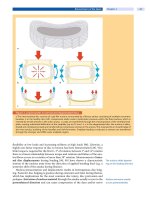
Figure 2. Sagittal balance
a The spine is sagittally balanced when the plumb line
from C7 touches the posterior edge of S1.
b Spinal imbal-
ance is positive when the line falls in front of this point.
c It
is negative when the plumb line falls behind this point.
Figure 3. Pelvic incidence (PI)
a = midpoint of the sacral endplate, 0 = center of the femo-
ral head.
Figure 2. Sagittal balance
770 Section Spinal Deformities and Malformations
alignment of the spine and pelvis [9, 36, 73]. As a rule of thumb, lumbar lordosis
is approximately 10 degrees greater than the pelvic incidence in normal individu-
als. However, no study has focused yet on any possible relationship between pel-
vic incidence and Scheuermann’s kyphosis.
Definition and Classification
According to Sörensen [65], the diagnostic criteria are wedging of more than
5 degrees in three consecutive vertebrae with typical endplate irregularities on a
lateral radiograph. A widely accepted definition isbasedonBradford[11]:
irregular vertebral endplates
narrowing of the intervertebral disc space
one or more vertebrae wedged 5 degrees or more
an increase of normal kyphosis beyond 40 degrees
Both Sörensen’s and Bradford’s definitions do have their shortcomings since they
are arbitrary. Sörensen’s criteria exclude deformities with less than three
deformed vertebrae. Bradford’s 40 degrees of thoracic kyphosis as the borderline
between normal and pathologic has its origin in an unpublished X-ray study by
Boseker, who found a range of 25–42 degrees in 121 normal children [8, 10]. This
is extremely low in comparison to the ranges for thoracic kyphosis in healthy
individuals reported later by other investigators (see above). Besides, it cannot be
generalized for the different regions of the spine. In the authors’ opinion, the
diagnosis should be based mainly on the typical pathologic vertebral and disc
changes. Bearing in mind the immense variability of thesagittal profile in healthy
persons, it seems inappropriate to base the diagnosis on a certain amount of
(hyper-)kyphosis measured in degrees (
Table 1)(Fig. 4a):
Table 1. Diagnostic criteria for juvenile kyphosis (Type I)
wedging of more than 5 degrees in one or more vertebrae in the
thoracic or thoracolumbar region
disc space narrowing
endplate irregularities increased thoracic or thoracolumbar kyphosis
Schmorl’s nodes
are not pathognomonic
Schmorl’s nodes areoftenassociatedwithjuvenilekyphosisbutarenotapatho-
gnomonic sign.
The classification of Scheuermann’s disease concerning its localization in the
spine is inconsistent in the literature. In the classic sense, it is a deformity of the
thoracic spine. Lindemann reported in 1933 four cases with affection of the lum-
bar spine and called the condition the “lumbar form of adolescent kyphosis”
[37]. Lumbar Scheuermann’s disease as a separate entity was described in more
detail by Edgren and Vaino [19]. Out of 900 radiographs of Scheuermann’s
patients, they found 30 cases with distinct radiographic features in the lumbar
spine. During the growth period (initial stage), they recognized a typical local
defect in the spongiosa in the ventral part of the endplates of one or several verte-
bral bodies (
Fig. 4c). After the end of growth (final stage), the contours of the ver-
tebral endplates were uneven. Schmorl’s nodes and disc prolapses dislocating the
border of the vertebra were seen. Intervertebral disc spaces were narrowed. A
slight angular kyphosis was present, and the sagittal diameter of the vertebral
bodies was increased. Clinically, the patients showed flattening of the lumbar lor-
dosis or a slight kyphosis, stiffness, and tenderness of the lumbar spine. No root
symptoms were seen. They coined the term “osteochondrosis juvenilis lumba-
lis” (atypical juvenile kyphosis) (
Table 2).
Juvenile Kyphosis (Scheuermann’s Disease) Chapter 28 771
abc
Figure 4. Types of juvenile kyphosis
a Standing lateral radiographs of juvenile kyphosis Type I changes in the thoracic spine in an 18-year-old male and b tho-
racolumbar area in a 52-year-old male. Scheuermann’s Type II changes from L1 to L4 in an 18-year-old female gymnast.
The thoracolumbar junction is slightly kyphotic.
c Note the decrease in thoracic kyphosis.
Table 2. Diagnostic criteria for juvenile kyphosis (Type II, “lumbar”)
Obligatory Facultative
endplate irregularities in one or several vertebral bodies of the lumbar
or thoracolumbar area
apophyseal separation
increased sagittal diameter of vertebral bodies loss of lumbar lordosis or slight kyphosis
disc space narrowing Schmorl’s node
Blumenthal et al. defined cases with involvement from T10 to L4 as lumbar juve-
nile kyphosis. They proposed three different types:
I: “classic” juvenile kyphosis (three or more consecutive vertebrae each
wedged over 5 degrees)
IIa: “atypical” juvenile kyphosis (endplate irregularities, anterior Schmorl’s
nodes, disc space narrowing)
IIb: acute traumatic intraosseous disc herniation (after acute vertical com-
pression injury) [7]
772 Section Spinal Deformities and Malformations
Wenger proposes a distinction between Type I (thoracic, with wedging), being
the most common form, and Type II (thoracolumbar, lumbar), developing at a
slightly later age and being more commonly painful. A mechanical overloading is
thought to be its basis. Murray et al., in their natural history study, divided the
patients according to the apex level of the kyphosis into “cephalad” (apex at T1–
T8) and “caudad” (apex at T9–T12) [44].
The confusion arising from these different classifications seems to be mainly
due to the fact that localization and pathoanatomical picture are mingled. Typi-
cal wedging (classical juvenile kyphosis, Type I) occurs usually in the thoracic
spine but it may also cross the thoracolumbar junction and reach into the upper
lumbar spine (
Fig. 4b). Endplate impressions, disc narrowing, and increased sag-
ittal diameter of the vertebral bodies without significant wedging (lumbar
“atypical” juvenile kyphosis, Type II), as described by Edgren and Vainio, seem
to occur only in the lumbar spine up to the thoracolumbar junction (
Table 2).
Severe wedging does
not develop in the lordotic
lumbar spine
Possibly both types are expressions of the same pathology. Severe wedging does
not develop in the primarily lordotic lumbar spine due to the fact that the loading
conditions are different from those in the primarily kyphotic thoracic spine [37].
Type II Scheuermann’s disease is commonly attributed to mechanical overload-
ing [23, 40, 74]. However, in the reports of Edgren and Vainio as well as Blumen-
thal et al., the majority of patients had not been involved in heavy physical activ-
ity [7, 19]. Obviously, there is an idiopathic form due to an “intrinsic” factor and
a secondary form caused by mechanical overloading and endplate damage as
seen in certain sports disciplines (weight lifting, gymnastics, motocross).
For the purposes of clear communication, we propose to define the condition
primarily according to the vertebral changes as Type I or Type II, respectively. If
deemed necessary, one can then add the vertebral level(s) for specification.
Clinical Presentation
History
In the initial phase of the disease posture changesare not visible yet but back pain
maybepresent.
The cardinal symptoms of juvenile kyphosis are:
back pain
cosmetic disturbance
Back pain is activity
dependent
Usually, juvenile kyphosis is detected first by caretakers or the school nurse or
doctor (
Case Introduction) when a visible deformity has already developed. Dur-
ing adolescence, pain in the region of the kyphosis may occur during exercise or
prolonged sitting.Inlateradulthood,secondarycervicalandlumbarhyperlor-
dosismaycausepainsymptomsalsointhecervicaland/orlumbarregion.Seg-
mental thoracic pain or lower extremity root pain has not been described. Back
pain symptoms occur mainly during the day and under loading. They are more
common in Type II as compared to Type I [7, 19, 23, 40, 74]. Murray et al. found
in Type I that pain interfered significantly more with life if the kyphosis was more
Back pain is related
to curve size and location
severe and the apex more cephalad (T1–T8). But job activity level and pain
intensity were not dependent on the level of the apex of the kyphosis [44].
Patients with Type II Scheuermann’s disease are prone to develop lumbar spinal
stenosis [70]. As these patients often have a genetic predisposition,oneshould
focus on the existence of a family history of a deformity. Previous fractures,
infections and neurological disorders should be ruled out.
Juvenile Kyphosis (Scheuermann’s Disease) Chapter 28 773
a
b
c
d
e
f
Figure 5. Clinical appearance of juvenile kyphosis
a Normal harmonic kyphosis of the spine in flexion. b, c A 16-year-old female with a thoracic hyperkyphosis of
88 degrees, apex T8.
d, e A 20-year-old male with a low thoracic hyperkyphosis of 79 degrees, apex T10. f A 19-year-old
male with Scheuermann’s Type II; the upper lumbar spine is slightly kyphotic.
Physical Findings
Rigid thoracic
hyperkyphosis is the
cardinal physical finding
When an adolescent patient presents with a thoracic or thoracolumbar hyperky-
phosis, the diagnosis can be suspected at first glance. The hyperkyphosis is fre-
quently accompanied by compensatory hyperlordosis of the cervical and/or
lumbar spine (
Fig. 5). The spine is balanced in the coronal plane but usually in a
negative balance in the sagittal plane. The clinical examination aims to assess the
rigidity of the curve. Asking the patient to lift the head and extend the spine in
the prone position best assesses this aspect. Mild secondary scoliosis with mini-
mal or no rotation may be present. The muscles in the region of the kyphosis or
in hyperlordotic areas above (shoulder-neck region) or below (low back) the
main deformity may be painful on palpation. Hamstring tightness is common.
Neurology should be assessed carefully. Pathologic neurological findings, how-
ever, are very rare.
Distinguish juvenile
kyphosis from idiopathic
roundback
UsuallyitiseasytodistinguishScheuermann’skyphosis(TypeI)fromidio-
pathic roundback. In the latter, the hyperkyphosis is harmonic also in flexion.
Moreover, it corrects well in extension.
774 Section Spinal Deformities and Malformations


