Spinal Disorders: Fundamentals of Diagnosis and Treatment Part 83 pdf
Bạn đang xem bản rút gọn của tài liệu. Xem và tải ngay bản đầy đủ của tài liệu tại đây (215.97 KB, 10 trang )
from 16–20 weeks. However, spina bifida may be missed, particularly in the
L5–S2 region [24, 44].
Magnetic Resonance Imaging
MRI is the modality of
choice for prenatal imaging
Since its advent, MRI has become the imaging modality of choice. While ultraso-
nography is an excellent screening procedure, it requires considerable expertise
to interpret, whereas MRI is definitive. Prenatal MRI can also be used to charac-
terize the Chiari II and other associated malformations [24]. Prenatal imaging
studies help to predict neurological deficits.
Postnatal Diagnostic Tests
Imaging Studies
For evaluation of the spinal cord malformations and tethered cord syndrome, the
most helpful diagnostic images are obtained by MRI, which provides excellent
details of anatomy and characterization of soft tissue anomalies [39, 58]. Other
imaging studies, including standard radiographs and CT, may also be helpful.
Plain radiographs will show vertebral anomalies. A CT scan is particularly useful
for the evaluation of bony anomalies and split cord malformations [34, 39].
Magnetic Resonance Imaging
The best demonstration of the entire craniospinal axis is made by MRI and
should be performed after the birth whenever possible. The T1- and T2-weighted
MR images in the sagittal and axial planes provide excellent demonstrations of
the anatomopathological characterization of the components of the malforma-
tion, i.e. relationship between placode and nerve roots and other associated
sequences (Chiari II, hydrocephalus, hydromyelia) [32].
Investigate the entire neural
axis when spinal malfor-
mations are suspected
Before theMRIera, it had been assumed that after untethering, there would be
upwardmigrationofthespinalcord,whichinfactdoesnotoccurinmostcases
[19]. Postoperative follow-up MRI almost always shows low-lying conus and
should not be confused with a “retethering” [10]. The diagnosis of retethering
and decision for untethering requires clinical judgment. Attempts to improve
conventional MRI techniques, including the use of prone positioning [10],
upright MRI and dynamic phase MRI, have been investigated but await valida-
tion through further studies [19].
Urodynamic Studies
Urodynamic studies may show low bladder capacity and overflow incontinence,
and may serve as a baseline for postoperative follow-up [15].
Treatment
It is important to recognize tethering of the spinal cord as early as possible. Once
the neurological deficits have occurred, many patients will not have recovery of
lost functions.
Tethered cord should be
treated as soon as possible
Although the underlying causes of tethered cord vary, thesigns and symptoms
of tethering are generally the same. Individuals with spinal malformations need
both surgical and medical lifelong management which should be provided by a
multidisciplinary team.
Malformations of the Spinal Cord Chapter 29 815
Asymptomatic patients with tethered cord should be instructed to avoid the fol-
lowing activities because of the risk of a potential sudden neurological deteriora-
tion [57]:
deep bending (touching the toes, high leg kicking)
holding any weight while standing that causes back and leg pain
sitting position such as the Buddha pose
sitting in a slouching position
horse riding
skiing at high altitude (might produce spinal hypoxia)
Valsalva-type maneuvers to prevent spinal venous congestion
In Utero Treatment
Fetalsurgeryforspinal
dysraphism is feasible
After a diagnosis of fetal spinal dysraphism, there are two choices: either termi-
nation or fetal surgery [24]. The period of legal termination differs between
countries. The first cases of in utero open spinal dysraphism repair were done in
1994 but proved unsatisfactory [3]. In 1997, in utero repair by hysterotomy was
reported [3, 20]. Up to 2004, more than 200 in utero, open spinal dysraphism clo-
sures are estimated to have been done [20]. Urodynamic and lower extremity
function seem to besimilar in infants treated in utero and postnatally [20]. Com-
pared with historical controls, infants treated in utero have a lower incidence of
Chiari II andhydrocephalus requiring shunting [3, 20]. Delivery via cesarean sec-
tion is preferred [28].
Postnatal Surgery
Open spinal dysraphisms must be treated surgically as early as possible (Table 6):
Table 6. General aims of surgery
untether the spinal cord prevent infections
repair of the dural/cutaneous defect restore normal anatomy as far as possible
Closure of the spinal lesions is usually done within 48–72 h of birth [20, 28, 58].
If there are signs of hydrocephalus, a shunt isplaced at the same time as the lesion
is closed.
There are some standard rules for closure of open spinal dysraphism, but in
many cases the surgeon must vary the technique on the basis of individual anat-
omy. The surgical microscope should assist in defining distorted anatomy and
associated pathologies in great detail. The interested reader is referred to repre-
sentative articles in the literature and textbooks [26, 28, 31, 50, 58].
Open Spinal Dysraphism
After careful and extensive dissection of the sac from the neural placode, neural
tissue is repositioned into the dural sac to preserve functional neural tissue.
There is no proven technique for closure of myelomeningocele at the time of the
original surgery that will prevent retethering. However, there are some tech-
niques that may minimize the amount of retethering that occur: The neural pla-
code can be folded over and anatomically made into a tube by suturing the edges
of theopen placode together. It does not prevent retethering, butitseems to make
the surgery for untethering easier. Sometimes the use of vascularized flaps may
be necessary.
816 Section Spinal Deformities and Malformations
Closed Spinal Dysraphism
In the cases of closed spinal dysraphisms, the associated lesions need careful dis-
section. In split cord malformation, after opening the dura, complete excision of
the bony spur or fibrous septum is performed. A thickened or fatty filum termi-
nale is cut and also released to detether the cord. Sometimes, closure of the dura
is a problem. In these cases, it is necessary to use fascia lata or synthetic dura sub-
stitutes to repair the dural deficiency. Wound closure is done in multiple layers in
order to prevent liquor leak.
Tethered Cord Syndrome
Surgery for tethered cord
must be early
In open spinal dysraphism, short- and long-term survival has increased with
improvements in medical and surgical management. Surgical intervention for
tethered spinal cord must be as early as possible to prevent progressive neuraltis-
sue damage. Once neurological function is lost it may never recover. The value of
early prophylactic surgical intervention in tethered cord is evident in the litera-
The only effective treatment
is surgical untethering
ture [16, 35, 48].Theonly effective treatment is surgical untethering of the spinal
cord from theunderlying cause. The goal of the untethering surgery is to stop any
further neurological deterioration [35, 48]. One of the current controversies with
respect to tethered cord management includes the untethering of the spinal cord
in asymptomatic patients. The majority of authors recommend prophylactic sur-
gery [16, 48].
The decision about the surgical technique should be made individually on a
case-by-case basis. The special details of the various surgical techniques are
beyond the scope of this chapter. Several excellent textbooks exist in the field of
spinal malformations–tethered cord surgery. Interested readers are referred to
representative articles in the literature andthese textbooks and atlases [26,28, 31,
50, 58].
Untethering is generally a safe surgical procedure in experienced hands [16].
Complications include infection, bleeding, and damage to the functional part of
the spinal cord. Although the causes of tethered cord vary, the general principles
of the surgery are similar.
The operating microscope and microsurgical technique are necessary for bet-
ter visualization and precise dissection. Different instrumentations are used to
perform the dissection including endoscopy, ultrasonic aspirator, and lasers; one
method is not necessarily better than the others, and the surgeon usually has her
or his own preference based upon their experience [8, 10, 48].
Intraoperative neuromoni-
toring and the microscope
are invaluable intraoperative
aids
Various intraoperative monitoring techniques such somatosensory evoked
potentials (SSEPs), lower extremity and anal sphincter EMGs, external anal
sphincter monitoring and nerve root stimulation studies are helpful to identify
functional elements [15, 58]. But it remains valid that the most important factor
for a good postoperative result is the experience of the surgeon in handling these
complex anomalies [12]. Retethering remains a risk and requires reexploration if
signs of tethered cord syndrome are seen.
In secondary tethered cord the untethering procedure usually involves open-
ing and dissecting the scar from the prior closure.
Malformations of the Spinal Cord Chapter 29 817
Recapitulation
Epidemiology.
Neural tube defects are the most
common congenital abnormalities of the central
nervous system.
Classification. Spinal cord malformations can be
classified based on the pathomorphological pre-
sentation as presenting with and without a back
mass. A secondary discriminator is related to the
coverage with skin in the presence of a back mass.
The vast majority of spinal cord malformations re-
sult in a tethering of the spinal cord. We differenti-
ate primary tethered cord as a result of spinal mal-
formations and secondary tethered cord which re-
sults from a surgical intervention.
Pathogenesis. Spinal cord malformations (=spinal
dysraphism) arise from defects occurring in the em-
bryological stages of gastrulation (weeks 2 – 3),
neurulation (weeks 3–6) and caudal regression.
There is an increased risk of spinal malformations
in pregnant women who are taking certain drugs.
An increased risk of spinal malformation is associat-
ed especially with exposure to valproic acid or carb-
amazepine. Patients with myelomeningocele and
myelocele almost always have associated Chiari II
malformation. Hydromyelia may occur in as many
as 80% of these patients, and may be localized or
extend through the whole cord. It may cause rapid
development of scoliosis if left untreated. Classical-
ly tethered cord is defined as having the tip of the
conusbelowtheL2discspaceand a pathologically
elongated spinal cord. However, in the medical lit-
erature, there are many publications of tethered
cord syndrome with the conus in a normal position.
Clinical presentation. Tethered cord–spinal cord
malformations are usually diagnosed at birth or
early infancy (open spinal dysraphism, closed spi-
nal dysraphisms with back mass) but sometimes
are discovered in older children or adults. Tethered
spinal cord should be highly suspected and consid-
ered in the differential diagnosis of patients who
present with cutaneous midline abnormalities,
low back pain, lower extremity and foot deformi-
ties, subtle neurological deficits, and bladder and
sexual dysfunctions. Irreversible neuronal damage
canoccurwhenthereissuddenstretchingoftheal-
ready chronically tethered conus.
Diagnostic work-up. The prenatal examination en-
compasses maternal serum -fetoprotein examina-
tion and ultrasound. The advent of diagnostic mo-
dalities such as MRI has increased the number of
tethered spinal cord diagnoses and will require
awareness and prompt multidisciplinary manage-
ment of the syndrome before neuronal loss ad-
vances. Since multiple tethering lesions and cere-
bral anomalies coexist in a significant number of
cases, it is absolutely necessary to investigate these
patients with craniospinal MRI to screen the entire
neuroaxis.
Prenatal treatment. It is important to counsel
women of childbearing age about the need to take
dietary supplements containing foliate before be-
coming pregnant. Up to 70 % of spina bifida cases
can be prevented by periconceptional folic acid
supplementation. Intrauterine surgery is possible
but superiority over postpartum surgery needs to
be established.
Postnatal treatment. Individuals with spinal malfor-
mations need both surgical and medical lifelong
management which should be provided by a multi-
disciplinary team. Open spinal dysraphism requires
immediate surgery (within 2– 3 days postpartum).
Main goal of surgery is to untether the spinal cord,
prevent infections, repair the dural/cutaneous de-
fect, and restore normal anatomy as far as possible.
Mainly the goal of the untethering is to stabilize the
progressive neurological deterioration but some
authors recommend a prophylactic untethering
procedure for asymptomatic patients. Early unteth-
ering, when minimum or mild symptoms are detect-
ed,isessentialfortetheredcordsyndrometreat-
ment. Surgical intervention for tethered cord in-
volves identification of the tethering lesion, release
of the spinal cord and reconstruction of the normal
anatomy as soon as possible. The operating micro-
scope and microsurgical technique are necessary
for better visualization and precise dissection. Intra-
operative neuromonitoring is useful.
818 Section Spinal Deformities and Malformations
Key Articles
Yamada S (1996) Tethered cord syndrome. The American Association of Neurological
Surgeons,ParkRidge,Illinois
This isa first andexcellent textbook on tethered cord syndrome. There are 16chapters on
embryology,pathophysiology,diagnosis,imaging,andtherapythatcoverallaspectsof
the syndrome. All chapters are superb didactically not only for neurosurgeons but also
for orthopedic surgeons, neurologists, pediatricians, and urologists.
Pang D (1995) Disorders of the pediatric spine. Raven Press, New York
This book covers perfectly all aspects of childhood spine, beginning with a section on
embryology and biomechanics, and bridging the philosophies of orthopedic surgeons
and neurosurgeons by including chapters written by these two specialties. Alarge section
is devoted to the many congenital malformations with deeply detailed definitions, nice
photos and drawings of operative techniques.
Tortori-Donati P, Rossi A, Cama A (2000) Spinal dysraphism: a review of neuroradiolog-
ical features with embryological correlations and proposal fo r a new classification. Neu-
roradiology 42(7):471 – 91
This paper presents the correlation between anatomy, embryology, neuroradiology and
clinical findings of spinal dysraphism and formulates a working classification of these
malformations.
Mitchell LE, Adzick NS, Melchionne J, Pasquariello PS, Sutton LN, Whitehead AS (2004)
Spina bifida. Lancet 364:1885 – 1895
This is an excellent review which highlights the key features of spina bifida.
References
1. Arai H, Sato O, Okuda O, Miyajima M,Hishii M, Nakanishi H,Ishii H (2001) Surgical experi-
ence of 120 patients with lumbosacral lipomas. Acta Neurochir (Wien) 143:857–864
2. Boop FA, Russell A, Chadduck WM (1992) Diagnosis and management of the tethered cord
syndrome. J Arkansas Med Soc 89(7):328–331
3. Bruner JP, Tulipan N, Reed G, Davis GH, Bennett K, Luker KS, Dabrowiak ME (2004) Intra-
uterine repair of spina bifida. Am J Obstetrics Gynecol 190:1305–12
4. Bulsara KR, Zomorodi AR, Villavicencio AT, Fuchs H, George TM (2001) Clinical outcome
differences for lipomeningoceles, intraspinal lipomas and lipomas of the filum terminale.
Neurosurg Rev 24:192–194
5. Chopra S, Gulati MS, Paul SB, Hatimota P, Jain R, Sawhney S (2001) MR spectrum in spinal
dysraphism. Eur Radiol 11(3):497–505
6. Dias MS, Pang D (1995) Human neural embryogenesis. In: Pang D (ed) Disorders of the
pediatric spine. Raven Press, New York, pp 1–26
7. GuggisbergD,SmailHE,VineyC,BodemerC,BrunelleF,ZerahM,Pierre-KahnA,ProstY
(2004) Skin markers ofoccult spinal dysraphism inchildren. Arch Dermatol 140:1109–1115
8. Haberl H, Tallen G, Michael T, Hoffmann KT, Bennedorf G, Brock M (2004) Surgical aspects
and outcome of delayed tethered cord release. Zentralbl Neurochir 65:161–167
9. Hoffman HJ (1996) Indications and treatment of the tethered spinal cord. In: Yamada S (ed)
Tethered cord syndrome. The American Association of Neurological Surgeons, Park Ridge,
Illinois, pp 21–28
10. Hudgins RJ, Gilreath CL (2004) Tethered spinal cord following repair of myelomeningocele.
Neurosurg Focus 16:E7
11. Iskandar BJ, Oakes WJ (1999) Occult spinal dysraphism. In: Albright AL, Pollack IF, Adelson
PD (eds) Principles and practice of pediatric neurosurgery. Thieme, New York, pp 321–351
12. Iskandar BJ, Fulmer BB, Hadley MN, Oakes WJ (2001) Congenital tethered spinal cord syn-
drome in adults. Neurosurg Focus 10:e7
13. Knierim DS (1996) Epidermoid and dermoid tumors associated with tethered spinal cord.
In: Yamada S (ed) Tethered cord syndrome. The American Association of Neurological Sur-
geons, Park Ridge, Illinois, pp 125–133
14. Kumar R, Singh SN (2003) Spinal dysraphism: Trends in northern India. Pediatr Neurosurg
38:133–145
15. Lapsiwala SB, Iskandar BJ (2004) The tethered spinal cord syndrome in adults with spina
bifida occulta. Neurol Res (7):735–740
Malformations of the Spinal Cord Chapter 29 819
16.
van Leeuwen R, Notermans NC, Vandertop P (2001) Surgery in adults with tethered cord syn-
drome: Outcome study with independent clinical review. J.Neurosurg (Spine 2) 94:205–209
17. Manfredi M, Donati E, Magni E, Salih S, Orlandini A, Beltramello A (2001) Spinal dysra-
phism in an elderly patient. Neurol Sci 22:405–407
18. McLone DG, Dias MS (1991) Complications of meningomyelocele closure. Pediatr Neuro-
surg:17:267–73
19. Michelson DJ, Ashwal S (2004) Tethered cord syndrome in childhood: Diagnostic features
and relationship to congenital anomalies. Neurol Res 7:745–753
20. Mitchell LE, Adzick NS, Melchionne J, Pasquariello PS, Sutton LN, Whitehead AS (2004)
Spina bifida. Lancet 364:1885–1895
21. Moore KL (1977) The nervous system. In: The developing human: clinically oriented
embryology. WB Saunders Co., Philadelphia, pp 327–357
22. Naidich TP, Zimmerman RA, McLone DG, et al. (1996) Congenital anomalies of the spine
and spinal cord. In: Atlas SW (ed) Magnetic resonance imaging of the brain and spine, 2nd
edn. Lippincott-Raven, Philadelphia, pp 1265–337
23. O’Rahilly R, Müller F (1987) Developmental stages in human embryos. Carnegie Institution
Washington, Washington, DC
24. Oi S (2003) Current status of prenatal management of fetal spina bifida in the world. Childs
Nerv Syst 19:596–599
25. Pang D, Wilberger JF, Jr (1982) Tethered cord syndrome in adults. J Neurosurg 57:32–47
26. Pang D (1995) Disorders of the pediatric spine. Raven Press, New York
27. Park TS (1999) Myelomeningocele. In: Albright AL, Pollack IF, Adelson PD (eds) Principles
and practice of pediatric neurosurgery. Thieme, New York, pp 291–320
28. Perry VL, Albright AL, Adelson PD (2002) Operative nuances of myelomeningocele closure.
Neurosurgery 51:719–724
29. Piatt JH (2004) Syringomyelia complicating myelomeningocele: review of the evidence.
J Neurosurg (Pediatrics 2) 100:101–109
30. Ratliff J, Mahoney PS, Kline DG (1999) Tethered cord syndrome in adults and children.
Southern Med J 92:1119–1203
31. Rengachary SS, Wilkins RH (eds) (1991–2000) Neurosurgical operative atlas, vols 1–9. The
American Association of Neurological Surgeons. Williams and Wilkins, Baltimore, pp
1991–2000
32. Rossi A, Biancheri R, Cama A, Piatelli G, Ravegnani M, Tortori-Donati P (2004) Imaging in
spine and spinal cord malformations. Eur J Radiol 50:177–200. Review
33. Sarwar M, Virapongse C, Bihimani S (1984) Primary tethered cord syndrome. AJNR 5:
235–242
34. Schijman E (2003) Split spinal cord malformations. Childs Nerv Syst 19:96–103
35. Schmidt DM, Robinson B, Jones DA (1990) The tethered spinal cord, etiology and clinical
manifestations. Orthopaed Rev XIX(10):870–876
36. Schneider S (1996) Tethered cord syndrome: The neurological examination. In: Yamada S
(ed) Tethered cord syndrome. The American Association of Neurological Surgeons, Park
Ridge, Illinois, pp 49–54
37. Selcuki M, Coskun K (1998) Management of tight filum terminale syndrome with special
emphasis on normal level conus medullaris. Surg Neurol 50:318–322
38. Tortori-Donati P, Rossi A, Biancheri R, Cama A (2001) Magnetic resonance imaging of spi-
nal dysraphism. Top Magn Reson Imaging 12(6):375–409. Review.
39. Tortori-Donati P, Rossi A, Cama A (2000) Spinal dysraphism: a review of neuroradiological
features with embryological correlations and proposal for a new classification. Neuroradiol-
ogy 42:471–91. Review
40. Tubbs RS, Oakes WJ (2004) Can the conus medullaris in normal position be tethered? Neu-
rol Res 26(7):727 –731
41. Tubbs RS, Wellons III JC, Grabb P,Oakes WJ (2003) Chiari II malformation and occult spinal
dysraphism. Pediatric Neurosurg 39:104–107
42. Tubbs RS, Wellons III JC, Grabb P, Oakes WJ (2003) Lumbar split cord malformation and
Klippel-Feil syndrome. Pediatric Neurosurg 39:305–308
43. Verhoef M, Barf HA, Post MWM, van Asbeck FWA, Gooskens RHJM, Prevo AJH (2004) Sec-
ondary impairments in young adults with spina bifida. Dev Med Child Neurol 46:420–427
44. Verity C, Firth H, Constant CF (2003) Congenital abnormalities of the central nervous sys-
tem. J Neurol Neurosurg Psychiatry 74(Suppl I):i3–i8
45. Wakhlu A, Ansari NA (2004) The prediction of postoperative hydrocephalus in patients
with spina bifida. Childs Nerv Syst 20:104–106
46. Warder DE, Oakes WJ (1993) Tethered cord syndrome and the conus in a normal position.
Neurosurgery 33(3):374–378
47. Warder DE (2001) Tethered cord syndrome and occult spinal dysraphism. Neurosurg Focus
10:E1
48. Warder DE (2001) Tethered cord syndrome and occult spinal dysraphism. Neurosurg Focus
10:1–9
820 Section Spinal Deformities and Malformations
49. Wenger M, Hauswirt CB, Brodhage RP (2001) Undiagnosed adult diastematomyelia associ-
ated with neurological symptoms following spinal anesthesia. Anaesthesia 56:764–776
50. Yamada S, Iacono RP, Douglas CD, Lonser RR, Shook JE (1996) Tethered cord syndrome in
adults. In: Yamada S (ed) Tethered cord syndrome. The American Association of Neurologi-
cal Surgeons, Park Ridge, Illinois, pp 139–165
51. Yamada S, Iacono RP, Yamada BS (1996) Pathophysiology of the tethered cord. In: Yamada
S (ed) Tethered cord syndrome. The American Association of Neurological Surgeons, Park
Ridge, Illinois, pp 29–48
52. Yamada S, Knerium DS, Mandybur GM, Schulz RL, Yamada BS (2004) Pathophysiology of
tethered cord syndrome and other complex factors. Neurol Res 26(7):722–726
53. Yamada S, Won DJ, Kido DK (2001) Adult tethered cord syndrome. Neurosurg Q 11:260–275
54. Yamada S, Won DJ, Siddiqi J, Yamada SM (2004) Tethered cord syndrome: overview of diag-
nosis and treatment. Neurol Res 26:719–721
55. Yamada S, Won DJ, Yamada SM (2004) Pathophysiology of tethered cord syndrome: correla-
tion with symptomatology. Neurosurg Focus 16(2):E6
56. Yamada S, Won DJ, Yamada SM, Hadden A, Siddiqi J (2004) Adult tethered cord syndrome:
relative to spinal cord length and filum thickness. Neurol Res (7):732–734
57. Yamada S, Yamada SM, Mandybur GM, Yamada BS (1996) Conservative versus surgical
treatment and tethered cord syndrome prognosis. In: Yamada S (ed) Tethered cord syn-
drome. The American Association of Neurological Surgeons, Park Ridge, Illinois, pp
183–202
58. Yamada S (1996) Tethered cord syndrome. The American Association of Neurological Sur-
geons, Park Ridge, Illinois
59. Yamada S (1996) Introduction to tethered cord syndrome. In: Yamada S (ed) Tethered Cord
Syndrome. The American Association of Neurological Surgeons, Park Ridge, Illinois, pp
1–4
60. Yamada S (2004) Tethered cord syndrome in adults and children. Neurol Res 26(7):717–718
61. Youmans JR (1982) Neurological surgery, 2nd edn. WB Saunders, Philadelphia, pp
1237–1346
62. Zipper SG, Neumann M (2001) Conus cauda syndrome after spinal anesthesia. Anasthesio
Intensivmed Nottfallmed Schmerzther 36(6):384–7
Malformations of the Spinal Cord Chapter 29 821
30
Cervical Spine Injuries
Michael Heinzelmann, Karim Eid, Norbert Boos
Core Messages
✔
Cervical spine injuries account for about one-
third of all spinal injuries and the most com-
monly injured vertebrae are C2, C6 and C7
✔
A neurological deficit occurs in about 15% of
all spinal injuries
✔
Atlas burst fractures result from axial compres-
sion in slight extension, dens fractures are due
to a combination of horizontal shear and verti-
cal compression, and traumatic spondylolisthe-
sis is caused by an extension-distraction injury
✔
The flexed lower cervical spine is susceptible to
ligamentous injuries without fractures on axial
loading, which can result in bilateral facet sub-
luxation or luxation. Additional rotation leads
to unilateral dislocations
✔
Whiplash associated disorders, which fre-
quently result from rear-end collisions, tend to
become chronic in about half of injured indi-
viduals. Late whiplash disorders have strong
similarities with chronic pain syndrome
✔
The assessment of vital and neurological func-
tions is a priority in cervical injuries
✔
Polytraumatized and head injury patients are at
very high risk of having sustained a cervical
injury
✔
Standard radiography is indicated in cervical
injuries according to the Canadian C-Spine Rule
or NEXUS criteria
✔
CT is the imaging modality of choice for the
evaluation of cervical fracture/dislocation but
MRI can add important information with regard
to neural compromise and injury to the disco-
ligamentous complex
✔
Patients with a cervical sprain/strain or whip-
lash injury should be treated with reassurance
about the absence of serious pathology (after
diagnostic assessment), education about the
prognosis, early return to normal activities and
physical exercises (if needed)
✔
Fracture reduction by traction and/or urgent
decompression is recommended in patients
with progressive or incomplete SCI and persis-
tent spinal cord compression
✔
Traction must not be applied before ruling out
atlanto-occipital or discoligamentous dislocation
✔
Occipital condyle fractures, atlanto-occipital
dislocation and atlantoaxial instabilities are rel-
atively rare after trauma but must not be over-
looked
✔
Unstable burst (Jefferson) fractures of the atlas
must be treated by rigid external fixation or
surgery (C1/2 or Judet screw fixation)
✔
Type I and III dens fractures can be treated non-
operatively by rigid external fixation but Type II
fractures require a surgical approach because
of the high non-union rate
✔
Type II dens fractures are treated by anterior
screw fixation or posterior atlantoaxial instru-
mented fusion in cases with delayed union or
advanced age
✔
Traumatic spondylolisthesis of the axis can be
treated non-operatively in Type I fractures,
while Type II and III require anterior or posterior
instrumented fusion
✔
Lower cervical spine fractures can be classified
into Type A (compression), Type B (distraction)
and Type C (rotation) injuries
✔
Type A injuries are usually treated conserva-
tively in the absence of severe anterior column
involvement and neurological deficits
✔
Type B and Type C injuries should be treated
operatively by anterior or posterior instru-
mented fusion
✔
Most lower cervical spine injuries can be
treated successfully by an anterior approach
✔
Facet dislocation injuries require closed or open
reduction and adequate fixation with rigid
external or internal fixation
Fractures Section 825
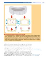
ab c
d
e
f
g
hi
Case Introduction
This 20-year-old male patient had a mo-
tor vehicle accident with a polytrauma.
Extraspinal injuries included a closed
head injury (Glasgow Coma Scale 6) with
shearing injuries and consecutive intra-
cranial pressure monitoring for 2 weeks, a
thorax injury with lung contusions, bilat-
eral hematopneumothorax, manubrium
sterni fracture, and multilevel spinal inju-
ries with fractures of the vertebrae T6, T8,
T10, T12 and L3. The thoracolumbar spinal fractures were treated conservatively. In addition, a traumatic spondylolisthe-
sis C2 (Type Effendi II) was initially treated conservatively (
a). After 6 weeks, the instability of the C2 injury became obvi-
ous, as shown in the standard lateral radiographs (
b)andtheCTscan(c). The small bony fragment indicates a rupture of
the disc C2/C3. The fractures of the pedicles C2 are shown in the CT scan (
d, e). The ruptured disc C2/C3 was removed and
replaced with a tricortical iliac crest bone graft. Subsequently, the cervical spine was stabilized with an anterior plate. The
lateral views demonstrate the radiographs/CT scan taken during the operation (
f), postoperatively (g, h), and after
9months(
i). Note that the fractures of the arc/pedicles healed after 9 months.
826 Section Fractures
Epidemiology
Themostcommonlyinjured
vertebrae are C2, C6, and C7
Cervical spine injuries
account for one-third
of all spinal injuries
Cervical spine injuries account for about one-third of all spinal injuries. Gold-
berg et al. [89] prospectively studied 34069 patients with blunt trauma undergo-
ing cervical spine radiographs at 21 institutions to accurately assess the preva-
lence, spectrum, and distribution of cervical spine injury after blunt trauma. Of
these patients, 818 (2.4%) had a total of 1496 distinct cervical spine injuries. The
second cervical vertebra was the most common (24.0%) level of injury, one-third
of which were odontoid fractures. In the subaxial spine, C6 and C7 were the most
frequently affected levels (40%). The most frequent fracture site was the verte-
bral body. Nearly two-thirds of all injuries (71%) were considered clinically sig-
nificant.
A neurological injury occurs
in about 15% of spine
trauma patients
In order to evaluate the true incidence of spinal column and cord injury, Hu et
al. [108] used the database of the Manitoba Health Services Insurance Plan
(1981–1984) to identify all patients who had spinal injuries. The annual inci-
dence rate of all spinal fractures was 64 per 100000. A total of 2063 patients were
identified, 944 of whom were admitted to hospital. There were two incidence
peaks, one occurring in young men and the other in elderly women. Of the hospi-
talized patients, 182 had cervical injury, 286 had thoracic fracture, and 403 had
injury in the lumbosacral spine. Associated injuries occurred in 38% of hospital-
ized patients. Neurological injury occurred in 122 patients (13%).
A low GCS indicates
a high risk for a concomitant
cervical injury
In a retrospective review of 14577 blunt trauma victims in a tertiary referral
center in Baltimore, 614 (4.2%) had cervical spine injuries. In a series of 14755
trauma cases in Los Angeles [64], 292 (2%) patients had cervical spinal injuries.
Of these, 86% had fractures, 10% had subluxations and 4% had an isolated spi-
nal cord injury without fracture or obvious ligamentous damage. Importantly,
the incidence of cervical injuries increased in patients with a low Glasgow Coma
Scale (GCS) score, indicating that patients with a relevant head injury are at risk
of having concomitant cervical injuries. The combination of head injury and cer-
vical spine injury represents a difficult diagnostic problem due to the lack of con-
sciousness in these patients. In a consecutive study of 447 patients with head
injuries [106], 24 (5.4%) patients suffered a cervical spine injury. Of these, 14
(58%) sustained spinal cord injuries. Furthermore, patients with a GCS of less
than 9 have an almost 3 times higher risk of sustaining a cervical injury [64]. Sim-
ilarly, patients involved in motor vehicle accidents – either as passengers or as
pedestrians – are at high risk of sustaining cervical spine injuries. Alker et al.
examined 312 victims from traffic accidents and found cervical spine injuries in
24.4%. Of these, 93% affected the upper cervical spine [15].
A specific entity of cervical injuries (sprains and strains)isrelatedtorear-end
or side impact motor vehicle collisions [184], but can also occur during diving or
other mishaps [201]. In the United States, neck strain/sprain is the most com-
mon type of injury to motor vehicle occupants treated in US hospital emergency
departments, with an annual incidence of 328 per 100000 inhabitants [158]. The
impact during the motor vehicle collision may result in bony or soft-tissue inju-
ries (whiplash injury), which in turn may lead to a variety of clinical symptoms
(whiplash-associated disorders, WAD) [184].
Injury mechanism and
symptoms after rear-end
collision must be
differentiated
The unfortunate term “whiplash” was introduced into the literature by Crowe
in 1928 [55]. This expression was intended to be a description of a motion, but it
has been accepted by physicians, patients and attorneys as the name of a disease.
This misunderstanding has led to its misapplication by many physicians and oth-
ers over the years [55].
The incidence of WAD is
substantially increasing
Reliable epidemiological data on this type of injury is hampered by the fact
that definitions are largely variable [181]. Depending on the definition of whip-
Cervical Spine Injuries Chapter 30 827


