Spinal Disorders: Fundamentals of Diagnosis and Treatment Part 103 ppt
Bạn đang xem bản rút gọn của tài liệu. Xem và tải ngay bản đầy đủ của tài liệu tại đây (453.1 KB, 10 trang )
ab
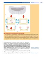
Figure 1. Pathomechanism of spinal infections
a The richly vascularized vertebral bodies with their valveless venous plexus (Batson) predispose to infection in this ana-
tomic region.
b Hematogenous seeding from peripheral ulcers, genitourinary infection, or pulmonary infection can
result in an outbreak of the infection close to the vertebral endplates and affect the intervertebral disc.
Pathogenesis
The richly vascularized
vertebral bodies predispose
to spinal infections
Spinal infections are assumed to start from the disc space in children, in whom
the intervertebral disc is still vascularized. In contrast, the disease appears to
start from the vertebral endplates in adults. However, this strict distinction has
recently been questioned by Ring et al. [41], who consider it more a continuous
disease. The blood supply to the vertebral bodies and intervertebral disc
remains a key issue in the predilection of spinal infections. The most frequent
pathomechanism is a hematogenous spread of microorganisms via the blood
vessels, resulting from urogenital, pulmonary, or diabetic foot infections
(
Fig. 1
). Batson [2] assumed that the valveless venous plexus and the slow
blood flow within predisposes to spinal infections of the vertebral body. Wiley
andTrueta[50]haveprovidedevidencefrominjectionstudiesthatthearterial
routeisofsignificantrelevance.Todayitisassumedthatbothmechanisms
play a role. With the increased frequency of spinal interventions, direct inocu-
lation of microorganisms has become an additional relevant pathomechanism
[3,4,10].
Classification
Spinal infections can be classified according to the causative organism.Clas-
sically, we differentiated between specific and so-called non-specific infec-
tions. Today, it is more appropriate to differentiate tuberculosis from pyo-
genic (e.g., Staphylococcus,Streptococcus,E.coli), fungal (e.g., Aspergillus,
Cryptococcus neoformans), parasitic (e.g., Echinococcus) and postoperative
infections.
Infections of the Spine Chapter 36 1023
Table 1. Classifcation of spinal infections
Causative organism Spatial location
pyogenic infections
tuberculosis
parasitic infections
fungal infections
vertebrae (spondylitis)
intervertebral disc (discitis)
epidural abscess
paravertebral abscess
A different approach is to classify the spinal infection according to the anatomic
region within the spine, i.e., anterior spine, spinal canal, or posterior spine. More
reasonable is differentiation with regard to the involvement of specific compart-
ments, i.e., vertebral body, intervertebral disc, epidural, intradural or paraverte-
bral (e.g., psoas muscle, retropharyngeal) extension (
Table 1).
Clinical Presentation
History
Diagnosis of spinal infection
is often delayed
Clinical presentation
is dependent on virulence,
host immunocompetence
and duration
The key feature of the history is the delayed diagnosis (Case Introduction). In an
extensive literature review, Sapico and Montgomerie [43] found that only 20% of
patients had a symptom duration of less than 3 weeks, 20% had complaints for 3
weeks to 3 months, and the remaining 50% of individuals had symptoms for
more than 3 months prior to diagnosis. The clinical presentation is related to the
virulence of the organism, immunocompetence of the host, and duration of the
infection. In this setting, Louis Pasteur’s maxim,“The organism is nothing, the
environment is everything,” has to be kept in mind. In general, the history of
patients with spinal infections is highly variable and non-specific.
The cardinal symptoms are:
slowly progressive, continuous, and localized back pain
pain exacerbation during rest and at night
back pain and gibbus (in spinal tuberculosis)
Additional but less frequent findings may be:
muscle spasm (e.g., torticollis)
weight loss
“feeling sick”
pain exacerbation with movement and weight bearing (as signs of instability)
pain in the loin, groin, or buttocks (due to an abscess)
symptoms of radiculopathy and myelopathy (late)
Search for
predisposing factors
Although the source of infection remains unidentified in more than one-third of
cases [43], predisposing f actors should be specifically sought:
diabetes mellitus
intravenous drug abuser
immune deficiency states
preexisting paraplegia
dental granuloma
soft tissue ulcers
urinary tract infections
previous septic conditions
Cardinal symptoms in
children and adults
are similar
In children,spinalinfectionsmost frequently occur in the first decade of
life. The mean age at presentation appears to be lower in children with discitis
1024 Section Tumors and Inflammation
compared to vertebral osteomyelitis (2.8 vs 7.5 years of age) [15]. The presenta-
tion of similar spinal infection in children can differ from that in adults, while
the cardinal symptoms remain very similar, i.e., slowly progressing symptoms
with a general aspect of appearing ill. Frequent findings in children are [15, 16,
49]:
refusaltowalk
back pain and abdominal pain
“appearing ill”
fever (in cases of vertebral osteomyelitis)
Physical Findings
Physical findings
are non-specific
Although clinical examination is seldom helpful in making the diagnosis, the
most frequent findings are:
local tenderness (less specific)
positive psoas sign
pain provocation by flexion, rotation, and percussion
limping (in children)
Triad of Pott: gibbus, spinal
abscess, paraparesis
A thorough neurolog ic al examination is mandatory to diagnose neural com-
pression syndromes, in particular to rule out early para/tetraparesis.
The classic clinical presentation of spinal tuberculosis includes back pain and
a gibbus and in later stages symptoms caused by an epidural abscess and devel-
oping neurologic deficits [23]. In Western industrialized countries, patients
today present with less specific symptoms and often have an underlying general
illness (e.g., HIV, diabetes). The prevailing symptoms in a study by Fam and
Rubenstein were back pain and weight loss [13].
Diagnostic Work-up
Key to diagnosis is
“consider it”
The most important aspect of diagnosing spinal infection is to include this diag-
nosis in the differential diagnosis. The diagnostic work-up is apparently clear
when spinal infection is considered as a cause of the patient’s symptoms and con-
sists of laboratory investigations, imaging studies, and biopsy.
Laboratory Investigations
BSR, CRP and WBC
are frequently elevated
The most helpful laboratory investigations are:
elevated blood sedimentation rate (BSR)
C-reactive protein (CRP)
white blood cell count (WBC)
Infection parameters are
sensitive but not specific
These inflammation markers are sensitive but non-specific and are more helpful
in terms of the temporal course rather than as absolute (single) values. The
parameters can reliably be used to monitor treatment response. The white blood
cell count is only elevated in about half of the patients and depends on the nutri-
tional state of the patient. The determination of antibody titers for putative bac-
teria is valuable in identifying certain causative organisms.
In the presence of a septic state, blood cultures should be obtained, but the hit
rate is low. It can be increased if more than one blood sample (three to five recom-
mended) is taken from different veins.
Inputativetuberculosis,theMantouxortuberculinskintestishelpfulto
investigate present or past exposure to Mycobacterium tuberculosis. Direct evi-
Infections of the Spine Chapter 36 1025
ab
Figure 2. Radiographic findings in spinal infection
The classical radiographic signs of spinal infection consist of a loss of vertebral endplate definition, b decrease of disc
height, gradual development of osteolysis, development of a paravertebral soft tissue mass, and reactive changes with
sclerosis.
dence can seldom be obtained from examination of material aspirated from an
abscess.
Imaging Studies
Modern imaging modalities have substantially improved accuracy in diagnosing
spinal infection. However, standard radiographs are still very helpful because they
allow an overview of the osseous destruction and resulting deformity.
Standard Radiographs
Radiographic diagnosis is
hampered by a delay in the
appearance of alterations
The major drawback of standard radiography is the delay in the appearance of
radiographic signs (
Fig. 2). The sequence of changes demonstrable on radio-
graphs is [48]:
loss of vertebral endplate definition (at earliest 10–14 days after onset)
reduction of disc height
gradual development of endplate osteolysis
development of a paravertebral soft tissue mass
reactive changes with sclerosis and new bone formation (at earliest 4–6
weeks after onset)
vertebral collapse (late) with spinal deformity (kyphosis/scoliosis)
Magnetic Resonance Imaging
MRI is the imaging study
of choice
Today MRI has become the imaging modality of choice in diagnosing spinal
infection. Recent comparisons with bone scans have demonstrated that MRI is as
accurate and sensitive [48].
Characteristic findings (
Fig. 3) suggestive of spinal infections are [11]:
decreased vertebral endplate signal intensity on T1-weighted images (95%)
1026 Section Tumors and Inflammation
ab c
Figure 3. MRI characteristics of spinal infections
a The predominant features of spinal infections are decreased vertebral body signal intensity on T1-weighted images,
b loss of endplate definition and increased disc signal on T2-weighted images, increased vertebral body signal intensity
on T2-weighted images and increased signal intensity on T1-weighted fat-suppressed images after injection of gado-
pentetate.
c Note the retrovertebral epidural spinal abscess (arrow).
loss of endplate definition (95%)
increased disc signal on T2-weighted images (95%)
increased vertebral endplate signal intensity on T2-weighted images (56%)
contrast enhancement of the disc and vertebral body (94%)
The increased signal intensity is more obvious on short tau inversion recovery
(STIR) or frequency-selective fat-suppressed T2-weighted spin echo sequence,
but with the depiction of less anatomical detail [11].
In appropriate cases, the diagnosis of spinal tuberculosis (
Fig. 4) can be made
by MRI with high diagnostic accuracy [46]. Loke et al. [28] have reported that the
most common site is the lumbar spine, often with involvement of more than one
Contrast enhancement
is helpful in differentiating
spinal TB from other
granulomatous infections
vertebra. Contrast enhancemen t is helpful in differentiating spinal tuberculosis
from other granulomatous infections [46]. Frequent findings [28] suggestive of
spinal tuberculosis are:
paraspinal soft-tissue masses (73%)
vertebral destruction and collapse (73%)
epidural abscess (53%)
posterior element involvement (40%)
intraosseous abscess (20%) with contrast enhancement
Computed Tomography
CT demonstrates bony
destruction better than MRI
The predominance of computed tomography in diagnosing spinal infections has
been surpassed by MRI because of its spatial resolution, multiplanar capabilities
and tissue contrast. However, CT still has a role with regard to the assessment of the
osseous destruction, which is important for the choice of treatment (i.e., non-oper-
Infections of the Spine Chapter 36 1027
a bc d
Figure 4. Radiographic features of spinal tuberculosis
Spinal tuberculosis can be diagnosed with satisfactory accuracy using standard radiographs and MRI. The key findings
include paraspinal soft-tissue masses, vertebral destruction and collapse, epidural abscess, posterior element involve-
ment, and intraosseous abscess.
ative vs surgical) and planning of the surgical approach and technique. It is also
invaluable in patients unsuitable for an MRI scan (e.g., because of a pacemaker).
Radionuclide Studies
Bone scan and FDG-PET
arehelpfulin
making the diagnosis
Because of the comparable diagnostic accuracy of MRI, technetium-99m labeled
methylene-diphosphonate (Tc-99m MDP) bone scintigraphy is today more infre-
quently used in the diagnosis of spinal infections. However, an indication for a
bone scan is still the search for a focus lesion, e.g., dental granuloma and osteo-
myelitis.
Confusion may arise with regard to the differential diagnosis of a degenerative
endplate abnormality and spinal infections. Positron emission tomography
(PET) with fluorine-18 fluorodeoxyglucose (FDG) (
Fig. 5) has been used in sus-
pected spinal infection [45]. In a recent study, FDG-PET has been shown to be
helpful in differentiating spinal infection from disc degeneration because the lat-
ter condition generally does not show FDG uptake [47].
Biopsy
Biopsy is a “must”
prior to treatment
The isolation of the causative organism is of utmost importance and must be
attempted in every case. While a biopsy can be performed under image intensi-
fier control, CT guidance [7, 34, 39] is preferable because of the accurate spatial
resolution,whichisimportanttodocumentthatthebiopsywasactuallytaken
from within the lesion. This is particularly valid in areas that are difficult to
access, such as the sacrum or sacroiliac joints and upper thoracic or cervical
region [48].
Percutaneous needle biopsy provides a definitive diagnosis ranging from 57%
to 92% [7, 34, 39] and depends on previous antibiotic treatment.
The most frequently found organisms are:
Staphylococcus aureu s (30–55%)
gram-negative organisms (e.g.,E.coli,Salmonella,Enterococcus,Proteus)
Pseudomonas a eruginosa (in 65% of drug abusers)
Streptococcus viridans, epidermatitis
Proprionibacterium acnes
1028 Section Tumors and Inflammation
ab
Figure 5. Radionuclide study of spinal infection
Positron emission tomography with FDG demonstrates uptake at the level of L4/5 (same patient as in Fig. 3), strongly
indicative of spinal infection.
Tuberculosis can
mimic tumor
Differentiation of tuberculosis from tumor may sometimes be difficult and a cul-
ture takes considerable time. In the clinical situation it is not possible to await the
results from the culture and the diagnosis has to rely on the imaging findings.
Non-operative Treatment
Do not start treatment
prior to isolation of the
causative organism
(if possible)
In the absence of a life-threatening condition, treatment of spinal infections
should not be started without vigorous attempts to isolate the causative organ-
ism. It is mandatory to obtain the causative organism prior to antibiotic treat-
ment because of the substantially reduced likelihood of a secondary diagnosis
(
Case Introduction). In the absence of a causative organism and progressing
infection despite (non-specific) antibiotic treatment, high-dose broad-spectrum
double or triple drug chemotherapy is often required. However, subsequent
severe pharmacological side effects may limit the use of high-dose antibiotics
and may result in a life-threatening situation if the infection is not controlled.
This holds true for conservative as well as surgical treatment.
Table 2. General objectives of treatment
eradicate the infection
prevent recurrence
relieve pain
prevent or reverse a neurologic deficit
restore spinal stability
correct spinal deformity
Non-operative therapy
is still the gold standard
for uncomplicated cases
The choice of treatment is related to the chances of achieving the general objec-
tives of treatment with the respective therapy (
Table 2). While radical debride-
ment, internal fixation, and appropriate antibiotic treatment have become the
gold standard in the treatment of osteomyelitis of long bones, the mainstay for
Infections of the Spine Chapter 36 1029
Table 3. Favorable indications for non-operative treatment
single disc space infection (discitis)
known causative organism
absence of gross bony destruction and instability
mobile patients with only moderate pain
absence of relevant neurologic deficit
rapid normalization of inflammation parameters
the treatment of spinal infection is still non-operative (Table 3). However, the
trend in the literature is to support more aggressive treatment of spinal infections
even in situations where non-operative treatment can be successful. This trend is
because of a shorter hospitalization and recovery time.
The mainstay of treatment
is chemotherapy
The mainstay for the treatment of bacterial and parasitic infection is still rest
and intravenous antibiotics for a minimum of 4–6 weeks, depending on the
extent of the infection and organism (
Case Study 1). As outlined above, specific
chemotherapy is mandatory. Depending on the resistance of the organism and
the bone penetration of the respective antibiotic drug, administration by the oral
route may be appropriate for the post-primary treatment. We strongly recom-
mend that the antibiotic treatment be discussed with an infection specialist to
ab c
d
e
Case Study 1
A 70-year-old woman presented with an infected great toe and was treated with antibiotics for 3 weeks after a biopsy
was taken. The biopsy revealed Proteus mirabilis and Pseudomonas aeruginosa as the responsible germs. Two months
later the patient developed severe neck pain, which became worse with movement. There were no radicular symptoms
or neurologic deficits. The radiographic evaluation of the cervical spine demonstrated blurred endplates and somewhat
narrowed disc space (
a). The MRI showed strong evidence of a spinal infection at the level of C3/4 (b, c). Note the contrast
enhancement from C2 to C5 (
d). There was no epidural abscess or spinal cord compromise. A CT-guided needle biopsy
did not reveal a positive result, but allowed the exclusion of a tumor. This case exemplifies the notion that detection of
a germ after previous antibiotic treatment is unlikely. Bone scintigraphy provided further evidence of an infection (
e).
The patient was treated with double chemotherapy and a hard collar. In the absence of a neurologic deficit, severe pain
or substantial deformity, non-operative treatment was successful. The patient recovered completely from her symptoms
within 2 months.
1030 Section Tumors and Inflammation
allow for the most specific (narrow) drug therapy with the least chances of phar-
macological side effects.
According to Pertuiset et al. [35], there appears to be a consensus that the ini-
tial antituberculous treatment should consist of a triple (isoniazid, rifampin, and
pyrazinamide) or quadruple chemotherapy (plus ethambutol) given for 2–3
months. After this period, chemotherapy should be continued with isoniazid and
rifampin in the absence of resistance or side effects. There is still debate on the
optimal duration of antituberculous chemotherapy required for complete recov-
ery. While a minimum of 12 months is favored by the majority of experts, no con-
vincing evidence can be derived from the literature [35].
Early ambulation
is attempted
While bedrest may be indicated for the initial treatment, early mobilization of
the patient with an orthosis is recommended. The need for cast immobilization,
including neck or thigh extension, has to be determined on an individual basis and
depends on the location of the infection, general condition, and age of the patient.
CRP is helpful
in monitoring healing
of infection
It is imperative to monitor the treatment success by regular determination of
the inflammation parameters (i.e., SR, CRP, and WBC). Follow-up imaging stud-
ies should be done in the case of persistent symptoms and in the absence of
decreasing inflammation parameters. In general, antibiotic treatment should be
continued for at least 4–6 weeks because of a high recurrence rate in pyogenic
spinal infections. Antibiotic treatment should only be ceased after normalization
of the CRP.
Indication for a change from non-operative to operative treatment is the per-
sistence of the infection despite adequate antibiotic treatment or in the presence
of pharmacological side effects (e.g., kidney or liver dysfunction) limiting the
further use of specific antibiotics in adequate dosage. A recent study has demon-
strated a favorable outcome by surgical treatment in this situation [8].
Operative Treatment
General Principles
Although the majority of cases with spinal infections can be successfully treated
non-operatively, surgery may become necessary in about one-third of the
patients (
Table 4):
Table 4. Indications for surgery
disease progression despite adequate antibiotic treatment
progressive spinal deformity and instability
neurological compromise
incapacitating pain
Increasing evidence is presented in the literature [32] that radical debridement
and bone grafting of specific (TB) spinal infections are superior to non-operative
treatment [30, 33]. Less information is available from the literature with regard to
the treatment of pyogenic infections. On the other hand, no evidence is presented
that the spinal infection responds differently to radical debridement and bone
grafting than to long bone osteomyelitis. No reports indicate that this approach
is ill-advised in cases where conservative treatment does not result in rapid reso-
lution of the infection and recovery of the patient.
Surgical Techniques
The surgical approach is largely dependent on the extent and location of the
infection, spinal destruction, neurologic deficits, health status, and comorbidity
of the patient (
Fig. 6).
Infections of the Spine Chapter 36 1031
ab
cd
Figure 6. Surgical treatment of spinal infections
The key to the treatment of spinal infections is radical debridement of the infected spine. a Often spinal infections are
associated with disc space collapse, instability, and kyphotic deformity. In cases of thoracolumbar spondylodiscitis, an
accepted standard for the treatment of spinal infection today is posterior instrumentation, followed by anterior radical
debridement. In a first step, the spine is exposed by a posterior approach. Pedicle screws are inserted in the vertebrae
adjacent to the infection. If a kyphotic deformity is present, a lordic prebent rod is first inserted and connected to the dis-
tal screws.
b By levering the rod into the distal screws, the deformity is corrected. c In a second stage, the spine is
approached anteriorly. With curets and pituitary forceps, the infected area is debrided to the bleeding bone. The inter-
vertebral disc is resected as completely as possible.
d The anterior column is reconstructed with a tricortical iliac bone
graft and additional circumferential cancellous bone.
Percutaneous Debridement and Drainage
In discitis with suspicion of abscess formation, percutaneous debridement and
drainage is the preferred treatment [17, 18]. It can be performed using local
anesthesia, sufficient material can be obtained for culture, and it allows for
debridement and drainage of the infection.
1032 Section Tumors and Inflammation


