Spinal Disorders: Fundamentals of Diagnosis and Treatment Part 106 pptx
Bạn đang xem bản rút gọn của tài liệu. Xem và tải ngay bản đầy đủ của tài liệu tại đây (226.99 KB, 10 trang )
Recapitulation
Epidemiology.
Approximately 40% of patients with
rheumatoid arthritis show pathology in the cervical
spine, mainly the atlantoaxial segment.
Pathogenesis. The translational instability between
axis and atlas might be painful and leads in the long
term to myelopathic changes duetochronictrau-
matization of the myelon. Ongoing osseous resorp-
tion of the lateral masses of the atlas causes upward
migration of the dens into the foramen magnum. In
the subaxial cervical spine, the inflammatory process
causes instability and deformity.
Clinical presentation. The instability and deformity
are mostly associated with the corresponding clini-
cal symptoms: pain and neurological signs in dif-
ferent stages. However, it has to be kept in mind
that these patients are used to tolerating pain and
that often other problems of the joints are more
prominent. The pathology of the cervical spine may
progress unnoticed in these cases.
Diagnostic work-up. Every patient with RA should
have a lateral flexion radiography of the cervical
spine performed as a screening investigation at
least every 3–5 years (according to the aggressivity
of the disease). In cases of manifest instability or de-
formity, a neurophysiological work-up and MRI
should be performed.
Non-operative treatment. If surgery is not indicat-
ed, the patient should be given regular observation
with neurophysiological examinations, radiographs
and MRI.
Operative treatment. Neck pain is the most com-
mon indication for surgery, but neurological symp-
toms with myelopathy or radicular deficits might be
the primary cause for surgery. It should be kept in
mind that clinical assessment in rheumatoid patients
might be extremely difficult since previous surgery
on various articulations of the extremities makes in-
terpretation of clinical findings difficult. Neurophysi-
ological investigation is a suitable means for obtain-
ing objective results. Stabilization of the atlantoaxial
segment is the most common procedure for treat-
ment of atlantoaxial instability. It is performed by
screw fixation technique from a posterior approach.
In the case of severe occipitocervical dislocation, the
fixation should be extended to the occiput. Persis-
tent dislocation or compression by the dislocated
dens should be treated by transoral decompression.
In the subaxial spine, instabilities may be treated by
posterior plate fixation with lateral mass screws or
pedicle screws. Concomitant narrowing of the spinal
canal should be approached by anterior decompres-
sion with corpectomy and/or posterior laminecto-
my. The timing of surgery in rheumatoid patients is
crucial to obtaining satisfactory clinical results.
Key Articles
Boden SC, Dodge LD, Bohlmann HH, Recht ine GL (1993) Rheumatoid arthritis of the
cervical spine. Long term analysis with predictors of paralysis and recovery. J Bone Joint
Surg 75-A(9):1282 –1297
The authors report their experience in treating 73 patients with rheumatoid arthritis with
an average follow-up of 7 years. The authors highlight that the most important predictor
of the potential for neurological recovery after the operation was the preoperative poste-
rior atlanto-odontoid interval. In patients who had paralysis due to atlantoaxial subluxa-
tion, no recovery occurred if the posterior atlanto-odontoid interval was less than 10 mm,
whereas recovery of at least one neurological class always occurred when the posterior
atlanto-odontoid interval was at least 10 mm. If basilar invagination was superimposed,
clinically important neurological recovery occurred only when the posterior atlanto-
odontoid interval was at least 13 mm. All patients who had paralysis and a posterior
atlanto-odontoid interval or diameter of the subaxial canal of 14 mm had complete motor
recovery after the operation.
Crockard HA, Pozo JL, Ransford AO, Stevens JM, Kendall BE, Essigman WK (1986)
Transoral decompression and posterior fusion for rheumatoid atlanto-axial subluxa-
tion. J Bone Joint Surg 68B(3):350 – 356
In this landmark paper, Crockard et al. describe a surgical technique for transoral ante-
rior decompression and posterior occipitocervical fusion, which removes both bony and
soft-tissue causes of compression and allows early mobilization without major external
fixation.
1054 Section Tumors and Inflammation
Key Articles
Dvorak J, Grob D, Baumgartner H, Gschwend N, Grauer W, Larsson S (1989)Functional
evaluation of the spinal cord by magnetic resonance imaging in p atients with rheuma-
toid arthritis and instability of upper cervical spine. Spine 14(10):1057 – 1064
This study describes the imaging findings in patients with atlanto-axial instability due to
rheumatoid arthritis and provides recommendations for surgical treatment.
Matsunaga S, Sakou T, Onishi T, Hayashi K, Taketomi E, Sunahara N, Komiya S (2003)
Prognosis of patients with up per cervical lesions ca used by rheumatoid arthritis: com-
parisonofoccipitocervicalfusionbetweenC1 laminectomy and nonsurgical manage-
ment. Spine 15(28):1581 – 1587
In a matched controlled comparative study, non-surgical treatment and occipitocervical
fusion associated with C1 laminectomy were evaluated in patients with upper cervical
lesions caused by rheumatoid arthritis. The authors concluded that occipitocervical
fusion associated with C1 laminectomy for patients with rheumatoid arthritis is useful
for decreasing nuchal pain, reducing myelopathy, and improving prognosis.
CombeB,LandeweR,LukasC,BolosiuHD,BreedveldF,DougadosM,EmeryP,Ferrac-
cioli G, Hazes JM, Klareskog L, Machold K, Martin-Mola E, Nielsen H, Silman A, Smolen
J, Yazici H (2007) EULAR recommendations for the management of early arthritis:
report of a task force of the European Standing Committee for International Clinical
Studies Including Therapeutics (ESCISIT). Ann Rheum Dis 66:34 – 45
Excellent review on the conservative treatment of rheumatoid arthritis with recommen-
dations on the management of early rheumatoid arthritis
References
1. Almeida Mdo S, et al. (2005) Epidemiological study of patients with connective tissue dis-
eases in Brazil. Trop Doct 35(4)206–9
2. Boden SC, et al. (1993) Rheumatoid arthritis of the cervical spine. Long term analysis with
predictors of paralysis and recovery. J Bone Joint Surg 75A(9):1282–1297
3. Brooks AL, Jenkins EG (1978) Atlanto-axial arthrodesis by the wedge compression method.
J Bone Joint Surg 60A:279–284
4. Crockard HA, et al. (1986) Transoral decompression and posterior fusion for rheumatoid
atlanto-axial subluxation. J Bone Joint Surg 68B(3):350–356
5. Dvorak J, et al. (1989) Functional evaluation of the spinal cord by magnetic resonance imag-
ing in patients with rheumatoid arthritis and instability of upper cervical spine. Spine
14(10):1057–1064
6. Dvorak J, et al. (1993) Clinical validation of functional flexion/extension radiographs of the
cervical spine. Spine 18(1):120–127
7. Edwards CJ, et al. (2005) The changing use of disease-modifying anti-rheumatic drugs in
individuals with rheumatoid arthritis from the United Kingdom General Practice Research
Database. Rheumatology (Oxford) 44(11)1394–1398
8. Gallie WE (1939) Fractures and dislocations of the cervical spine. Am J Surg 46A:495–499
9. Goel A, Laheri V (1994) Plate and screw fixation for atlanto-axial subluxation. Technical
report. Acta Neurochir 129:47–53
10. Grob D (2000) Atlantoaxial immobilization in rheumatoid arthritis: a prophylactic proce-
dure? Eur Spine J 9:404–409
11. Grob D, et al. (1992) Biomechanical evaluation of four different posterior atlantoaxial fixa-
tion techniques. Spine 17(5):480–490
12. Grob D, et al. (1994) The role of plate and screw fixation in occipitocervical fusion in rheu-
matoid arthritis. Spine 19:2545–2551
13. Grob D, Schütz U, Plötz G (1999) Occipitocervical fusion in patients with rheumatoid
arthritis. Clin Orthop 366:46–53
14. Harms J, Melcher RP (2001) Posterior C1–C2 fusion with polyaxial screw and rod fixation.
Spine 26(22):2467–71
15. Kraus DR, et al. (1991) Incidence of subaxial subluxation in patients with generalized rheu-
matoid arthritis who have had previous occipital cervical fusions. Spine 16(10S):486–489
16. Magerl F, Seemann P (1986) Stable posterior fusion of the atlas and axis by transarticular
screw fixation. Cervical Spine 1:322–327
17. Mannion AF, Elfering A (2006) Predictors of surgical outcome and their assessment. Eur
Spine J 15(Suppl 1)93–108
Rheumatoid Arthritis Chapter 37 1055
18. Matsunaga S, et al. (2003) Prognosis of patients with upper cervical lesions caused by rheu-
matoid arthritis: comparison of occipitocervical fusion between C1 laminectomy and non-
surgical management. Spine 15(28):1581–1587
19. Ono K, Ebara S, Fuji T (1987) Myelopathy hand. J Bone Joint Surg 69B:215–219
20. Ranawat CS, et al. (1979) Cervical spine fusion in rheumatoid arthritis. J Bone Joint Surg
61A:1003–1010
21. Shimizu T, Shimada H, Shirakura K (1993) Scapulohumeral reflex (Shimizu). Its clinical sig-
nificance and testing maneuver. Spine 18(15):2182–2190
1056 Section Tumors and Inflammation
38
Ankylosing Spondylitis
Thomas Liebscher, Kan Min, Norbert Boos
Core Messages
✔
Ankylosing spondylitis (AS) is a systemic,
inflammatory, seronegative rheumatoid disease
✔
Ankylosing spondylitis in 90 % of cases is asso-
ciated with HLA-B27
✔
The male/female ratio is 2– 7:1
✔
The onset of the disease is usually between 15
and 35 years of age, and it can take up to
10 years before the diagnosis is made
✔
The imaging modalities of choice are standard
radiographs and MRI. Computed tomography is
useful for diagnosing occult fractures and for
preoperative planning
✔
Ankylosing spondylitis is treated non-opera-
tively by analgesics, anti-inflammatory drugs
and physiotherapy
✔
Spinal surgery is only indicated if conservative
treatment has failed to prevent spinal deformi-
ties and instabilities or in the case of disc space
infections
✔
The surgical techniques for treating spinal
deformity, instabilities and infections depend
on the localization and etiology of the pathol-
ogy
✔
Surgical techniques include lumbar closing
wedge (pedicle subtraction) osteotomies, mul-
tisegmental posterior wedge osteotomy, cervi-
cal opening or closing wedge osteotomies
✔
Meticulous preoperative planning of the osteo-
tomy is mandatory
✔
Unstable fractures with neurological dysfunc-
tions at the cervical spine are stabilized from a
combined anterior and posterior approach. In
the lumbar spine, the surgery is most fre-
quently done from posterior
✔
Surgical interventions for ankylosing spondyli-
tis are prone to complications
Epidemiology
Spondyloarthropathies
are chronic systemic
inflammatory rheumatic
disorders
Spondyloarthropathies (SPAs) are systemic and chronic inflammatory rheu-
matic disorders with involvement of the axial skeleton or asymmetrical arthritis
of large joints of the lower extremities.
SPAs are divided into five subcategories:
ankylosing spondylitis
psoriatic arthritis
reactive arthritis
inflammatory bowel disease related arthritis
undifferentiated spondyloarthropathy
Ankylosing spondylitis is the
most common form of SPA
Ankylosing spondylitis (AS) is the most common form of SPAaffecting the whole
spine [7, 17, 20, 105]. The final result is a kyphosis of the whole column with sag-
ittal imbalance (
Case Introduction). Besides spinal ankylosis, inflammatory
lesions, bony erosions, discitis and loss of bone mineral density (BMD) can occur
during the process of this disease. AS was described for the first time by Vladimir
von Bechterew in 1893 [9]. The description was initially based on clinical symp-
Tumors and Inflammation Section 1057
a b
Case Introduction
A 42-year-old male had suffered from ankylosing spondylitis for over 10 years and developed a progressive ankylosis of
the entire spine. Despite intensive physiotherapy, the patient developed an increasing sagittal deformity and loss of his
verticalgaze(
a). When shaking hands, he was unable to look at his counterpart, which was quite disturbing in his job. The
standing lateral radiograph demonstrates a significant loss of lumbar lordosis (
b). Since the pathology was predomi-
nantly located in the lumbar spine, a lumbar closing wedge osteotomy at L3 was suggested and carried out.
toms and the spinal deformity. With the advance of radiography, it was possible
to document the articular changes. AS is associated with chronic inflammation
of the:
sacroiliac joints
vertebral column
osteoarthritis of the large joint (hip, knee and shoulder joints)
extra-articular disorders including enthesitis and uveitis
AS more frequently
occurs in males
Ankylosing spondylitis occurs more frequently in the male population with a
ratio of between 2 and 7 to 1 [28, 31, 43, 49, 53, 79, 105]. The prevalence rate in
Europe and North America ranges between 0.1 and 1.4/100000 and regionally
1058 Section Tumors and Inflammation
c d
e
Case Introduction (Cont.)
Postoperative radiographs (c, d) demonstrate an excellent correction and alignment of the spine with recreation of lum-
bar lordosis. At a 2-year follow-up, the patient was very satisfied with the result, able to look straight ahead and fully func-
tional in his job (
e).
The onset of disease
is usually between 15
and 35 years of age
can rise up to 8.2/100000 [87]. The onset of disease is usually between 15 and
35 years. Up to 10 years can pass before the diagnosis is made [40, 43, 49, 79].
This delay in diagnosis is due to the initially non-specific clinical symptoms
(e.g., low back pain) and lack of early pathognomonic imaging findings. During
the later disease stage, inflammatory spinal lesions can be found which most
commonly occur in the thoracic and lumbar spine [8, 105]. Aseptic spondylodis-
AS is characterized by
progressive kyphosis with
segmental instability, asep-
tic discitis and osteoporosis
citis is an erosive lesion of the disc and vertebral body without infection or
trauma, first described by Andersson in 1937 [2]. Clinical and radiographic find-
ings demonstrate a progressive vertebral and discovertebral kyphosis with seg-
mental instability [99, 103]. The prevalence of aseptic discitis is about 18% of
patients with AS [61]. Almost half (40–50%) of the patients with mild AS exhibit
osteopenic or osteoporotic lumbar vertebrae [6, 94, 107]. Severe complications of
osteoporosis and loss of trabecular bone are spinal fractures subsequent to
The prevalence of spinal
fractures is about 5 %
and increases with age
minor trauma. The prevalence of spinal fracture is about 5% and increases with
age [40]. It reaches about 15% at the age of 42 years and older [40]. Unilateral
Ankylosing Spondylitis Chapter 38 1059
AS also frequently affects
hips, knee and shoulder
joints
inflammationoflargediarthrodialjointssuchaships,kneesandshouldersisa
common symptom of SPA. Hip joints are affected in 57% of patients [37]. The
prevalence of unilateral shoulder arthritis in patients with AS is estimated to be
between 30% and 58%. Approximately 25% of AS patients even suffer from bilat-
eral shoulder arthritis [37, 38, 43]. Besides changes in physical function, other
areas also affect the quality of life such as [12]:
psychological domain [67]
social domain
economic aspects
A disease duration of
15 years is associated with
a 50% inability to work
Afteradiseasedurationof15years,about50%ofpatientsareusuallynolonger
able to work full time [43]. Up to 80% of patients suffer from daily pain and more
than 60% need to take painkillers daily [43]. In addition, anxiety and depression
are correlated with the degree of disorder [45, 67].
Pathogenesis
Genetic factors play
a key role
Despite intensive research, the pathogenesis of AS is not yet clear [19]. There is
increasing evidence that AS is genetically linked. The association of AS and the
The pathogenesis of AS
is not clear
HLA -B27 gene is well known. HLA-B27 can be found in up to 90% of patients
with AS [49, 79, 105]. The HLA-B27 gene is mapped to the major histocompatibil-
ity complex (MHC) class I region on the short arm of chromosome 6 [55]. There
are24subtypesofHLA-B27 [54, 55]. The subtype HLA-B27 05 is most common
worldwide. Twin studies have shown that AS is passed on to the next generation
with a higher incidence for monozygotic than for dizygotic or even heterozygotic
parent-child pairs [24, 49]. Since 80–90% of all HLA-B27 carriers do not develop
AS, it is widely assumed that more genetic factors are involved [87]. HLA subtype
carriers B27 06 (found in the Southeast Asian population) and B27 09 (Sardinian
population) do not develop AS [54, 55], which also strongly indicates the exis-
tence of other genetic factors. Whole genome mapping and within-family studies
have demonstrated a link between AS and other non-HLA -B27 genes mainly on
the short arm of chromosome 6 [23, 62, 89, 93].
Bacterial infections
may trigger autoimmune
responses
An infection-based pathogenesis of AS has been the subject of critical debate
[19, 41, 66, 96]. Antigenic peptides are thought to derive from bacterial proteins
(P.aeruginosa,E.coliand Bacillus megaterium) which have a similar alignment
of amino acids like peptides inside articular joints [41, 66]. HLA-B27 restricted
CD8-T lymphozytes are suspected of identifying the bacterial protein as a target
and thereafter could also aim at peptide structures inside the sacroiliac joint or
vertebral column resulting in an autoimmune reaction with inflammatory signs.
The finding that reactive arthritis is triggered by genitourinary infections with
Chlamydia trachomatis or by enteritis caused by gram-negative enterobacteria
(e.g., Shigella, Salmonella, Yersinia andCampylobacter) supports this hypothesis,
but the evidence for triggering infections in other spondylarthopathies is limited
[19].
Inflammatory reactions
play a key role
in the pathogenesis
The detailed pathogenetic mechanisms have yet to be elucidated for associ-
ated bone mineral density loss, bony lesions as well as the formation of new bone
material ending up in ankylosis. It is assumed that new bone formations are
independent of local inflammatory processes [66]. On the other hand, there is
some evidence that persistent inflammation might be an etiologic factor of bone
lossinAS[65].Consequencesofbonelossare(occult)fracturesandpseudar-
throsis, in which microscopically necrotic bone material and cartilage can be
observed besides vascular fibrous tissue [39]. The existence of an aseptic discitis
supports an inflammatory origin for bony changes. CD3+ lymphocytes and IgA
1060 Section Tumors and Inflammation
positive plasma cells have been identified in vertebral bones and the surrounding
soft tissue affected by aseptic discitis [76]. Bloodmarkers for inflammation(CRP,
ESR) are found elevated in aseptic discitis as well [61, 76]. After local inflamma-
tory processes, disc replacing fibrous tissue and cartilaginous nodules have been
identified in later stages of aseptic discitis [27, 61]. Bone marrow from zygapo-
physeal joints demonstrates persistent inflammation even in those patients with
long-standing disease. The findings of increased numbers of T cells and B cells
and neoangiogenesis suggest that these features play a role in the pathogenesis of
AS [3].
Stages of pathological
changes include inflamma-
tory responses, proliferative
bone sclerosis and ankylosis
with increasing deformity
Pathological changes ofthevertebralcolumnduetoASoccurinthreeconsecu-
tive or side by side stages: First, there is an inflammatory process with bony ero-
sions and destruction of vertebrae and discs. The development of square vertebral
bodies is shown to be based on a combination of a destructive osteitis and repair
[5]. These changes initially are noted in the whole spine yet more frequently are
seen in the lower thoracic spine [8, 105]. Second, a proliferatory bone sclerosis
develops followed by a reactive bone formation with syndesmophytes. These
changes are slow in growth throughout the whole spine followed by kyphotic defor-
mation and progressive sagittal imbalance of the spine. Third, the spine deformity
will increase to an ankylosing process and end in a so-called bamboo spine.
The rationale of conservative therapy is to protract the consequences of
inflammation and osteoporosis and defer structural damage to the affected
bones. The finding of abundant tumor necrosis factor (TNF)- message in
affected joints provides the rationale for the therapeutic use of TNF- inhibitors
[18, 19]. A strategy of continuous use of non-steroidal anti-inflammatory drugs
(NSAIDs) has been shown to reduce radiographic progression in symptomatic
patients with AS, without increasing toxicity substantially [102]. Early treatment
therefore appears essential for a good clinical outcome [15, 71].
Clinical Presentation
History
The diagnosis
is often delayed
Ankylosing spondylitis predominantly affects the mobility of the vertebral col-
umn, joint function and pain. This entity is sometimes difficult to diagnose par-
ticularly during the onset of the disease. Quite often the diagnosis is therefore
delayed.
It is important to consider the diagnosis of AS in patients who present with
early symptoms such as:
morning stiffness
pain in the pelvic region (sacroiliac joints)
pain at night
decreasing pain during movement
musculoskeletal pain at varying locations
fatigue
loss of body weight
subfebrile temperature
When AS has become manifest, the disease affects the function and mobility of
the spine and diarthrodial joints and results in pain. The cardinal sy mptoms are:
Inflammatory back pain
is a hallmark
“inflammatory” back pain
typical arthritis pain (pain at night and stiffness in the morning)
progressive spinal stiffness
progessive hyperkyphosis (inability to look straight ahead)
Ankylosing Spondylitis Chapter 38 1061
Table 1. Criteria for inflammatory back pain
morning stiffness > 30 min improvement in back pain with exercise
but not with rest
awakening because of back pain during
thesecondhalfofthenight
alternating buttock pain
The criteria are fulfilled if at least two of four of the parameters are present [80]
Inflammatory pain is among the first symptoms and the key clinical sign
of AS. The criteria [80] for inflammatory back pain in younger patients
(<50 years) are shown in
Table 1.
Rudwaleit reported that none of the single parameters sufficiently differenti-
ated AS from mechanical low back pain. Several sets of combined parameters
proved to be well balanced between sensitivity and specificity. If at least two of
the aforementioned four parameters were fulfilled (positive likelihood ratio 3.7),
a sensitivity of 70.3% and a specificity of 81.2% was found. If at least three of the
four parameters were fulfilled, the positive likelihood ratio increased to 12.4 [80].
Additional symptoms are:
enthesitis (e.g., Achilles tendon, plantar fascia)
anterior uveitis
pulmonary, cardial and bowel inflammation
Typical concomitant disorders or extra-articular manifestations have been
observed to be part of AS: painful tendinopathy, acute anterior uveitis (AAU),
pulmonary and cardial inflammation, e.g., aortitis, and bowel disease. The fre-
quency, duration and intensity of these concomitant disorders varies individu-
AS is a systemic disease ally. The prevalence of AAU is between 33% and 49% [21, 25, 43]. AS is perceived
as a systemic disease.
Physical Findings
Ankylosing spondylitis is a potentially progressive disease. The first symptoms
of AS are mild and non-specific.
The physical findings
are often non-specific
Frequent physical findings are:
pain provocation of sacroiliac joints (positive Mennell test)
decreased spinal mobility (Schober and Ott test)
anterior sagittal imbalance (plumbline falling in front of the hip joint)
coronal spinal imbalance (less frequently)
reduced chest expansion during inspiration and expiration after a chronic
progression
loss of body height
A neurological examination of the upper and lower extremities is mandatory to
diagnose neural compression. In the presence of severe back pain, it is manda-
Rule out spinal instability
or an occult fracture in cases
of severe back pain
tory to rule out a spinal instability or an occult fracture [34, 42] in order to pre-
vent neurological deterioration due to epidural bleeding or secondary fracture
displacement [77, 78]. Compensatory balance adjustment occurs in the cranial
segments of the cervical spine as a direct consequence of the AS associated col-
umn stiffening. Furthermore, an increased force effect for the small vertebral
joints can be observed with the risk of atlanto-occipital subluxation or even a ver-
tebral dislocation. Pain, stiffness and reduced range of motion in peripheral
joints can occur at any stage of the disease. A thorough examination of the large
diarthrodial joints and the search for enthesopathies is compulsory in addition to
the mandatory clinical examination of the spine [37, 38].
1062 Section Tumors and Inflammation
Diagnostic Work-up
Early diagnosis can improve
treatment outcome
The ultimate goal is to diagnose AS as early as possible so as to start an appropri-
ate therapy. When AS is suspected, a thorough diagnostic assessment must be
enforced because early diagnosis can improve treatment outcome. A positive
familyhistoryandreportsoftypicalarthritissymptomssuchaspainatnightand
stiffness in the morning can be helpful. In addition to the physical examination,
the diagnostic work-up comprises laboratory investigations, including HLA-B27
determination and imaging studies.
Laboratory Investigations
The most important laboratory investigations are:
C-reactive protein (CRP)
elevated erythrocyte sedimentation rate (ESR)
white blood cell count (WBC)
determination of HLA-B27 only in symptomatic patients
Inflammatory markers are
sensitive but non-specific
These inflammation markers are sensitive but non-specific [35, 36, 68, 69]. Occa-
sionally, a light anemia can be observed. The sensitivity of HLA-B27 determina-
tion is about 90% but the specificity is low since up to 80% of HLA-B27 carriers
do not suffer from AS [43, 49, 54]. The laboratory examination could evolve to a
better diagnostic tool through the identification of non-major histocompatibility
complex (n-MHC) “genetic susceptibility factors” in AS using gene mapping
techniques [23, 55, 62].
Imaging Studies
Besides the typical clinical signs and laboratory investigations, the imaging stud-
ies are essential for the early diagnosis of AS. However, imaging findings of acute
inflammation, or bony alterations of sacroiliac joints (SI joints) or vertebral col-
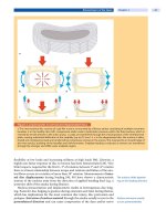
umn, can be absent in the early stages of AS (
Fig. 1). Imaging studies of the spine
are essential to:
make the diagnosis of AS
exclude fractures, spondylolisthesis or Andersson lesions
assess sagittal imbalance
monitor progress of the disease
assess the treatment effect
Signs of acute inflammation
and bony alterations can
be absent in early stages
Clinical examinations are complemented by various imaging studies (X-ray, CT,
MRI and bone scan). However, whole-body MR imaging will more and more be
used to monitor inflammatory spinal lesions at an early or an active stage of dis-
ease. The possibility of evaluating shoulder and hip joints together with the axial
skeleton is the major advantage of whole-body MRI [105].
Standard Radiographs
Standard radiography
remains the mainstay
of diagnostic imaging
Standard radiographs of the spine and sacroiliac joints (SIJs) remain the main-
stay of diagnostic imaging for AS (
Fig. 1a).
The hallmark of AS is a sacroiliitis and at a later stage ankylosis of the SIJs
(
Fig. 1a–c). Radiologic alterations of the SIJs are differentiated by the modified
New York classification [97] into four grades (
Table 2).
Ankylosing Spondylitis Chapter 38 1063


