medical biochemistry human metabolism in health and disease
Bạn đang xem bản rút gọn của tài liệu. Xem và tải ngay bản đầy đủ của tài liệu tại đây (20.72 MB, 439 trang )
M
ED
I
CAL
B
I
OC
H
E
M
I
ST
RY
This Page Intentionally Left Blank
M
E
D
I
CAL
B
I
OC
H
E
M
I
STRY
Human Metabolism in
Health and Disease
MIRIAM
D.
ROSENTHAL
ROBERT
H.
GLEW
@
WILEY
A
JOHN WILEY
&
SONS, INC., PUBLICATION
Copyright
0
2009 by John Wiley
&
Sons, Inc. All rights reserved.
No
part of this publication may be reproduced, stored in a retrieval system,
or
transmitted in
any
form
or
by any means, electronic, mechanical, photocopying, recording, scanning, or otherwise, except
as
permitted under Section 107
or
108 of the 1976 United States Copyright Act, without either the prior
written permission of the Publisher,
or
authorization through payment of the appropriate per-copy fee to
the Copyright Clearance Center, Inc., 222 Rosewood Drive, Danvers, MA
0
1923,978-750-8400,
fax 978-750-4470,
or
on the web at www.copyright.com. Requests to the Publisher for permission
should be addressed to the Permissions Department, John Wiley
&
Sons, Inc.,
11
1
River Street, Hoboken,
NJ 07030,201 -748-601
1,
fax 201-748-6008,
or
online at
Limit of Liability/Disclaimer of Warranty: While the publisher and author have used their best efforts in
preparing this book, they make no representations
or
warranties with respect
to
the accuracy
or
completeness of the contents of this book and specifically disclaim any implied warranties of
merchantability
or
fitness for
a
particular purpose.
No
warranty may be created
or
extended by sales
representatives
or
written sales materials. The advice and strategies contained herein may not he suitable
for
your
situation.
You
should consult with a professional where appropriate. Neither the publisher nor
author shall be liable for any
loss
of profit or any other commercial damages, including but not limited to
special, incidental, consequential, or other damages.
For
general information on
our
other products and services or for technical support, please contact our
Customer Care Department within the United States at 877-762-2974, outside the United States at
317-572-3993
or
fax
317-572-4002.
Wiley
also
publishes its books in
a
variety
of
electronic formats. Some content that appears in print may
not be available in electronic format.
For
more information about Wiley products, visit
our
web site at
www.wiley.com
Library
of
Congress Cataloging-in-Publication Data:
Rosenthal, Miriam D.
Miriam D. Rosenthal and Robert
H.
Glew.
Medical biochemistry
:
Human metabolism in health and disease
/
p.
;
cm.
Includes index.
ISBN
978-0-470-1 2237-2
[DNLM:
1.
Metabolism.
2.
Metabolic Diseases. QU I20 R8 1st 20091
QP171.R65 2009
612.3’94~22 2008029609
1.
Metabolism. 2. Metabolism-Disorders.
I.
Glew, Robert H.
11.
Title.
Printed in the United States of America
10
9
8
7 6
5
4
3 2
I
CONTENTS
PREFACE
ACKNOWLEDGMENTS
THE AUTHORS
1
INTRODUCTION TO METABOLISM
2
ENZYMES
vii
ix
xi
1
11
3 DIGESTION AND ABSORPTION 38
4
GLYCOLYSIS 58
5
PYRUVATE DEHYDROGENASE AND THE TRICARBOXYLIC
ACID CYCLE 77
6
ELECTRON TRANSPORT AND OXIDATIVE
PHOSPHORYLATION
89
7 THE PENTOSE PHOSPHATE PATHWAY 102
8
GLYCOGEN 112
9
GLUCONEOGENESIS 126
10 FATTY ACID OXIDATION AND KETONES 141
11
FATTY ACID SYNTHESIS 162
12 TRIACYLGLYCEROL TRANSPORT AND METABOLISM 177
V
vi
CONTENTS
13
14
15
16
17
18
19
20
21
22
23
24
25
ETHANOL
PHOSPHOLIPIDS AND SPHINGOLIPIDS
EICOSANOIDS
GLYCOLIPIDS AND GLYCOPROTEINS
CHOLESTEROL SYNTHESIS AND TRANSPORT
STEROIDS AND BILE ACIDS
NITROGEN HOMEOSTASIS
AMINO ACIDS
SULFUR AMINO ACID METABOLISM
FOLATE AND VITAMIN
612
IN ONE-CARBON METABOLISM
PURINES AND PYRIMIDINES
HEME AND IRON
INTEGRATION OF METABOLISM
191
199
21 8
231
246
271
290
305
325
335
351
372
393
INDEX
41 1
PREFACE
Human metabolism is a key component of the basic science knowledge that un-
derlies the practice of medicine and allied health professions. It is fundamental
to
understanding how the body adapts to physiologic stress, how defects in metabolism
result in disease, and why data from the clinical chemistry laboratory are useful
to
diagnose disease and monitor the efficacy of treatment. Over the more than three
decades that each of the authors has been teaching biochemistry to medical students,
we have found students increasingly overwhelmed with details that tend to obscure
rather than elucidate principles of human metabolism.
Our main aim in writing this book was to provide students in the health pro-
fessions with a concise resource that will help them understand and appreciate the
functions, constituent reactions, and regulatory aspects of the core pathways that
constitute human metabolism and which are responsible for maintaining homeosta-
sis and well-being in humans. We have tried to accomplish this by emphasizing
function, regulation, and disease processes, while minimizing discussion of reaction
mechanisms and details of enzyme structure.
Each chapter is organized in a consistent manner beginning with an explanation of
the main functions of the pathway under discussion. Next comes a brief accounting
of the cells, tissues, and organs in which the pathway is expressed and the conditions
under which the normal function of the pathway is especially important. The bulk
of each chapter is devoted to the reactions that account for the function of the
pathway, with emphasis on key steps in the pathway. The next section of each chapter
discusses the ways in which the activity of the pathway is regulated by hormones,
genetic factors, or changes in the intracellular concentration of key metabolites. Each
chapter concludes with a discussion of the more common and illustrative diseases
that result from defects in or derangements
of
regulation of the pathway.
vii
Viii
PREFACE
This volume is deliberately modest in size. Instead of providing exhaustive cover-
age of all the reactions that human cells and tissues are capable of executing, we have
chosen examples that illustrate the physiologic and pathophysiologic significance
of the topic. The authors’ expectation is that each chapter will be read for com-
prehension rather than to provide abundant fact and detail. During their subsequent
education and professional careers, the readers will undoubtably have need for more
information on many topics discussed in this book. We hope that this book will pro-
vide them with the tools to comprehend and appreciate the detailed resources-both
print and electronic-that contain the ever-expanding body of knowledge on human
metabolism in health and disease.
MIRIAM
D.
ROSENTHAL
ROBERT
H.
GLEW
ACKNOWLEDGMENTS
We are grateful to our colleagues and friends who generously devoted time to reading
selected chapters and provided the authors with invaluable feedback: William L.
Anderson, Suzanne
E.
Barbour, Alakananda Basu, David G. Bear, Edward
J.
Behrman, Frank
J.
Castora, Anca
D.
Dobrian, Diane M. Duffy, Venkat Gopalan,
Maurice Kogut, William Lennarz, Robert B. Loftfield, Gerald
J.
Pepe,
Karl
A.
Schellenberg, David
L.
Vanderjagt, Dorothy
J.
Vanderjagt, and Howard D. White.
A
special thanks to Mary
H.
Hahn and Charles D. Varnell, Jr., at Eastern Virginia
Medical School, who provided the students’ perspective of the book, for their insights
on
clarity and accessibility. We also appreciate the perceptive critiques provided by
the University of New Mexico Medical School class of
201
1.
The authors are indebted to Lucy Hunsaker, who drafted the figures. Her uncom-
mon
patience and good judgment in making the many revisions required
to
get the
figures into final form are greatly appreciated.
We also thank the helpful people at John Wiley
&
Sons: Darla Henderson
who
championed our initial proposal, and Michael Foster, Rebekah Amos, Anita
Lekhwani, and Rosalyn Farkas who shepherded the book all the way to publication.
ix
This Page Intentionally Left Blank
THE
AUTHORS
Miriam
D.
Rosenthal,
Ph.D., is Professor of Biochemistry at Eastern Virginia Med-
ical School. She received her B.A. in biology from Swarthmore College in 1963,
followed by M.S. (1968) and Ph.D. (1974) degrees in biology from Brandeis Uni-
versity. Since 1977, Dr. Rosenthal has developed curricula, provided instruction, and
conducted assessment of medical and other health professions students in biochern-
istry, molecular biology, cell biology, and human genetics. She has served
as
Course
Director of Medical Biochemistry since 1997.
Robert
H.
Glew,
Ph.D., is Emeritus Professor of Biochemistry and Molecular Biol-
ogy at the University of New Mexico School of Medicine, where he was chair of the
department from 1990 to 1998. He received
a
B.S. in food science from the Univer-
sity
of
Massachusetts, Amherst in 1962,
M.S.
in nutrition and food science from the
Massachusetts Institute of Technology in 1964, and Ph.D. in biochemistry from the
University of California, Davis in 1968. Dr. Glew has taught medical biochemistry
at half
a
dozen medical schools and teaching hospitals in the United States and West
Africa.
Drs. Rosenthal and Glew previously coedited
Clinical Studies in Medical Biochem-
istry
(3rd ed., 2006, Oxford University Press, New York). The book uses case pre-
sentations to develop the contextual basis of human metabolism, nutrition, and the
molecular bases of disease.
xi
This Page Intentionally Left Blank
CHAPTER
1
INTRODUCTION TO METABOLISM
1.1
INTRODUCTION
Intermediary metabolism
is the name given to the sequences
of
biochemical reac-
tions that degrade, synthesize, or interconvert small molecules inside living cells.
Knowledge of the core metabolic pathways and their interrelations is critical
to
un-
derstanding both normal function and the metabolic basis of most human diseases.
Rational interpretation and application of data from the clinical chemistry laboratory
also requires a sound grasp of the major metabolic pathways. Furthermore, knowl-
edge of key biochemical reactions in the two dozen or
so
core metabolic pathways
in
humans is essential for an understanding
of
the molecular basis of drug action,
drug
interactions, and the many genetic diseases that are caused by the absence of
the activity of a particular protein or enzyme.
1.1.1
Metabolic Pathways
Metabolism occurs in small discrete steps, each
of
which is catalyzed by
an
enzyme.
The term
metabolic
pathway
refers to a particular set of reactions that carries out a
certain function or functions. The pathway
of
gluconeogenesis or glucose synthesis,
for
example, operates mainly during a period
of
fasting, and its primary function is
to maintain the concentration of glucose in the circulation at levels that are required
by
glucose-dependent tissues such as the brain and red blood cells. Another example
of a metabolic pathway is the tricarboxylic acid (TCA) cycle, which oxidizes the two
Medical Biochemistry: Human
Metabolisni
in
Health and Disease
Copyright
0
2009
John Wiley
&
Sons,
Inc.
By Miriam
D.
Rosenthal and Robert
H.
Glew
1
2
INTRODUCTION
TO
METABOLISM
carbons of acetyl-coenzyme A (acetyl-CoA) to CO2 and water, thus completing the
catabolism of carbohydrates, fats (fatty acids), and proteins (amino acids).
1.1.2 Metabolic Intermediates
Biochemical pathways are comprised of organic compounds called
metabolic inter-
mediates,
all of which contain carbon, hydrogen, and oxygen. Some metabolic in-
termediates also contain nitrogen or sulfur.
In
most instances, these compounds
themselves have
no
function. An exception would be a compound such as citric acid,
which is both an intermediate in the TCA cycle and a key regulator of other pathways,
including oxidation of glucose (glycolysis) and gluconeogenesis.
1.1.3 Homeostasis
Homeostasis
refers to an organism’s tendency or drive to maintain the normalcy of
its internal environment, including maintaining the concentration of nutrients and
metabolites within relatively strict limits.
A
good example is glucose homeostasis.
In
the face of widely varying physiological conditions, such as fasting or exercise, both
of which tend to lower blood glucose, or following the consumption of a carbohydrate
meal that raises the blood glucose concentration, the human body activates hormonal
mechanisms that operate to maintain blood glucose within rather narrow limits,
80
to
100
mg/dL (Fig.
1-1).
Hypoglycemia (low blood glucose) stimulates the release
of
gluconeogenic hormones such as glucagon and hydrocortisone, which promote
the breakdown of liver glycogen and the synthesis of glucose in the liver (gluconeo-
genesis), followed by the release of glucose into the blood.
On
the other hand,
hyperglycemia (elevated blood glucose) stimulates the release of insulin, which pro-
motes the uptake of glucose and its utilization, storage as glycogen, and conversion
to fat.
Maintenance of the blood calcium concentration between strict limits is another
example of homeostasis. The normal total plasma calcium concentration is in the
range
8.0
to
9.5
mg/dL. If the calcium concentration remains above the upper limit
of
normal for an extended period of time, calcium may deposit, with pathological
con-
sequences in soft tissues such as the heart and pancreas. Hypocalcemia (a.k.a. tetany)
can result in muscle paralysis, convulsions, and even death; chronic hypocalcemia
causes rickets in children and osteomalacia in adults. The body uses vitamin
D
and certain hormones (e.g., parathyroid hormone, calcitonin) to maintain calcium
homeostasis.
1.2 WHAT
DO
METABOLIC PATHWAYS ACCOMPLISH?
1.2.1 Generation
of Energy
The primary dietary fuels for human beings are carbohydrates and fats (triacyl-
glycerols). The human body also obtains energy from dietary protein and-for some
WHAT
DO
METABOLIC PATHWAYS ACCOMPLISH?
3
200
180
160
2
140
E
s
h
v
g
120
-
g
100
3
w
P
-
m
80
60
Hypoglycemia
Fasting
0
123
4
Hours
.t
Carbohydrate
intake
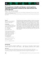
FIGURE
1-1
Changes that occur in the blood glucose concentration in a healthy adult,
a
person with type
I1
diabetes mellitus, and a person experiencing fasting hypoglycemia.
Following ingestion of a carbohydrate-containing meal, there are three features that distinguish
the glucose vs. time curve for the person with type
I1
diabetes relative to the healthy adult:
(1) the initial blood glucose concentration is higher (approx. 135 vs.
90
mg/dL), (2) the rise
in in the glucose level following the meal is greater; and (3) it takes longer for the glucose
concentration to return to the fasting glucose level.
people+thanol. Metabolism of these fuels generates energy, much of which is cap-
tured as the high-energy molecule adenosine triphosphate (ATP) (Fig.
1-2).
The ATP
can be used for biosynthetic processes (e.g., protein synthesis), muscle contraction,
and
active transport of ions and other solutes across membranes.
1.2.2
Degradation or Catabolism of Organic
Molecules
Catabolic pathways usually involve cleavage
of
C-0,
C-N,
or
C-C
bonds. Most
intracellular catabolic pathways are oxidative and involve transfer of reducing equiv-
alents (hydrogen atoms) to nicotinamide-adenine dinucleotide (NADf)
or
flavine-
adenine dinucleotide (FAD). The reducing equivalents in the resulting
NADH
or
4
INTRODUCTION
TO METABOLISM
OH
OH
FIGURE
1-2
Structure
of
adenosine triphosphate.
FADH2 can then be used in biosynthetic reactions or transferred to the mitochondria1
electron-transport chain for generation
of
ATP.
1.2.2.1 Digestion.
Before dietary fuels can be absorbed into the body, they must
be broken down into simpler molecules. Thus, starch is hydrolyzed to yield glucose,
and proteins are hydrolyzed to their constituent amino acids.
1.2.2.2
Glycolysis.
Glycolysis is the oxidation of glucose into the three-carbon
compound pyruvic acid.
1.2.2.3 Fatty Acid Oxidation.
The major route of fatty acid degradation is
P-oxidation, which accomplishes stepwise two-carbon cleavage of fatty acids into
acetyl-Co
A.
1.2.2.4 Amino Acid Catabolism.
Breakdown
of
most
of
the
20
common amino
acids is initiated by removal of the a-amino group of the amino acid via transamina-
tion. The resulting carbon skeletons are then further catabolized to generate energy or
are used
to
synthesize other molecules (e.g., glucose, ketones). The nitrogen atoms of
amino acids can be utilized for the synthesis of other nitrogenous compounds, such
as heme, purines, and pyrimidines. Excess nitrogen is excreted in the form
of
urea.
1.2.3
Synthesis
of
Cellular Building Blocks
and
Precursors
of
Macromolecules
1.2.3. 1 Gluconeogenesis: Synthesis
of
Glucose.
This pathway produces
glucose from glycerol, pyruvate, lactate, and the carbon skeletons of certain (gluco-
genic) amino acids. Gluconeogenesis is crucial to maintaining an adequate supply of
glucose to the brain during fasting and starvation.
1.2.3.2 Synthesis
of
Fatty Acids.
Excess dietary carbohydrates and the carbon
skeletons of ketogenic amino acids are catabolized to acetyl-CoA, which is then
utilized for the synthesis of long-chain
(C16
and C18) fatty acids. Storage of these
fatty acids as adipocyte triacylglycerols provides the major fuel source during the
fasted state.
WHAT DO METABOLIC PATHWAYS ACCOMPLISH?
5
7.2.3.3 Synthesis of Heme.
Heme is a component of the oxygen-binding pro-
teins hemoglobin and myoglobin. Heme also functions as part of cytochromes, both
in the mitochondria1 electron transport chain involved in respiration-dependent ATP
synthesis and in certain oxidation-reduction enzymes, such as the microsomal mixed-
function oxygenases (e.g., cytochrome P450). Although most heme synthesis occurs
in
hemopoietic tissues (e.g., bone marrow), nearly all cells of the body synthesize
heme for their own cytochromes and heme-containing enzymes.
1.2.4
Storage
of Energy
Cells have only a modest ability to accumulate ATP, the major high-energy molecule
in human metabolism. The human body can store energy in various forms, described
below.
7.2.4.7 Creatine Phosphate.
Most cells, especially muscle, can store a limited
amount of energy in the form of creatine phosphate. This is accomplished by a
reversible process catalyzed by creatine kinase:
ATP
+
creatine
+
creatine phosphate
+
ADP
When a cell’s need for energy is at
a
minimum, the reaction tends toward the right.
By
contrast, when the cell requires ATP for mechanical work, ion pumping, or as
substrate in one biosynthetic pathway or another, the reaction tends to the left, thereby
making ATP available.
7.2-4.2 Glycogen.
Glycogen is the polymeric, storage form of glucose. Nearly
all
of the body’s glycogen is contained in muscle (approximately
600
g) and liver
(approximately
300
g), with small amounts in brain and type
I1
alveolar cells in
the lung. Glycogen serves two very different functions in muscle and liver. Liver
glycogen is utilized to maintain a constant supply of glucose in the blood. By contrast,
muscle glycogen does not serve as a reservoir for blood glucose. Instead, muscle
glycogen is broken down when that tissue requires energy, releasing glucose, which
is subsequently oxidized to provide energy for muscle work.
1.2.4.3 Fat or Triacylglycerol.
Whereas the body’s capacity to store energy
in the form of glycogen is limited, its capacity for fat storage
is
almost limitless.
After a meal, excess dietary carbohydrates are metabolized to fatty acids in the liver.
Whereas some of these endogenously synthesized fatty acids, as well as some of the
fatty acids obtained through the digestion of dietary fat, are used directly as fuel by
peripheral tissues, most of these fatty acids are stored in adipocytes in the form of
triacylglycerols. When additional metabolic fuel is required during periods of fasting
or exercise, the triacylglycerol stores in adipose are mobilized and the fatty acids are
made available to tissues such as muscle and liver.
6
INTRODUCTION TO METABOLISM
1.2.5 Excretion of Potentially Harmful Substances
7.2.5.7
Urea Cycle.
This metabolic pathway takes place in the liver and synthe-
sizes urea from the ammonia (ammonium ions) derived from the catabolism of amino
acids and pyrimidines. Urea synthesis is one of the body’s major mechanisms for
detoxifying and excreting ammonia.
7.2-5.2
Bile
Acid
Synthesis.
Metabolism of cholesterol to bile acids in the liver
serves two purposes:
(1)
it provides the intestine with bile salts, whose emulsifying
properties facilitate fat digestion and absorption, and (2) it
is
a
mechanism for dis-
posing of excess cholesterol. Humans cannot break open any of the four rings of
cholesterol, nor can they oxidize cholesterol to carbon dioxide and water. Thus, bil-
iary excretion of cholesterol-both
as
cholesterol per se and
as
bile salts-is the only
mechanism the body has for disposing of significant quantities of cholesterol.
7.2.5.3
Heme Catabolism.
When heme-containing proteins (e.g., hemoglobin,
myoglobin) and enzymes (e.g., catalase)
are
turned over, the heme moiety is oxi-
dized to bilirubin, which after conjugation with glucuronic acid is excreted via the
hepatobiliary system.
1.2.6 Generation
of
Regulatory Substances
Metabolic pathways generate molecules that play key regulatory roles. As indicated
above, citric acid (produced in the TCA cycle) plays
a
major role in coordinating
the activities of the pathways of glycolysis and gluconeogenesis. Another example
of a regulatory molecule is 2,3-bisphosphoglyceric acid, which is produced in a side
reaction off the glycolytic pathway and modulates the affinity of hemoglobin for
oxygen.
1.3 GENERAL PRINCIPLES COMMON TO METABOLIC PATHWAYS
1.3.1 ATP Provides Energy for Synthesis
Anabolic or synthetic pathways require input of energy in the form
of
the high-energy
bonds of ATP, which is generated directly during some catabolic reactions (such
as
glycolysis)
as
well
as
during mitochondria1 oxidative phosphorylation.
1.3.2 Many Metabolic Reactions Involve Oxidation or Reduction
During catalysis, oxidative reactions transfer reducing equivalents (hydrogen atoms)
to cofactors such
as
NAD+, NADP+ (nicotinamide-adenine dinucleotide phosphate)
or
FAD. Reduced NADH and FADH2 can then be used to generate ,4TP through
oxidative phosphorylation in mitochondria. NADPH is the main source of reducing
equivalents for anabolic, energy-requiring pathways such as fatty acid and cholesterol
synthesis.
GENERAL PRINCIPLES COMMON TO METABOLIC PATHWAYS
7
pKG-p-m
andKETONES
+
-
Fattyacid
-
f'
synthesis
t\
AMINO
I
ACIDS
1
/
Svnthesis
of
Fatty acid nonessential
amino acids
and ketogenesis
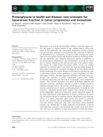
FIGURE
1-3
Possible interconversions of the three major metabolic
fuels
in
humans. Note
that
glucose
and
amino acids cannot
be
synthesized
from
(even-carbon) fatty acids.
1.3.3 Only Certain Metabolic Reactions Occur in Human Metabolism
It is important to appreciate that although humans possess the machinery to intercon-
vert many dietary components, not all interconversions are possible. Thus, humans
can convert glucose into long-chain fatty acids, but they cannot convert even-carbon-
numbered long-chain fatty acids into glucose (Fig.
1-3).
1.3.4 Some Organic Molecules Are Nutritionally Essential
to
Human Health
Certain key cellular components cannot be synthesized in the body and must therefore
be provided preformed in the diet and are therefore designated as
essential.
These
molecules include two polyunsaturated fatty acids (linoleic and a-linolenic) and the
carbon skeletons of some
of
the amino acids. They also include the vitamins (such
as
thiamine and niacin), most of which serve as components of enzymatic cofactors.
By contrast, other important compounds, such as glucose and palmitic acid, are not
essential in the diet. Glucose, whose blood levels are crucial to homeostasis, can be
synthesized from glycerol, lactate, pyruvate, and the carbon skeletons of glucogenic
amino acids when dietary glucose is not available.
1.3.5
Some Metabolic Pathways Are Irreversible or Contain
Irreversible Steps
One example of an irreversible pathway
is
glycolysis, the multistep catabolic pathway
that oxidizes glucose to pyruvate or lactate. Gluconeogenesis is essentially the reverse
of
glycolysis and is the process by which pyruvate (or a number
of
other molecules
such as lactate and the carbon skeleton of the amino acid alanine) can be used to
synthesized glucose. Although glycolysis and gluconeogenesis share many enzymes,
8
INTRODUCTION
TO
METABOLISM
specific gluconeogenic enzymes are required to bypass the steps in glycolysis that
are irreversible under physiological conditions.
1.3.6 Metabolic Pathways Are Interconnected
The initial step in glycolysis is the phosphorylation of glucose to form glucose
6-phosphate. Glucose 6-phosphate is
also
utilized in two other key metabolic path-
ways: glycogen synthesis and the pentose phosphate pathway (a.k.a. the hexose
monophosphate shunt), which generates ribose 5-phosphate and NADPH.
1.3.7 Metabolic Pathways Are Not Necessarily Linear
Both the tricarboxylic acid (TCA) cycle and the urea cycle are circular pathways. In
each case the pathway is initiated by addition of
a
small molecule to
a
key metabolic
intermediate (oxaloacetate in the TCA cycle and ornithine in the urea cycle). At the
end of one cycle, the key intermediate is regenerated and available to participate
in
another turn of the cycle. Although the TCA and urea cycles can be depicted
as
simple
circular pathways, metabolites can enter into-or be removed from-the pathways
at intermediate steps. For example, the amino acid glutamate can be used to generate
a-ketoglutarate,
a
key intermediate in the TCA cycle.
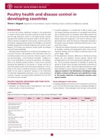
1.3.8 Metabolic Pathways Are Localized to Specific
Compartments Within the Cell
Many metabolic pathways occur within the mitochondria, including 6-oxidation of
fatty acids, the TCA cycle, and oxidative phosphorylation (Fig.
1-4).
Other pathways
are cytosolic, including glycolysis, the pentose phosphate pathway, and fatty acid
synthesis. Still others, including the urea cycle and heme synthesis, utilize both
mitochondria1 and cytosolic enzymes at different points in the pathways.
1.3.9 A Different Repertoire of Pathways Occurs in Different Organs
All cells are capable of oxidizing glucose to pyruvate via glycolysis to generate ATP.
However, since red blood cells lack mitochondria, they cannot further oxidize the
resulting pyruvate to COz and water via pyruvate dehydrogenase and the TCA cycle.
Instead, the pyruvate is converted to lactate and released from the red blood cells.
Most cells and organs can
also
utilize fatty acids
as
fuels. Although neural cells do
contain mitochondria, they do not oxidize fatty acids. The brain is therefore dependent
on
a
constant supply of glucose to provide energy. The gluconeogenesis pathway that
provides glucose for the brain occurs in the liver and to
a
lesser extent in the renal
cortex.
GENERAL PRINCIPLES COMMON TO METABOLIC PATHWAYS
9
Golgi complex
Glycoprotein oligosaccharide-
chain synthesis
Mitochondrion
Tricarhoxylic acid cycle
Fatty acid P-oxidation
*
Heme synthesis
Smooth endoplasmic
*
Urea synthesis
reticulum
Triacylglycerol synthesis
Mixed function oxygenases
Phospholipid synthesis
Rihosomes and rough
enplasmic reticulum
Protein synthesis
Lysosome
Peroxisome
Degradation
of
Bile salt synthesis glycosphingolipids,
Oxidation fatty acids
of
and very phytanic long-chain
3
acid Fatty Glycolysis acid synthesis macromolecules and other
Cytosol
mucopol ysaccharides,
Pentose phosphate pathway
*
Urea synthesis
*
Heme synthesis
FIGURE
1-4
A
liver cell, showing where various metabolic pathways occur.
An
asterisk
indicates
a
pathway, portions
of
which occur
in
more
than
one intracellular compartment.
1.3.10 Different Metabolic Processes Occur in the Fed State
Than During Fasting
After a meal, metabolic pathways are utilized to process the digested foods and store
metabolites for future utilization. Postprandially, glucose is plentiful and utilized both
for energy generation and to replenish glycogen stores (primarily in muscle and liver).
Excess glucose
is
metabolized to fatty acids in liver and fat cells and the resulting
triacylglycerols are stored in adipocytes.
By contrast, when a person is fasting there is a need to generate energy from
endogenous fuels. Consequently, the metabolic pathways involved in fuel metabolism
are
regulated in such a way as to promote the oxidation of stored fuels, including
the fatty acids stored in adipose tissue in the form of triacylglycerols and, to a lesser
extent, glycogen stored in liver and muscle.
In
fact, during a fast, most of the body's
energy needs are satisfied by the oxidation of fatty acids.
1.3.1
1
Metabolic Pathways Are Regulated
All this specialization of organs and coordination of metabolism in the fed or fasted
states
is
a highly regulated process with several levels
of
regulation.
One
level
of
10
INTRODUCTION TO METABOLISM
regulation is gene transcription and translation, which determines which enzymes are
actually present within a cell.
A
second level of control is substrate-level regulation,
whereby concentrations of key metabolites activate or inhibit enzymatic reactions.
A
metabolite that acts to regulate several pathways is citrate, which both inhibits
glycolysis and activates the first step in the pathway of fatty acid synthesis.
Hormones represent yet another level of control. Hormones act to coordinate
processes between the organs of complex, multicellular organisms. For example,
insulin, the main hormonal signal of the fed state, regulates both enzyme activity (at
the level of enzyme dephosphorylation) and gene transcription.
1.4
WHAT
IS
THE BEST WAY TO COMPREHEND AND RETAIN
A
WORKING KNOWLEDGE
OF
INTERMEDIARY METABOLISM?
Before learning about the various enzyme-catalyzed reactions and intermediates that
comprise a particular metabolic pathway, one should appreciate the major functions
which that pathway serves in the body and how the pathway relates to other pathways.
Particularly in the context of medical biochemistry, it is also important to understand
how the pathway is regulated and how it affects (or is affected by) disease processes.
As
you go through this book you will find that each chapter is organized
so
as to
answer the following questions:
1.
Why does the pathway exist? That is, what are its functions?
2.
Where does the pathway take place (i.e., what organ, tissue, cell. subcellular
compartment, or organelle)?
3.
When does the pathway operate, and when is it down-regulated: during the
fasted state or the fed state; during rest or extreme physical activity; during a
particular stage
of
development (e.g., the embryo, the neonate, old age)?
4.
What are the actual steps of the pathway, and what cofactors does it require?
5.
How is the pathway regulated?
6.
What can go wrong? Problems can include hormonal dysregulation (e.g., dia-
betes mellitus), inborn errors of metabolism (e.g., adrenoleukodystrophy), and
nutritional deficiencies (e.g., protein<alorie malnutrition, iron-deficiency ane-
mia). Normal metabolic homeostasis is also profoundly altered by toxins and
during infections.
CHAPTER
2
ENZYMES
2.1
THE
NATURE
OF ENZYMES
Enzymes are catalysts that greatly increase the rate of chemical reactions and thus
make possible the numerous and diverse metabolic processes that occur in the human
body. Catalysts increase the rate of a reaction without affecting its equilibrium.
Enzymes can increase the rate of physiological reactions by as much as 10"'-fold.
They accomplish this feat by decreasing the amount of energy required for activation
of the initial reactants (substrates), thereby increasing the percentage
of
substrate
molecules that have sufficient energy to react (Fig.
2-1).
With the exception of a few ribonucleic acid (RNA) molecules (ribozymes) that
catalyze reactions involving nucleic acids, enzymes are proteins. Every enzyme has an
active site that is composed of specific amino acid side chains which are brought into
close proximity when the enzyme is folded into its active conformation. During the
course of the reaction that it catalyzes, the enzyme's active site stabilizes the transition
state, which is an intermediate conformation between substrates and products. The
interaction between active site and substrate(s) is thus responsible for the catalytic
efficiency of the enzyme as well as its substrate specificity. After the reaction occurs,
the products are released from the enzyme and the active site is available to bind
additional substrate molecules.
Medical Biochemisrry: Human MPtaholism in Health and
Disease
By
Miriam
D.
Rosenthal and Robert
€1.
Glew
Copyright
0
2009
John
Wiley
&
Sons, Inc.
11
12 ENZYMES
I
x
f
w
Activation energy
(uncatal yzed)
Initial state
[substrate(s)]
Time
-
FIGURE
2-1
Activation energy
of
a chemical reaction.
2.2
TYPES
OF
ENZYMES
There are more than
2500
different enzymes in the human body. It is useful
to
group
them into six major classes based on the type
of
reaction they catalyze.
2.2.1
Oxidoreductases
Oxidative reactions remove electrons, usually one or two electrons per molecule
of
substrate, while reductive reactions accomplish the converse. The substrate in
an oxidation-reduction reaction may be a metal,
as
in the case
of
the one-electron
oxidation
of
the ferrous ion of hemoglobin to the ferric ion of methemoglobin, or an
organic compound
as
illustrated by the two-electron, reversible oxidation of lactate
to pyruvate.
Oxidoreductases transfer electrons from one compound to another, thus changing
the oxidation state of both substrates. Some oxidoreductases, such
as
lactate de-
hydrogenase, catalyze the removal of two hydrogen atoms
(two
electrons plus
two
hydrogen ions) to an acceptor molecule such as nicotinamide-adenine dinucleotide
(NADf) as illustrated by the lactate dehydrogenase reaction (Fig.
2-2):
lactate
+
NAD+
+
pyruvate
+
NADH
+
H'