Chapter 088. Hepatocellular Carcinoma (Part 9) pot
Bạn đang xem bản rút gọn của tài liệu. Xem và tải ngay bản đầy đủ của tài liệu tại đây (44.14 KB, 5 trang )
Chapter 088. Hepatocellular
Carcinoma
(Part 9)
Table 88-
6 Some Novel Medical Treatments for Hepatocellular
Carcinoma
EGF receptor antibody
Erlotinib, Gefitinib
Kinase antagonists, Sorafenib
Vitamin K
IL-2
131
I – ethiodol (Lipiodol)
131
I – Ferritin
90
Yttrium microspheres
166
Holmium
Three-dimensional conformal radiation
Proton beam high-dose radiotherapy
Anti-angiogenesis strategies, Bevacizumab
Note: EGF, epidermal growth factor; IL, interleukin.
Summary
Most Common Modes of Patient Presentation
1. A patient with known history of hepatitis, jaundice, or
cirrhosis, with an abnormality on ultrasound or CT scan, or rising AFP or
DCP (PIVKA-2)
2. A patient with an abnormal liver function test as part of a
routine examination
3. Radiologic workup for liver transplant for cirrhosis
4. Symptoms of HCC including cachexia, abdominal pain, or
fever.
History and Physical Examination
1. Clinical jaundice, asthenia, itching (scratches), tremors, or
disorientation
2. Hepatomegaly, splenomegaly, ascites, peripheral edema, skin
signs of liver failure.
Clinical Evaluation
1. Blood tests: full blood count (splenomegaly), liver function
tests, ammonia levels, electrolytes, α-fetoprotein and DCP (PIVKA-2), Ca
2+
and Mg
2+
; hepatitis B and C serology (and quantitative HBV DNA or
HCV RNA, if either is positive); neurotensin (specific for fibrolamellar
HCC)
2. Triphasic dynamic helical (spiral) CT scan of liver (if
inadequate, then follow with an MRI); chest CT scan; upper and lower
gastrointestinal endoscopy (for varices, bleeding, ulcers); and brain scan
(only if symptoms suggest)
3. A core biopsy: of the tumor and separately of the underlying
liver.
Therapy
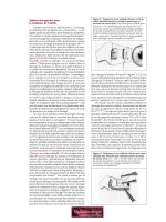
(See also Fig. 88-1)
1. HCC < 2 cm: RFA ablation, PEI, or resection
2. HCC > 2 cm, no vascular invasion: liver resection, RFA, or
OLTX
3. Multiple unilobar tumors or tumor with vascular invasion:
TACE
4. Bilobar tumors, no vascular invasion: TACE with OLTX for
patients whose tumors have a response
Extrahepatic HCC or elevated bilirubin: Phase I and II studies.