Báo cáo nghiên cứu khoa học: "THEORETICAL STUDY OF THE SPIN DENSITY DISTRIBUTION OF THE GREEN TEA CATECHIN RADICALS" doc
Bạn đang xem bản rút gọn của tài liệu. Xem và tải ngay bản đầy đủ của tài liệu tại đây (423.13 KB, 9 trang )
TẠP CHÍ PHÁT TRIỂN KH&CN, TẬP 9, SỐ11 -2006
Trang 69
THEORETICAL STUDY OF THE SPIN DENSITY DISTRIBUTION OF THE
GREEN TEA CATECHIN RADICALS
Pham Thanh Quan, Le Thanh Hung, Tran Thi Ha Thai
University of Technology, VNU-HCM
(Manuscript Received on June 20
th
, 2006, Manuscript Revised November 2
nd
, 2006)
ABSTRACT: The geometries and spin densities of green tea catechin radicals are
calculated using unrestricted B3LYP hydrid density functional calculations. The radicals which
have the smallest value of Maximum Spin Density (MSD) are referred to the most stable
radical species deriving from the absolute minimum energy of investigated compounds. In this
study, we have calculated these values in gas phase and water solution. For green tea
catechins, their stable radicals have MSD values lying at 0.37 – 0.38 in gas phase, and 0.33-
0.34 in water. Calculating some flavonoid stable radicals, it was found that the MSD values lie
at 0.31 – 0.37 in gas phase, and 0.29 – 0.37 in water solution. These calculated data were
compared with experimental data and were found to be in good agreement in predicting the
stability of radicals. On the basic of the computed MSD values, the stability of radicals can be
explored and give a relative trend of the activity and scavenging of antioxidant radicals.
Key words: antioxidants, phenolics, flavonoids, catechins, spin density
1. INTRODUCTION
Tea (Camellia sinensis) is believed to have a wide range of pharmaceutical properties
including being antihypertensive, antioxidative, antiarteriosclerotic, anticarcinogenic and
hypochlolesterolemic. These diverse biological activities are thought to be attributed to a group
of polyphenol compounds, namely green tea catechins (GTCs), present in tea leaves. The
content of GTCs varies among green tea, black tea, and oolong tea. Green tea refers to a
nonfermented product in which GTCs are mostly preserved while black tea is oxidized during
manufacturing process. Oolong tea is a partially fermented product in which GTCs are partially
degraded [1].
The yield of crude GTC extracts was 7.4% of dry tea leaves and it mainly consisted of
51.2% (-) epigallocatechingallate (EGCG), 18.7% (-) epigallocatechin (EGC), 12.3% (-)
epicatechin (EC) and 11.8% (-) epicatechin gallate (ECG) [2]. Several studies have suggested
that the GTC extracts exhibited strong antioxidative effect.
Green tea polyphenols, i.e., EC, EGC, ECG, and EGCG belong to flavonoid. The basic
flavonoid structure is the flavan nucleus, which consists of 15 carbon atoms arranged in three
rings (C
6
-C
3
-C
6
), which are labeled A, B, and C (Figure 1).
The function of antioxidants is to intercept and react with the free radicals at a rate faster
than the substrate, and since free radicals are able to attack a variety of targets including lipids,
fats, and proteins, it is believed that they are implicated in number of important degenerative
diseases including aging itself.
There are two pathways for oxidation in which antioxidants can play a preventive role. The
first is H-atom transfer, illustrated below for the important case of lipid peroxidation [6, 7, 11]:
RH
→ R
.
(initiation) (1)
R
.
+ O
2
→ RO
2
.
(addition of O
2
) (2)
RO
2
.
+ RH
→ RH + R
.
(H-atom exchange) (3)
Science & Technology Development, Vol 9, No.11- 2006
Trang 70
Once a free radical R
.
has been generated, then reaction 2 and 3 form a chain reaction. As
the chain cycles through (2) and (3) many lipid molecules (R-H) are converted into lipid
hydroperoxide (ROOH), resulting in oxidation and rancidity of fats. Reaction 2 is very fast, ca.
10
9
M
-1
s
-1
, whereas (3) is much slower, typically 10
1
M
-1
s
-1
.
For the phenolic antioxidant (ArOH), the role of the antioxidant is to interrupt the chain
reaction according to
RO
2
.
+ ArOH
→ ROOH + ArO (4)
To be effective ArO
.
must be a relatively stable free radical, so that it reacts slowly with
substrate RH but rapidly with ROO
.
, hence the term “chain-breaking-antioxidant”.
The rate of reaction of ArOH with peroxyl radicals depends on the barrier height for
transfer of an H-atom from ArOH. It is clear that the Bond Dissociation Enthalpy (BDE) in
ArOH will be an important factor in determining the antioxidant capacity, since the weaker the
OH bond the faster will be the reaction with free radical.
Another possible mechanism by which an antioxidant can deactivate a free radical is
electron transfer:
ROO
.
+ ArOH
→ ROO
-
+ ArOH
.+
Again, the radical cation arising from the electron transfer must be stable, so it does not
react with substrate molecules. In this case, the ionization potential (IP) is the most significant
energetic factor for the scavenging activity evaluation.
In this work, we would like to introduce another parameter which can be used to predict the
stable radicals through calculating the spin density distribution. Spin density is the upaired
electron density at a position of interest, usually at carbon, in a radical. The electron density
ρ(1) at the position r
1
can be described as a sum of a density with α and β spin:
ρ(1) = ρ
α
(1) + ρ
β
(1)
(
ρ
α
(1), ρ
β
(1) corresponds to the probability density of finding an electron with α and β
spin at the position r
1
)
The radical will be stable as the spin densities distribute over radical structure. This is
synonym with the maximum spin density – MSD at every atom of radical is small. At the
doublet state, sum of spin densities is 1.
In this paper, we have investigated at the density functional level of the conformation of
four catechins: EC, ECG, EGC, and EGCG to predict activity of flavonoids by the MSD values
2. COMPUTATIONAL METHODS
All of the calculations reported in this study were performed using the Gaussian03 code [4].
The B3LYP exchange correlation potential was used for optimizing geometries in connection
with 3-21G* basic set. Harmonic vibrational frequencies were computed at HF/3-21G*. Single
point energy refinement on the 3-21G* optimized geometries was performed with use of the 6-
311++G** basic set.
The unrestricted open-shell approach was used for radical species. Spin contamination was
found in accepted limit for radicals, being the <s
2
> values about 0.75-0.78 in all cases.
Solvent (water) effects were computed in the framework of the self-consistent reaction field
polarized continuum model (SCRF-PCM) implemented on the Gaussian03 package, using the
UAHF set of solvation radii to build the cavity for the solute, in the gas equilibrium geometries.
TẠP CHÍ PHÁT TRIỂN KH&CN, TẬP 9, SỐ11 -2006
Trang 71
O
H
H
HO
OH
OH
OH
OH
3'
4'
2'
5'
6'
Epicatechin (EC)
45
7
6
8
3
2
O
H
H
HO
OH
OH
OH
OH
3'
4'
2'
5'
6'
Epigallocatechin (EGC)
45
7
6
8
3
2
OH
O
H
H
O
HO
OH
OH
OH
3'
4'
2'
5'
6'
Epicatechin gallate (ECG)
45
7
6
8
3
2
C
O
OH
OH
OH
O
H
H
O
HO
OH
OH
OH
3'
4'
2'
5'
6'
Epigallocatechin gallate (EGCG)
45
7
6
8
3
2
C
O
OH
OH
OH
OH
(A)
(B)
(C)
(B)
(B)
(B)
(A)
(A)
(A)
(C)
(C)
(C)
ψ
ψ
ψ
ψ
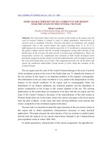
Figure 1. Structures of (-) epicatechin (EC), epicatechin gallate (ECG), epigallocatechin (EGC), and
epigallocatechin gallate (EGCG).
3.RESULTS AND DISCUSSION
The investigated compounds are depicted in Figure 1.
For clarity we will discuss separately the conformational properties and the relative
stabilities of radicals for each system and the MSD trend.
3.1.Conformations and Radical Stabilities
Green tea catechins represent the most common and active edible antioxidants. The
antioxidant ability was related to the number and mutural position of hydroxyl groups and to
conjugation and resonance effects [3].
Epicatechin (EC)
EC contains five phenolic groups (Figure 1) but the 3-OH group on ring C is an alcoholic
group to which has no antioxidant ability. The delocalization of the unpaired electron,
conjugation effects were determined by a dihedral angle C
3
-C
2
-C
1’
-C
2’
(ψ). The values of ψ
were from 71.81 to 82.77
o
(see Table 1). These values indicated that for EC there is no
possibility of conjugation between the rings, due to the saturation of the C ring.
Upon radicalization, EC can give four radicals of which relative energies were within
10kcal/mol (see Table 2). The most stable radical was the radical 4’-OH with the torsion angle
ψ was 82.77
o
. The 5-OH, 7-OH, and 3’-OH radicals lied at 4.76, 5.71, and 9.62 kcal/mol above
the 4’-OH.
Epigallocatechin (EGC)
EGC has the same EC structure but has three phenolic groups on B ring. EGC can give five
active radicals of which relative energies are within 10kcal/mol. The delocalization of the
unpaired electron, conjugation effects of EGC were stronger than those of EC radicals, the
values of
ψ were from 71.36 to 106.15
0
.
The most stable radical was the radical 3’-OH, close in energy to the 4’-OH one (0.00 and
0.94 kcal/mol). The dihedral angle
ψ of 106.15
o
was bigger than other EGC species. The other
isomers, generated by the loss the hydrogen atom from the 5’-OH, 5-OH, and 7-OH groups,
were found at 8.03, 5.05, and 6.79 kcal/mol, respectively (see Table 1 and Table 2).
Science & Technology Development, Vol 9, No.11- 2006
Trang 72
Table 1. Structures and total energies for both catechin radicals in gas phase. All calculation at
b3lyp/6-311++g**//b3lyp/3-21g*, freq at hf/3-21g*
Compound E (HF) ZPE E
total
ψ
5-OH -1031.0039214 0.275172 -1030.7562666 71.84
7-OH -1031.0010950 0.273713 -1030.7547533 71.81
3-OH -1030.9686978 0.276691 -1030.7196759 71.22
3’-OH -1030.9952564 0.274152 -1030.7485196 76.88
Epicatechin
(EC)
4'-OH -1031.0111722 0.274794 -1030.7638576 82.77
5-OH -1601.2330409 0.389749 -1600.8822668 99.10
7-OH -1601.2311256 0.389881 -1600.8802327 101.10
3'-OH -1601.2342982 0.389243 -1600.8839795 105.67
4'-OH -1601.2424672 0.388603 -1600.8927245 92.53
3''-OH -1601.2264416 0.389771 -1600.8756477 100.45
4''-OH -1601.2388684 0.390108 -1600.8877712 99.38
Epicatechin gallate
(ECG)
5''-OH -1601.2358019 0.388542 -1600.8861141 143.48
5-OH -1106.2526477 0.279607 -1106.0010014 71.41
7-OH -1106.2499998 0.279751 -1105.9982239 72.60
3-OH -1106.2203770 0.283265 -1105.9654385 71.36
3'-OH -1106.2609896 0.279937 -1106.0090463 106.15
4'-OH -1106.2599331 0.280420 -1106.0075551 78.79
Epigallocatechin
(EGC)
5'-OH -1106.2478936 0.279597 -1105.9962563 74.27
5-OH -1676.4834357 0.391586 -1676.1310083 91.52
7-OH -1676.4811357 0.391702 -1676.1286039 92.04
3'-OH -1676.4920751 0.392247 -1676.1390528 85.62
4'-OH -1676.4915579 0.392508 -1676.1383007 97.56
5'-OH -1676.4768603 0.391379 -1676.1246192 99.29
3''-OH -1676.4753531 0.391478 -1676.1230229 91.63
4''-OH -1676.4884617 0.392062 -1676.1356059 93.56
Epigallocatechin
gallate
(EGCG)
5''-OH -1676.4872895 0.391961 -1676.1345246 94.29
Epicatechin gallate (ECG)
ECG is a gallate ester moiety at the 3-position of EC, concludes 3,4,5-trihydroxyphenyl
group. This has effects on torsion angle
ψ and makes ECG have stranger properties than EC.
The values of
ψ were from 92.53 to 105.67
0
(see Table 1). Upon radicalization, ECG can give
seven active radicals of which relative energies were within 10kcal/mol. In gas phase, the
radical 4’-OH was the most stable one with the minimum dihedral angle
ψ was 92.53
0
. At 6.56,
7.84, 5.49, 10.72, 3.11, and 4.15 kcal/mol above the global minimum, we found the 5-OH, 7-
OH, 3’-OH, 3’’-OH, 4’’-OH, and 5’’-OH species, respectively.
Epigallocatechin gallate (EGCG)
EGCG makes up about 40% of the total catechin content and is widely accepted as the
major antioxidant ingredient in green tea [5]. EGCG is a gallate ester moiety at the 3-position
of EGC, contains 3,4,5-trihydroxyphenyl group. EGCG has 8 hydroxy groups and can give 8
active radicals of which relative energies were within 10 kcal/mol.
In gas phase, the most stable radical was the 3’-OH one, practically isoenergetic with the
radical 4’-OH (0.00 and 0.47 kcal/mol, respectively). It was similar to ECG, the most stable
TẠP CHÍ PHÁT TRIỂN KH&CN, TẬP 9, SỐ11 -2006
Trang 73
radical of EGCG was correlative with the minimum torsion ψ (85.62
0
). The radical 4’’-OH was
close to the 5’’-OH one (2.16 and 2.84 kcal/mol). The radical 5’-OH, 7-OH, 5’-OH, and 3’’-
OH lied at 5.05, 6.56, 9.06, and 10.06 kcal/mol, respectively (see Table 2).
3.2.Correlation between MSD values and relative energies.
Table 2 reports the relative energy and the maximum spin density (MSD) values in the gas
phase and water solution for all green tea phenolics.
For green tea stable radicals, the MSD lied at 0.37 – 0.38 in gas phase. A correlation (r =
0.95) was found between MSD values and relative energies for catechin radicals in gas phase
(Figure 2). The radical that had minimum in energy had the small in MSD. It could be observed
that the stable radical correlative with the smallest value of MSD.
However, not all compounds followed this trend in water due to the effect of solvent. We
found that the correlation coefficient between MSD values and relative energies is 0.75 in
water. All green tea catechin radicals EC and EGC radicals were more stable in solution than in
gas phase but ECG and EGCG radicals were not. We thought that the presence of 3,4,5-
trihydroxyphenyl group in structure of ECG and EGCG caused stranger properties in water.
Example, for EGCG, it was slightly different: the most stable radical has the MSD value
close to minimum in gas phase and water. In gas phase, the radical 3’-OH is the most stable one
in energy but the radical 4’’-OH is the most stable one in spin density (value is 0.38 and 0.37,
respectively). In water solution, the radical 4’-OH is the most stable one in energy but the
radical 3’-OH is the most stable one in spin density (value is 0.35 and 0.34, respectively).
Table 2. Relative energies and max spin values for both catechin radicals in gas phase and
water solution. All calculation at b3lyp/6-311++g**//b3lyp/3-21g*
Gase phase Water solution
Compound
E
relative
(kcal/mol)
Max spin s
2
E
relative
(kcal/mol)
Max spin s
2
5-OH 4.76 0.444 0.784 2.30 0.458 0.779
7-OH 5.71 0.439 0.786 1.18 0.438 0.778
3-OH 27.72 0.891 0.750 172.40 0.858 0.754
3'-OH 9.62 0.454 0.782 0.70 0.377 0.774
EC
4'-OH 0.00 0.374 0.769 0.00 0.332 0.767
5-OH 6.56 0.466 0.785 10.44 0.491 0.780
7-OH 7.84 0.439 0.786 10.98 0.438 0.783
4'-OH 0.00 0.367 0.769 0.00 0.332 0.767
3'-OH 5.49 0.380 0.773 5.61 0.331 0.769
3''-OH 10.72 0.465 0.778 9.66 0.404 0.773
4''-OH 3.11 0.370 0.774 4.34 0.340 0.772
ECG
5''-OH 4.15 0.419 0.773 4.13 0.384 0.769
5-OH 5.05 0.444 0.784 8.01 0.458 0.779
7-OH 6.79 0.437 0.785 7.79 0.433 0.779
3-OH 27.36 0.875 0.754 30.78 0.868 0.750
3'-OH 0.00 0.381 0.772 4.37 0.334 0.768
4'-OH 0.94 0.391 0.771 0.00 0.350 0.768
EGC
5'-OH 8.03 0.431 0.777 4.91 0.364 0.771
5'-OH 5.05 0.451 0.785 8.40 0.472 0.780
7-OH 6.56 0.438 0.786 8.81 0.433 0.779
3'-OH 0.00 0.382 0.771 5.32 0.342 0.768
4'-OH 0.47 0.396 0.771 0.00 0.353 0.768
5'-OH 9.06 0.442 0.779 5.31 0.400 0.773
3''-OH 10.06 0.469 0.779 8.06 0.405 0.773
4''-OH 2.16 0.374 0.774 3.12 0.355 0.772
EGCG
5''-OH 2.84 0.419 0.773 8.19 0.385 0.770
Science & Technology Development, Vol 9, No.11- 2006
Trang 74
0.20
0.30
0.40
0.50
0.60
0.00 2.00 4.00 6.00 8.00 10.00
E
rel
(kcal/mol)
MSD
Water solution
Gas phase
0.26
1.26
2.26
3.26
4.26
5.26
6.26
7.26
0.28 0.3 0.32 0.34 0.36 0.38 0.4
MSD
n
radical
/n
antioxidant
DPPH
Galvinoxyl
DPPH
Galvinoxyl
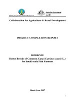
Figure 2. Correlation between MSD values and
relative energies in gas phase and water
solution. The correlation coefficient is 0.95 in
gas phase, 0.75 in water solution
Figure 4. Correlation between computed MSD and
experimental values. The correlation coefficient is -
0.93 for both DPPH radical and galvinoxyl radical.
3.3. Spin density and the activity of antioxidants
As mentioned before, phenolics can play their protective role by donating an H atom or
acting as electron donors. It is clear that as far as specific molecular properties are concerned,
the bond dissociation enthalpy (BDE) for the –OH bond and ionization potential (IP) are of
particular importance in deciding which the mechanism is the favored one for the radical
scavenging activity. Flavonoids with the dihydroxy functionality are the most active
compounds in donating an H atom, as confirmed by their low BDE and IP values [6, 7, 8].
Many studies in experiment showed that upon radicalization, the 4’-OH flavonoid radical
was the most stable radical. The antioxidant activity of flavonoid was represented by free
radical scavenging activity which is measured by the molar ratio (
n
radical
/n
atioxidant
). The bigger
molar ratio, the stronger antioxidant activity of flavonoid.
Calculating the MSD values, we found that all the values of MSD were referred to the most
stable radical species deriving from the minimum value of each antioxidant radical (see Table 1
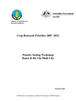
and Table 2). Then, we have calculated the MSD values of some 4’-OH flavonoid radicals
(Figure 3) in gas phase and water solution (see Table 3, 4). Our results were compared to
computed values [6, 7, 8] and experimental data [9, 10].
O
O
OH
OH
HO
OH
O
O
OH
OH
HO
OH
OH
OH
O
O
OH
OH
HO
OH
OH
O
O
OH
HO
OH
OH
O
O
OH
OH
HO
OH
OH
O
OOH
HO
OH
OH
keamferol quercetin myricetin
fisetin
taxifolin
epicatechin
O
OH
OH
HO
OH
OH
luteolin
morin
O
O
OH
OH
HO
OHHO
Figure 3. Structure of some studied flavonoids
TẠP CHÍ PHÁT TRIỂN KH&CN, TẬP 9, SỐ11 -2006
Trang 75
Table 3. Comparison between MSD values and BDE and IP values in gas phase for some 4’-
OH flavonoid radicals
(*)
. All MSD values were calculated at b3lyp/6-311++g**// b3lyp/3-21g*
Compound MSD (this work)
BDE (*)
kcal/mol
IP (*)
kcal/mol
Quercetin 0.31092 72.35 166.08
Fisetin
0.31042
- -
Luteolin 0.33199
74.54 174.44
Taxifolin 0.37441 74.73 182.82
Kaemferol 0.37454 80.94 167.99
Epicatechin 0.37395
73.72 170.85
Myricetin 0.32460 - -
Morin 0.37333
- -
(*) In reference 6, 7, 8
From Table 3, it could be observed that the compound of which stable radical has the low
MSD, BDE and IP value. Our results were in good agreement with other computed values:
quercetin could to be good candidate for active antioxidants.
Table 4. Comparison between the MSD values of 4’-OH flavonoid radicals and Free radical
scavenging activity (
n
radical
/n
atioxidant
) (**). All computed MSD values at b3lyp/6-311++g**//
b3lyp/3-21g*
Free radical scavenging activity (**)
n
radical
/n
atioxidant
Compound
MSD in water solutio
n
(this work)
DPPH radical Galvinoxyl radical
Quercetin
0.29686 6.74
3.27
Fisetin 0.29472 5.59
3.68
Luteolin 0.32690 4.73
3.24
Taxifolin 0.33698 4.09
2.82
Kaemferol 0.335169 1.87
1.84
Epicatechin 0.33175 -
2.96
Myricetin 0.29172 -
4.08
Morin 0.36718 -
1.83
(**) In reference 9, 10
In comparison between the computed MSD values and the molar ratio, a good correlation
was found (Table 4 and Figure 3). The correlation coefficient is -0.93 for both DPPH (2,2-
diphenyl-1-picrylhydrazyl) radical and galvinoxyl (2,6-di-tert-butyl-α-[3,5-ditert-butyl-4-oxo-
2,5-cyclohexadien-1-ylidene]-p-toly-loxy)
radical (Figure 3). It could be observed that the
strong antioxidant correlative with the smallest value of MSD. It also meant that all computed
values were excellent indicators of free radical scavenging activity. The flavonoid has strong
antioxidant activity for three criteria: the o-dihydroxy structure in the B ring, which confers
higher stability to the radical form and participates in electron delocalzation; the 2,3 double
bond in conjugation with a 4-oxo function in the C ring is responsible for electron
delocalization from the B ring; the 3- and 5-OH groups with 4-oxo function in A and C rings.
Myricetin and quercetin satisfy all the above mentioned determinants and they have strong
Science & Technology Development, Vol 9, No.11- 2006
Trang 76
antioxidant activity than others. From the table 4, the studied flavonoids appear to be good
candidates for active antioxidant as confirmed by their stable radicals has low MSD values,
which are less than 0.31 in gas phase and less than 0.29 in water solution.
Studying the MSD values between green tea catechin radicals, we found that the stable
radicals have similar values in MSD, so we could not compare their antioxidant activities using
MSD values. Because there is no electron delocalization between the A and B rings, the
antioxidant activity of green tea catechin responds broadly to the tenet that structure with the
most hydroxyl groups exert the greatest antioxidant activity. Therefore, the order of decreasing
effectiveness, EGCG
≈ ECG > EGC > EC.
4. CONCLUSIONS
In summary, a density functional - based method has been applied to study naturally
antioxidant compounds, especially green tea catechins. The study has concerned the
determination of the max spin density according to the stability of radicals and their scavenging
activity.
In solution and gas phase, the minimum of MSD values do not always follow the same
trends. In particular, some compounds that appear to be good candidates for H-atom transfer in
the gas phase are less active in water.
For green tea stable radicals, the MSD lie at 0.37 – 0.38 in gas phase, 0.33 – 0.34 in water.
The most active systems able to work through the H atom transfer mechanism are those with
the smallest value of max spin density (MSD). Besides, the antioxidant activity of green tea
catechins depends on the number of hydroxyl groups.
Studying the antioxidants by calculating the MSD values gives the same results as by
calculating the BDE and IP values. It is also in good agreement with experimental data. Thus,
the spin density distribution can now be further used to explore the reactivity and scavenging
activity of radicals.
NGHIÊN CỨU SỰ PHÂN BỐ MẬT ĐỘ ELECTRON ĐỘC THÂN CỦA CÁC
GỐC TỰ DO CATECHIN NHÓM TRÀ XANH
Phạm Thành Quân, Lê Thanh Hưng, Trần Thị Hà Thái
Trường Đại học Bách khoa, ĐHQG-HCM
TÓM TẮT: Sử dụng thuyết phiến hàm mật độ B3LYP tính tối ưu hóa cấu trúc và mật độ
phân bố electron độc thân của các gốc tự do catechin nhóm trà xanh. Electron độc thân càng
phân bố đều thì giá trị lớn nhất của spin density (Maximum Spin Density – MSD) trên từng
nguyên tử của nó càng nhỏ, năng lượng gốc tự do càng nhỏ và gốc tự do càng bền. Ở đây,
chúng tôi tiến hành tính toán giá trị MSD trong pha khí và trong nước. Đối với gốc tự do trà
xanh, giá trị MSD của các gốc tự
do bền nằm trong khoảng 0.37 – 0.38 tính trong pha khí và
0.33 – 0.34 tính trong nước. Đối với một số gốc tự do bền của hợp chất flavonoid, giá trị MSD
nằm trong khoảng 0.31 – 0.37 tính ở pha khí, 0.29 – 0.37 tính trong nước. Các giá trị MSD tính
toán đuợc so sánh với các giá trị thực nghiệm khác và chúng tôi thấy rằng kết quả như nhau về
dự đoán tính bền của các gốc tự do. Về cơ bản, tính toán giá trị MSD có thể biết độ bền các
gốc tự
do, dự đoán khả năng bắt giữ gốc tự do và hoạt tính của các chất kháng oxy hóa.
TẠP CHÍ PHÁT TRIỂN KH&CN, TẬP 9, SỐ11 -2006
Trang 77
REFERENCES
[1]. Quin Yan Zhu, Anqi Zhang, David Tsang, Yu Huang, and Zhen Yu Chen, Stability of
Green Tea Catechins, J.Agric.
Food Chem, 45, 4624-4628, (1997).
[2].
Z. Y. Chen, P. T. Chan, Antioxidative activity of Green Tea catechins in canola oil,
Chemistry and Physics of Lipid, 82, 163-172, (1996).
[3].
Pier-Giorgio Pietta, Flavonoids as Antioxidants (reviews), J. Nat. Prod, 63, 1035-1042,
(2000).
[4].
Frisch, M. J.; Trucks, G.W.; Schlegel, H. B.; et al., Gaussian; Gaussian, Inc.:
Pittsburgh, PA, 2003.
[5].
Navindra P. Seeram, Susanne M. Henning, et al., Catechin and Caffeine Content of
Green Tea Dietary Supplements and Correlation with Antioxidant Capacity
, J.Agric.
Food Chem, 54, 1599 – 1603, (2006).
[6].
Monica Leopodini, Tiziana Marino, Nino Russo, et al., Antioxidant Properties of
Phenolic Compounds: H-Atom versus Electron Transfer Mechanism
, J. Phys. Chem.
A, 108, 4916-4922, (2004).
[7].
Monica Leopodini, Nino Russo, and Marirosa Toscano, Gas and Liquid Phase Acidity
of Natural Antioxidants
, J. Agric. Food Chem, 54, 3078-3085, (2006)
[8].
Monica Lepoldini, Immaculada Prieto Pitarch, Nino Russo, et al., Structure,
Conformation, and Electronic Properties of Apigenin, Luuteolin, and Taxifolin
Antioxidants.
A First Principle Theoretical Study, J. Phys. Chem. A, 108, 92 – 96,
(2004)
[9].
D. I. Tsimogiannis, V.Oreopoulou, Free radical scavenging and antioxidant activity of
5, 7, 3’, 4’-hydroxy-substitued flavonoids
, Innovative Food Science and Emerging
Technologies 5, 523-528, (2004)
[10]. Donald B. McPhail, Richard C. Hartley, Peter T. Gardner, and Garry G. Duthie,
Kinetic and Stoichiometric,
Assessment of the Antioxidant Activity of Flavonoids by
Electron Spin Resonance Spectroscopy,
J. Agric. Food Chem. 51, 1684-1690, (2003)
[11].
James S. Wright, Erin R. Johnson, and Gino A. DiLabio, Predicting the Activity of
Phnolic Antioxidants:
Theoretical Method, Analysis of Substituent Effects, and
Application to Major Families of Antioxidants
, J. Am. Chem. Soc, 123, 1173 – 1183,
(2001).