Preimplantation Genetic Diagnosis pdf
Bạn đang xem bản rút gọn của tài liệu. Xem và tải ngay bản đầy đủ của tài liệu tại đây (181.35 KB, 18 trang )
15
Preimplantation Genetic Diagnosis
Mandy G. Katz-Jaffe
Colorado Center for Reproductive Medicine, Englewood, Colorado, U.S.A.
INTRODUCTION
In 1990, preimplantation genetic diagnosis (PGD) was introduced as an
experimental procedure to genetically screen human embryos during an in
vitro fertilization (IVF) cycle (1,2). More than a decade later, PGD has
become an established clinical procedure in assisted reproductive technolo-
gies with over 6500 PGD cycles performed worldwide, resulting in the birth
of well over 1000 healthy babies and a pregnancy rate per transfer of
approximately 24% (3). The safety of PGD is reflected in these comparable
pregnancy rates with conventional IVF, as well as the equivalent incidence
of birth abnormalities in the general population (4). PGD was initially per-
formed for preexisting Mendelian-inherited monogenic disorders including
X-linked disorde rs involving sex selection (1), cystic fibrosis (5), and Tay-
Sachs disease (6). With the development of interphase single-cell fluorescent
in situ hybridization (FISH) in the early 1990s, PGD has expanded to offer
screening for chromosomal disorders including aneuploidy detection for
clinically significant chromosomes (7,8) and translocations (9,10). PGD
involves the molecular analysis of genetic material derived from oocytes
or embryos during an IVF cycle. Only embryos identified as free of the
indicated genetic disorder or chromosomal error are selected for transfer to
the woman’s uterus. Consequently, an established pregnancy is expected
to be unaffected with respect to the indicated genetic testing.
313
SOURCE OF GENETIC MATERIAL
There are three different sources of genetic material potentially available for
PGD: polar bodies from the initial conception, blastomeres from early
cleaving embryos, and trophectoderm cells from the later stage blastocyst.
A biopsy is performed to remove these cells for subsequent genetic analysis
(11). Several procedures have been developed to create a hole in the zona
pellucida including mechanically by conventional partial zona dissection
(12), chemically using acid Tyrodes solution (13), or with the use of non-
contact laser technology (14,15). The biopsy of these cells is predominantly
performed under an inverted microscope with contrast optics using glass
holding, needle, or suction micropipettes, and a set of micromanipulators
attached to a pneumatic- or hydraulic-based system (11). The biopsy of
polar bodies extruded by the oocyte or blastomeres from cleava ge-stage
embryos is considered safe on the basis of implantation and pregnancy rates
reported in the literature that are comparable with conventional IVF (16) .
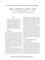
Polar body biopsy can be performed preconceptionally to remove the
first polar body or post-fertilization to remove either only the second polar
body or both polar bodies simultaneously (Fig. 1) (17,18). Polar bodies are
naturally extruded from the oocyte with no further role in the development
of the future embryo. They each have a set of chromosomes that are comp-
lementary to those present in the oocyte. The first polar body is formed
during meiosis (M) I of oogenesis and has a set of bivalent chromosomes,
whereas the second polar body is formed during MII, after fertilization,
and contains a haploid set of chromosomes. Both polar bodies give comp-
lementary diagnostic readouts and by deduction infer the genetic status
of the oocyte. The main advantages of polar body biopsy include the
Figure 1 Polar body biopsy.
314 Katz-Jaffe
extra-embryonic nature of the polar bodies and the additional amount of
time available for gene tic analysis prior to embryo transfer. However, the
major disadvantage of this technique is that the sex and paternal genotype
are not available for the analysis, thus precluding polar body biopsy for the
analysis of paternal mutations, gender determination, and chromosomal
abnormalities arising from paternal meiosis. In addition, polar bodies
undergo fragmentation rendering them often difficult to biopsy which can
potentially lead to misdiagnosis if the embryologist is unable to retrieve
all the polar body fragments.
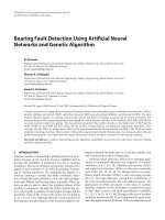
Currently, the preferred stage for obtaining cells for genetic diagnosis
is a blastomere biopsy of the cleavage-stage embryo performed at the
6–10-cell stage on day three post-fertilization (Fig. 2) (16). This allows for
the retrieval of a blastomere containing both the maternal and paternal gen-
omes. As these blastomeres are totipotent, the biopsy of 1–2 cells from the
developing embryo does not seem to drastically reduce either the mass or
contribution to the fetus, thereby not affecting the future viability of the
embryo (19,20). Reports of ongoing comparable clini cal pregnancy and
implantation rates with conventional IVF confirm these earlier studies (4).
The blastomere biopsy procedure can be performed by either gentle aspir-
ation (Fig. 2) or by the nudge of the flow of biopsy media. Even if the
embryo has begun compaction with gap junctions forming between
the blastomeres, a blastomere biopsy can be performed after a short
pre-incubation in calcium–magnesium-free media to reduce the cellular
apposition. The major disadvantage of blastomere biopsy is the invasive
nature of the procedure on the embryo itself along with the reduction in cell
Figure 2 Blastomere biopsy.
Preimplantation Genetic Diagnosis 315
number and the potential influence on further fetal development. There has
been debate in the PGD field as to whether one or two cells should be biop-
sied. Clearly, the removal of two cells further reduces the cellular mass of the
cleavage-stage embryo and may result in a reduction in developmental
potential (21). Only a handful of studies have comp ared the outcome of
one versus two biopsied cells, concluding that there were no decreases in
implantation rates (22) with potentially fewer misdiagnoses (23). Ongoing
prospective studies are underway to further address this question.
A human blastocyst, depending on the exact stage of development, can
contain over 100 cells. Hence, the biopsy of 6–10 cells from the outer layer
of trophectoderm is unlikely to have a detrimental effect on the blastocyst’s
mass or on the developing fetus that originates from the inner cell mass
(ICM) (24–26). Prior to biopsy, the position of the ICM is identified so that
the hole in the zona can be created on the opposite side of the blastocyst,
reducing possible developmental and ethical concerns (Fig. 3). The cells
are removed either mechanically by mild teasing using needles (24,27) or
after herniation of the trophectoderm allowing biopsy by laser (Fig. 3)
(28,29). The major advantage of this procedure is the larger amount of
material available for the genetic testing, thereby increasing the reliability
and accuracy of the diagnosis. However, the time for the analysis is limited
to no more than 24 hours, as the blastocyst needs to implant at this stage.
There are also uncertainties surrounding the genetic make up of the trophec-
toderm in relation to that of the ICM, the future-developing fetus. Due
to these issues and the fact that some clinics prefer not to culture to the
Figure 3 Trophectoderm biopsy.
316 Katz-Jaffe
blastocyst stage, only a limited number of IVF clinics perform blastocyst
biopsy routinely. However, these clinics have reported ongoing clinical preg-
nancies and healthy babies (28,29).
PGD FOR MONOGENIC DISORDERS
The current experience of PGD for monogenic disorders exceeds more than
1500 cycles comprising over 50 different conditions and the birth of more
than 300 unaffected children (3). PGD has been performed for autosomal-
recessive (e.g., cystic fibrosis), autosomal-dominant (e.g., Huntington
disease), and X-linked (e.g., Fragile X) inherited disorders (3). PGD has been
established as an acceptable form of early prenatal diagnosis with the spec-
trum of conditions expanding with patient demand. The main motives
behind couples seeking PGD are objection to potential therapeutic abortion
($47%), genetic risk coupled with low fertility ($32%), and repeated thera-
peutic abortions of identified affected fetuses ($26%) (30). In addition to
conventional monogenic disorders, PGD is now requested for conditi ons
such as late-onset predisposition disorders, blood group incompatibility,
and human leukocyte antigen (HLA) matching (3,31).
In routine genetic diagnostic procedures, a starting template of at least
10 ng of DNA is usually available. However, a single cell contains only 6 pg
of DNA (32) and has only two copies of each target locus. Some of the
requirements that need to be addressed in PGD for monogenic disorders
are the difficulties in the amplification of single-cell templates and the estab-
lishment of a procedure for high amplification efficiency and accuracy. A
considerable amount of time and resources are required for the development
of reliable and accurate single-cell diagnostic tests including the preliminary
mutation workup. Careful experimental practices and suitable facilities
including allocated equipment and vigilant quality control are essential (16).
Specific and individual mutation-detection systems have been developed to
capture and visualize the different DNA variants involved in monogenic
disorders, including single base pair substitutions, deletions, insertions,
duplications, and trinucleotide repeat expansions (3,4).
Polymerase chain reaction (PCR) is a common technique performed in
PGD for monogenic disorders. It is a rapid, highly sensitive, and specific
molecular technique that is capable of amplifying single copies of DNA tem-
plate into large numbers with high fidelity (33). In a PCR reaction, several
DNA sequences or loci can be independently amplified at the same time
using multiple primer pairs in a technique called multiplex PCR. Once the
DNA from the single cell has been amplified, there are numerous detection
methods available to visualize the PCR products for the presence or absence
of the specific DNA mutation or variant. The choice of the technique is
usually dependent on the nature of the specific DNA mutation or variant
and includes restriction endonuclease digestion (34,35), single-strand
Preimplantation Genetic Diagnosis 317
conformational polymorphism (36), denaturant gradient gel electrophoresis
(37), heteroduplex analysis (38), single nucleotide primer extension (39), and
analysis of DNA fragment size (40,41). Fluorescent technology has further
increased sensitivity of these detection methods resulting in the requirement
of fewer PCR amplification cycles and greater reliability.
Real-time PCR is a more recent molecular technique that allows the
mutation amplification and subsequent fluorescent detection procedure
to be carried out in the same tube. Fluorescently tagged probes directed to
either the normal or mutant sequence allow detection of the rate of amplifi-
cation product accumulation to be measured directly by associated computer
software as the PCR reaction proceeds (42,43). Unfortunate ly, there has
been a slow uptake of this technology in PGD labs due to the enormous costs
involved in purchasing the specialized equipment and consu mables.
Several misdiagnoses have been reported by clinics around the world
due to the complexity and sensitivity of single-cell PCR analysis (16). It is
of vital importance that PGD relies on a positive result from the biopsied cell,
thereby reducing the possibility of the transfer of affected embryos. The three
main sources of potential misdiagnosis in PGD include external DNA con-
tamination, complete amplification failure, and allele drop out (ADO).
External DNA contamination is a major problem due to the limited
starting template and the large number of PCR amplification cycles required.
Even at normal detectable levels, external DNA contamination may disguise
or overwhelm a single cell and cause a misdiagnosis. The main laboratory
contaminants include previously amplified PCR products accumulated in
the laboratory and skin cells from the technician. The implementation of
strict experimenta l practices and appropriate facilities will essentially mini-
mize this risk including the following examples: a dedicated laminar flow
hood with ultraviolet light to destroy any DNA by thymidine cross-linking,
the isolation of all equipment for PGD use only, filtration and autoclaving of
reagents, aliquots for storage, long sleeve lab gowns, caps and masks, and
frequent glove changes (44). Pre-testing of all reagents and solutions prior
to a clinical PGD case is essential to confirm the reliability of the test as well
as for contamination prevention. Other potential sources of contaminants
include the cumulus cells that surround the oocyte and excess sperm bound
to the zona pellucida at the time of fertilization. These cells could accidentally
be removed along with the polar body or blastomere during the biopsy pro-
cedure. It is therefore recommended for PGD of monogenic disorders that all
oocytes be completely striped of their cumulus complex and that intracyto-
plasmic sperm injection is chosen as the method for fertilization.
It is also possible to detect contamination by the simultaneous PCR
amplification of highly polymorphic DNA markers (45). This is similar to
DNA fingerprinting techniques whereby highly polymorphic DNA markers
allow the distinction between maternal and paternal alleles (46). As poly-
morphic DNA markers obey the laws of Mendelian inheritance, the
318 Katz-Jaffe
embryos of any couple can only inherit a specific combination of alleles at
any particular locus (47). Therefore, an incorrect combination of alleles or
the presence of extra foreign alleles is an indication that contamination
has occurred. It has become increasingly common for PCR-based single-cell
tests to include the amplification of several polymorphic DNA markers
alongside the mutation loci used for diagnosis. If intragenic markers are
chosen, they are linked in 100% disequilibrium to the gene of intere st and
are unlikely to be separated by recombination during meiosis. Hence, these
polymorphic DNA markers ha ve a dual purpose to recognize potential
extraneous DNA contamination and act as a positive control for amplifi-
cation (34,38).
Complete amplification failure of a mutat ion locus is observed in
5–10% of single cells. The source of the failure could be due to a number
of factors, including failure to transfer the single cell into the PCR tube,
an enucleate cell, degradation or loss of the target DNA sequence, and/or
inefficient cell lysis (48,49). In contrast, ADO is defined as the amplification
failure of either the maternal or paternal allele, giving the impression that a
locus is homozygous. ADO has been observed to affect both parental alleles
randomly. PGD involving dominant monogenic disorders is particularly
vulnerable to misdiagnosis from ADO by the transfer of affected embryos
that are incorrectly diagnosed as unaffected homozygous. One method to
prevent misdiagnosis by ADO is the simultaneous amplification of intra-
genic polymorphic DNA markers in combination with the gene of interest.
These markers would be inheri ted alongside the mutation locus providing
additional loci for the detection of affected embryos. It is improb able that
all loci amplified during a multiplex PCR reaction would be affected by
ADO (38,50). The factors that cause ADO are yet to be completely eluci-
dated; however, differences in PCR thermal cycling conditions, fragm ent
size, incomplete cell lysis, the degradation of target template sequence, freez-
ing and thawing, and poor specificity of primer pairs could possibly explain
the variability in observed ADO rates (51).
The development of new reliable single-cell strategies, often for only
one specific monogenic disorder, requires a major investment in resources,
staff, finances, and time. Obviously, more adaptable and univers al tech-
niques are required in PG D that will allow a wider range of mutations to
be concurrently investigated. One platform that may be able to achieve this
goal is microarray technology. Specific sequences of DNA incorporating dif-
ferent mutations would act as probes on a microarray slide or chip, allowing
hybridization between these known DNA probes and test DNA amplified
from the single biopsied cell. Initial development of a specific cystic fibrosis
deltaF508 array highlighted the diagnostic capability of microarrays for
PGD (52). However, for this technology to be offered clinically, several
issues need to be addressed including the reliable amplification of the whole
genome from a single biopsied cell, a reduction in the complexity and time
Preimplantation Genetic Diagnosis 319
for data analysis, and more cost-effective microarray platforms comprising
DNA probes for numerous common monogenic disorders.
Whole genome amplification (WGA) is a technique aimed at maximiz-
ing the amount of information that can be obtained from a single cell or
limited template. WGA theoretically involves the non-specific amplification
of the entire genome, thereby increasing the amount of template for sub-
sequent PCR reactions and multiple genetic analyses (53,54). There are
several types of WGA protocols that have been developed to amplify DNA
from small numbers of cells including primer extension preamplification
(55), degenerate oligonucleotide primer PCR (56), and multiple displacement
amplification (MDA) (57). Recently, MDA has been incorporated in clinical
PGD for cystic fibrosis and b-thalassaemia resulting in two pregnancies (58).
Some of the drawbacks to WGA methods include higher incidences of ADO,
inaccurate size fragments, and inconsistent amplification of the whole gen-
ome (59,60). It is paramount that any WGA protocol incorporated in clinical
PGD be reliable, accurate, and complete in the amplification of the entire
human genome from a single cell.
PGD is considered an early form of prenatal diagnosis allowing high-
risk couples to establish pregnancies free of the indicated genetic disorder.
This technology is viewed as a positive contribution to the field giving cou-
ples early reassurance and avoidance of therapeutic abortion. Indications
for PGD will con tinue to grow with patient demand and advancing
technology. The use of PGD for non- medical indications, including HLA
matching for siblings suffering lethal diseases such as leukemia and late-
onset diseases such as cancer predisposition, are also likely to become more
common. These non-medical indications have attracted media attention and
passionate public debate concerning the ethics of ‘‘designer bab ies.’’ In con-
trast, it has been argued that PGD for non-medical reasons highlights the
love and commitment of couples to treat and prevent disease in their chil-
dren and therefore should be viewed as an acceptable treatment.
CHROMOSOMAL ANEUPLOIDY SCREENING
Chromosomal analysis of human IVF embryos using single-cell inter-
phase fluorescent in situ hybridization (FISH) was first developed to screen
for embryo sex, allowing for the detection of the two sex chromosom es (7,61).
Over time, the number of chromosomes for detection has increased signifi-
cantly, allowing for screening of chromosomal aneu ploidy in up to 9–10
chromosomes (62,63). Studies have shown that fetal chromosomal abnor-
malities are associated with human implantation failure and pregnancy loss
(64,65). Therefore, chromosomal aneuploidy screening in PGD was
introduced for IVF patients who are considered to be at increased risk of
producing embryos with chromosomal abnormalities. These at-risk groups
include advanced maternal age (> 36 year), repeated miscarriages (RM), poor
320 Katz-Jaffe
IVF prognosis (>3 failed cycles), and couples who carry a chromosome
rearrangement (such as translocations and inversions). Chromosomal aneu-
ploidy screening of these patients’ embryos should identify euploid embryos
for the indicated chromosomes leading to a greater chance of implantation
and clinical pregnancy. PGD for aneuploidy screening now accounts for the
majority of PGD cycles worldwide, estimated at close to 5000 cycles (3). The
benefit of PGD for aneuploidy screening has been reported by several
groups showing an increase in implantation rates and decrease in miscar-
riage rates (31,62,63,66,67).
Single-cell interphase FISH is a rapid, reliable, and efficient technique
capable of detecting up to 9–10 chromosomes in two rounds of hybridiza-
tion on a single nucleus (62,63). Currently, fluorescent DNA probes for
chromosomes X, Y, 13, 14, 15, 16, 18, 21, and 22 are being used in PGD
for aneuploidy screening as they are involved in more than 50% of all
chromosomal abnormal miscarriages (65). In the cases of chromosomal
translocations, probes distal to the sites of chromosome breakage are used
in addition to centromeric and proximal probes (68). The technical dif-
ficulties encountered in regards to selecting appropriate FISH probes and
optimizing protocols for each couple’s specific chromosome rearrangement
are considerably time-consuming and expensive. However, these couples are
considered to be one of the most motivated groups of PGD for aneuploidy
screening due to their history of RM and infertility. A clear advantage has
been documented in more than 500 clinical cycles with a fourfold reduction
in miscarriage rates and an increase of live births (69,70).
Successful FISH involves annealing of the single-stranded fluorescent-
labeled DNA probes to its complementary target sequence on a specific
chromosome. The biggest limitation to this technique is the fact that only
one or two cells are available for analysis. The error rate for single-cell inter-
phase FISH has been recorded in several studies at frequencies betw een
5 and 15% (18,68,71). Numerous variables could be responsible including
signal overlap, signal splitting, cross-hybridization of FISH probes, and
the presence of chromosomal mosaicism (72) . A greater number of monoso-
mies have been diagnosed by single-cell FISH than trisomies. This could be
due to insufficient binding, loss of DNA, poor probe penetration, or an
overlap of chromosome signals due to the poor spread of the nucleus during
fixation (68). In an attempt to counteract the possibility of misdiagnosis, a
FISH scoring system has been implemented (68) to reduce the incidence
of false-positive and false-negative results. Nevertheless, several misdiag-
noses have been recorded, where aneuploid embryos were misdiagnosed
as normal, but on transfer resulted in aneuploid pregnancies that either
spontaneously aborted or were detected after prenatal diagnosis (16).
Interestingly, the data from FISH analysis of human IVF embryos
have revealed a high incidence of chromosomal mosaicism, with over 30%
containing a proportion of aneuploid cells (61,73,74). These high rates of
Preimplantation Genetic Diagnosis 321
chromosomal mosaicism observed in human IVF embryos are a major con-
cern in chromosomal aneuploidy screening, questioning the validity of the
test, with the possibi lity of transferring affected mosaic embryos (75,76).
Embryonic chromosomal mosai cism is the existence of two or more differ-
ent chromosomal complements in a single embryo. Mitotic cell division
errors post-fertilization, appear to be responsible for the observed chromo-
somal mosaicism in early human preimplantation development (77, 78). The
chances of detecting mosaicism would depend on the timing of the mitotic
cell division error, e.g., a non-disjunction event during the second cleavage
division would resul t in a 25% chance of biopsing an aneuploid blastomere.
Studies have also revealed frequent mosaicism in both the trophe ctoderm
and ICM of human blastocysts (79–81), with the significance at this stage
of human embryonic development still to be clarified. The current under-
standing of the normal dynamics and regulation of mitotic chromosomal
segregation during early embryonic cleavage divisions is critically insuf-
ficient. Hence, several clinics support the biopsy and analysis of two
blastomeres for chromosomal aneuploidy screening in order to reduce the
chance of misdiagnosis due to mosaicism (22,82). However, this approach
cannot completely overcome the possibility of a misdiagnosis, nor does it
address the problem of mosaicism in human IVF embryos. One approach
to gaining a better understanding of chromosomal mosaicism during human
preimplantation development is to determine the underlying mechanisms
causing this phenomenon includi ng the origin and nature of the cell division
errors (83). Such knowledge may translate into a revision of current clinical
and lab procedures to produce higher numbers of non-mosaic embryos
available for transfer, thereby potentially improving implantation rates.
Current single-cell interphase FISH methods are limited to the
analysis of less than half of the human chromosomal complement. It is
hypothesized that the development of a technique that can analyze all 23
pairs of human chromosomes will allow for the selection of entirely euploid
embryos for transfer, furth er improving pregnancy rates and decreasing
miscarriage rates for indicated couples. There are several alternatives that
are currently being investigated including metaphase nuclear conversion.
Customarily, cytogenetic techniques are performed on cells that are in meta-
phase, when the nuclear membrane has broken down and the chromosomes
are condensed allowing for the identification of each individual chromo-
some. However, blastomeres are typically observed to be in interphase,
when chromosomes are in an unrecognizable state. Metaphase nuclear con-
version is a technique that fuses blastomeres or second polar bodies with
enucleated or intact oocytes (mouse, bovine, or human) allowing for the
metaphase visualization of all 23 pairs of chromosomes to identify both
chromosomal aneuploidy and rearrangements. Several method s can then
be used on these metaphase converted chromosomes for enumeration
including G-banding, chromosome painting, and spectral imaging (84–86).
322 Katz-Jaffe
Metaphase nuclear conversion has been applied in 52 clinical PGD cases for
translocations with an 88% success rate and 38 transfers of chromosomally
balanced embryos (87) . Nevertheless, there are several concerns in produc-
ing these metaphase chromosome spreads from a single cell including labor
intensity, technical difficulties, and the ethical considerations in fusing
human blastomeres with enucleated oocytes from other species.
Another alternative method for enumerating all 23 pairs of chromo-
somes is comparative genomic hybridization (CGH) (88). This method
involves a test DNA sample (blastomere) and a normal control DNA sam-
ple differentially labeled with fluorochromes (red or green) and hybridized
simultaneously to a spread of normal metaphase chromosomes. If there is
a chromosomal imbalance in the test sample, then a deviation of the 1:1
fluorescence ratio will be observed for the particular chromosome affected
indicating aneuploidy. The major drawbacks to this method include the
inability to detect ploidy changes or balanced translocations, the extremely
lengthy labor-intensive procedure taking up to five days to complete, and a
substantial amount of starting DNA template (at least 200 ng) necessitating
the requirement of WGA for single cells. Several groups have attempted to
overcome these difficulties to develop CGH for aneuploidy screening in
PGD (89,90). Clinical experience of CGH has included a study of 20 poor
IVF prognosis patients resulting in 14 embryo transfers and three clinical
pregnancies after freezing and thawing of biopsied embryos (91). In this
study, only 54% of the embryos survived the freezing and thawing with at
least 50% of cells intact; therefore, to maximize the potential of CGH and
the analysis of all chromosomes, it would appear that cryopreservation
should be avoided. The biopsy of polar bodies followed by CGH and a
blastocyst transfer has also been clinically reported; however, with this
method only the maternal chromosomes are available for analysis (92) . In
its current state, single-cell CGH is a difficult, highly labor-intensive, and
complex technique that is unlikely to be widely accepted in clinical PGD
for aneuploidy screening.
A new promising technology that could transform chromosomal
aneuploidy screening in PGD merges single-cell CGH with a micr oarray
platform. Array CGH is a powerful technique that allows for genome-wide
analysis with high resolution and detection of DNA copy number variations
(93). Array CGH provides several advantages over conventional CGH,
including substantially increased resolution and the ability to directly corre-
late chromosomal abnormalities with the genomic sequence. Array CGH
has the potential to be a primary screening tool for cancer and genetic
disease detecting genomic imbalance in cells. It is anticipated that array
CGH will transform the practice of both medical genetics and clinical cyto-
genetics. Currently there are several hurdles that need to be overcome before
this technology can be introduced in PGD, including the length of time
required for analysis, production of comprehensive WGA products, greater
Preimplantation Genetic Diagnosis 323
sensitivity in detecting single copy number changes, adaptability for the IVF
clinical setting, and a reduction in the cost of the microarrays themselves.
A novel approach that has recently been developed involves whole human
chromosome-specific libraries spotted onto glass slides with a total analysis
time of just 30 hours (94). This array CGH format has yet to be tested on
human embryonic material, but initial results from aneuploid cell lines were
promising (94). Microarray technology, still currently in its infancy, has
the future capability of providing extremely detailed information on one
single platform including chromosome copy number and DNA sequence
variations (95).
In summary, PGD is recognized as a reliable and safe reproductive
option available to a range of indicated couples including those at risk of
passing on an inherited genetic disorder to their offspring and infertile cou-
ples with a high chance of producing chromosomally aneuploid embryos.
The future of this field will be driven by patient demand, with the number
and types of disorders continually expanding. Research and advancing tech-
nology will follow suit and involve improvements to the efficiency and
reliability of the procedures as well as advances in the techniques utilized
for the molecular analysis of a single cell.
REFERENCES
1. Handyside AH, Kontogianni EH, Hardy K, et al. Pregnancies from biopsied
human pre-implantation embryos sexed by Y-specific DNA amplification.
Nature 1990; 344:768–770.
2. Verlinsky Y, Ginsberg N, Lifchez A, Valle J, Moise J, Strom CM. Analysis of the
first polar body: preconception genetic diagnosis. Hum Reprod 1990; 5:826–829.
3. Kuliev A, Verlinsky Y. Thirteen years experience of preimplantation diagnosis:
the fifth international symposium on preimplantation genetics. Reprod Biomed
Online 1990; 8:229–235.
4. Anon. Report of the 11th Annual Meeting of International Working Group on
Preimplantation Genetics: preimplantation genetic diagnosis—experience of
3000 clinical cycles. Reprod Biomed Online 2001; 3:49–53.
5. Handyside AH, Lesko JG, Tarin JJ, et al. Birth of a normal girl after in vitro
fertilization and preimplantation diagnostic testing for cystic fibrosis. N Engl J
Med 1992; 327:905–909.
6. Snabes MC, Chong SS, Subramanian SB, et al. Preimplantation single-cell
analysis of multiple genetic loci by whole-genome amplification. Proc Natl Acad
Sci USA 1994; 91:6181–6185.
7. Munne S, Lee A, Rosenwaks Z, Grifo J, Cohen J. Diagnosis of major chromo-
some aneuploidies in human preimplantation embryos. Hum Reprod 1993; 8:
2185–2191.
8. Verlinsky Y, Cieslak J, Freidine M, et al. Pregnancies following pre-conception
diagnosis of common aneuploidies by fluorescent in situ hybridization. Hum
Reprod 1995; 10:1923–1927.
324 Katz-Jaffe
9. Conn CM, Harper JC, Winston RM, Delhanty JD. Infertile couples with
Robertsonian translocations: preimplantation genetic analysis of embryos
reveals chaotic cleavage divisions. Hum Genet 1998; 102:117–123.
10. Scriven PN, Handyside AH, Mackie Ogilvie C. Chromosome translocations:
segregation modes and strategies for preimplantation genetic diagnosis. Prenat
Diagn 1998; 18:1437–1449.
11. De Vos A, Van Steirteghem A. Aspects of biopsy procedures prior to preimplan-
tation genetic diagnosis. Prenat Diagn 2001; 21:767–780.
12. Cieslak J, Ivakhnenko V, Wolf G, Sheleg S, Verlinsky Y. Three-dimensional
partial zona dissection for preimplantation genetic diagnosis and assisted hatch-
ing. Fertil Steril 1999; 71:308–313.
13. Grifo JA, Tang YX, Munne
´
S, et al. Healthy babies from biopsied human
embryos. Hum Reprod 1994; 9:912–916.
14. Tadir Y, Neev J, Ho P, Berns MW. Lasers for gamete micromanipulation: basic
concepts. J Assist Reprod Genet 1993; 10:121–125.
15. Boada M, Carrera M, De La Iglesia C, et al. Successful use of laser for human
embryo biopsy in preimplantation genetic diagnosis: report of two cases. J Assist
Reprod Genet 1998; 15:301–306.
16. ESHRE PGD Consortium. Hum Reprod 2002; 20:35–48.
17. Verlinsky Y, Rechitsky S, Cieslak J, et al. Preimplantation diagnosis of single
gene disorders by two-step oocyte genetic analysis using first and second polar
body. Biochem Mol Med 1997; 62:182–187.
18. Verlinsky Y, Cieslak J, Ivakhnenko V, et al. Implantation diagnosis of common
aneuploidies by the first and second-polar body FISH analysis. J Assist Reprod
Genet 1998; 15:285–289.
19. Handyside AH, Pattinson JK, Penketh RJ, et al. Biopsy of human preimplanta-
tion embryos and sexing by DNA amplification. Lancet 1989; 1:347–349.
20. Hardy K, Martin KL, Leese HJ, et al. Human pre-implantation development in
vitro is not adversely affected by biopsy at the 8-cell stage. Hum Reprod 1990; 5:
708–714.
21. Tarin JJ, Conaghan J, Winston RM, et al. Human embryo biopsy on the 2nd
day after insemination for preimplantation diagnosis: removal of a quarter of
embryo retards cleavage. Fertil Steril 1992; 58:970–976.
22. Van de Velde H, De Vos A, Sermon K, et al. Embryo implantation after biopsy
of one or two cells from cleavage-stage embryos with a view to preimplantation
genetic diagnosis. Prenat Diagn 2000; 20:1030–1037.
23. Emiliani S, Gonzalez-Merino E, Englert Y, Abramowicz M. Comparison of the
validity of preimplantation genetic diagnosis for embryo chromosomal anoma-
lies by fluorescence in situ hybridization on one or two blastomeres. Genet Test
2004; 8:69–72.
24. Dokras A, Sargent IL, Ross C, et al. Trophectoderm biopsy in human blasto-
cysts. Hum Reprod 1990; 5:821–825.
25. Veiga A, Sandalinas M, Benkhalifa M, et al. Laser blastocyst biopsy for preim-
plantation diagnosis in the human. Zygote 1997; 5:351–354.
26. Veiga A, Gil Y, Boada M, et al. Confirmation of diagnosis in preimplantation
genetic diagnosis (PGD) through blastocyst culture: preliminary experience.
Prenat Diagn 1999; 19:1242–1247.
Preimplantation Genetic Diagnosis 325
27. Dokras A, Sargent IL, Gardner RL, et al. Human trophectoderm biopsy and
secretion of chorionic gonadotrophin. Hum Reprod 1991; 6:1453–1459.
28. de Boer KA, Catt JW, Jansen RP, Leigh D, McArthur S. Moving to blastocyst
biopsy for preimplantation genetic diagnosis and single embryo transfer at
Sydney IVF. Fertil Steril 2004; 82:295–298.
29. Kokkali G, Vrettou C, Traeger-Synodinos J, et al. Birth of a healthy infant
following trophectoderm biopsy from blastocysts for PGD of beta-thalassaemia
major: case report. Hum Reprod 2005; 20:1855–1859.
30. Wells D, Delhanty JD. Preimplantation genetic diagnosis: applications for
molecular medicine. Trend Mol Med 2001; 7:23–30.
31. Verlinsky Y, Cohen J, Munne S, et al. Over a decade of experience with preim-
plantation genetic diagnosis: a multicenter report. Fertil Steril 2004; 82:292–294.
32. Morton NE. Parameters of the human genome. Proc Natl Acad Sci USA 1991;
88:7474–7476.
33. Erlich HA, Arnheim N. Genetic analysis using the polymerase chain reaction.
Ann Rev Genet 1992; 26:479–506.
34. Kuliev A, Rechitsky S, Verlinsky O, et al. Preimplantation diagnosis of thalasse-
mias. J Assist Reprod Genet 1998; 15:219–225.
35. Strom CM, Rechitsky S, Wolf G, et al. Preimplantation diagnosis of autosomal
dominant retinitis pigmentosum using two simultaneous single cell assays for a
point mutation in the rhodopsin gene. Mol Hum Reprod 1998; 4:351–355.
36. Larsen LA, Christiansen M, Vuust J, et al. High-throughput single-strand con-
formation polymorphism analysis by automated capillary electrophoresis: robust
multiplex analysis and pattern-based identification of allelic variants. Hum Mut
1999; 13:318–327.
37. Vrettou C, Palmer G, Kanavakis E, et al. A widely applicable strategy for single
cell genotyping of b-thalassaemia mutations using DGGE analysis: application
to preimplantation genetic diagnosis. Prenat Diagn 1999; 9:1209–1216.
38. Ao A, Wells D, Handyside AH, et al. Preimplantation genetic diagnosis of inher-
ited cancer familial adenomatous polyposis coli. J Assist Reprod Genet 1998;
15:140–144.
39. Cram DS, Song B, Trounson AO. Preimplantation diagnosis of Lesch–Nyhan
using mini-sequencing primer extension. Reprod Biomed Online 2003; 7:
342–345.
40. Sermon K, De Vos A, Van de Velde H, et al. Fluorescent PCR and automated
fragment analysis for the clinical application of preimplantation genetic diag-
nosis of myotonic dystrophy (Steinert’s disease). Mol Hum Reprod 1998; 4:
791–796.
41. Sermon K, Goossens V, Seneca S, et al. Preimplantation diagnosis for
Huntington’s Disease (HD): clinical application and analysis of the HD expan-
sion in affected embryos. Prenat Diagn 1998; 18:1427–1436.
42. Pals G, Young C, Mao HS, et al. Detection of a single base substitution in a
single cell using the LightCycler. J Biochem Biophys Meth 2001; 47:121–129.
43. Vrettou C, Traeger-Synodinos J, Tzetis M, Palmer G, Sofocleous C, Kanavakis
E. Real-time PCR for single-cell genotyping in sickle cell and thalassemia
syndromes as a rapid, accurate, reliable, and widely applicable protocol for pre-
implantation genetic diagnosis. Hum Mutat 2004; 23:513–521.
326 Katz-Jaffe
44. Thornhill AR, deDie-Smulders CE, Geraedts JP, et al. ESHRE PGD Consor-
tium on ‘Best practice guidelines for clinical preimplantation genetic diagnosis
(PGD) and preimplantation genetic screening (PGS).’ Hum Reprod 2005; 20:
35–48.
45. Rechitsky S, Verlinsky O, Amet T, et al. Reliability of preimplantation diagnosis
for single gene disorders. Mol Cell Endocrinol 2001; 183(suppl 1):S65–S68.
46. Jeffreys AJ, Wilson V, Thein SL. Individual-specific ‘fingerprints’ of human
DNA. Nature 1985; 316:76–78.
47. Katz MG, Trounson AO, Cram DS. DNA fingerprinting of sister blastomeres
from human IVF embryos. Hum Reprod 2002; 17:752–759.
48. Hardy K, Winston RM, Handyside AH. Binucleate blastomeres in preimplanta-
tion human embryos in vitro: failure of cytokinesis during early cleavage.
J Reprod Fertil 1993; 98:549–558.
49. Ray PF, Handyside AH. Increasing the denaturation temperature during the
first cycles of amplification reduces allele dropout from single cells for preim-
plantation genetic diagnosis. Mol Hum Reprod 1996; 2:213–218.
50. Lewis CM, Pinel T, Whittaker JC, et al. Controlling misdiagnosis errors in pre-
implantation genetic diagnosis: a comprehensive model encompassing extrinsic
and intrinsic sources of error. Hum Reprod 2001; 16:43–50.
51. Piyamongkol W, Bermudez MG, Harper JC, et al. Detailed investigation of
factors influencing amplification efficiency and allele drop-out in single cell
PCR: implications for preimplantation genetic diagnosis. Mol Hum Reprod
2003; 9:411–420.
52. Salvado CS, Trounson AO, Cram DS. Towards preimplantation diagnosis of
cystic fibrosis using microarrays. Reprod Biomed Online 2004; 8:107–114.
53. Paunio T, Reima I, Syvanen AC. Preimplantation diagnosis by whole genome
amplification, PCR amplification and solid-phase minisequencing of blastomere
DNA. Clin Chem 1996; 42:1382–1390.
54. Sermon K, Lissens W, Joris H, et al. Adaptation of the primer extension pream-
plification (PEP) reaction for preimplantation diagnosis: single blastomere
analysis using short PEP protocols. Mol Hum Reprod 1996; 2:209–212.
55. Zhang L, Cui X, Schmitt K, et al. Whole genome amplification from a single cell:
implications for genetic analysis. Proc Natl Acad Sci USA 1992; 89:5847–5851.
56. Cheung VG, Nelson SF. Whole genome amplification using a degenerate oligo-
nucleotide primer allows hundreds of genotypes to be performed on less than one
nanogram of genomic DNA. Proc Natl Acad Sci USA 1996; 93:14676–14679.
57. Dean FB, Hosono S, Fang L, et al. Comprehensive human genome amplification
using multiple displacement amplification. Proc Natl Acad Sci USA 2002; 99:
5261–5266.
58. Hellani A, Coskun S, Tbakhi A, Al-Hassan S. Clinical application of multiple
displacement amplification in preimplantation genetic diagnosis. Reprod Biomed
Online 2005; 10:376–380.
59. Handyside A, Robinson MD, Sinpson RJ, et al. Isothermal whole genome
amplification from single and small numbers of cells: a new era for preimplanta-
tion genetic diagnosis of inherited disease. Mol Hum Reprod 2004; 10:767–772.
60. Sun G, Kaushal R, Pal P, et al. Whole-genome amplification: relative efficiencies
of the current methods. Leg Med (Tokyo) 2005; 7:279–286.
Preimplantation Genetic Diagnosis 327
61. Delhanty JD, Griffin DK, Handyside AH, et al. Detection of aneuploidy and
chromosomal mosaicism in human embryos during preimplantation sex determi-
nation by fluorescent in situ hybridisation (FISH). Hum Mol Genet 1993; 2:
1183–1185.
62. Gianaroli L, Magli MC, Ferraretti AP, et al. Preimplantation diagnosis for
aneuploidies in patients undergoing in vitro fertilization with a poor prognosis:
identification of the categories for which it should be proposed. Fertil Steril 1999;
72:837–844.
63. Munne
´
S, Sandalinas M, Escudero T, et al. Improved implantation after preim-
plantation genetic diagnosis of aneuploidy. Reprod Biomed Online 2003; 7:
91–97.
64. Boue A, Boue J, Gropp A. Cytogenetics of pregnancy wastage. Adv Hum Genet
1985; 14:1–57.
65. Hook EB. Chromosome abnormalities. In: Brock DJH, Rodek CH, Ferguson-
Smith MA, eds. Prenatal Diagnosis and Screening. Edinburgh: Churchill
Livingstone, 1992.
66. Verlinsky Y, Cohen J, Munne S, et al. Over a decade of experience with preim-
plantation genetic diagnosis. Fertil Steril 2004; 82:302–303.
67. Munne S, Chen S, Fischer J, et al. Preimplantation genetic diagnosis reduces
pregnancy loss in women aged 35 years and older with a history of recurrent
miscarriages. Fertil Steril 2005; 84:331–335.
68. Munne
´
S, Magli C, Bahce M, et al. Preimplantation diagnosis of the aneuploi-
dies most commonly found in spontaneous abortions and live births: XY, 13,
14, 15, 16, 18, 21, 22. Prenat Diagn 1998; 18:1459–1466.
69. Gianaroli L, Magli MC, Ferraretti AP, et al. Possible interchromosomal effect in
embryos generated by gametes from translocation carriers. Hum Reprod 2002;
17:3201–3207.
70. Verlinsky Y, Cieslak J, Evsikov S, et al. Nuclear transfer for full karyotyping and
preimplantation diagnosis for translocations. Reprod Biomed Online 2002; 5:
300–305.
71. Magli MC, Sandalinas M, Escudero T, et al. Double locus analysis of chromo-
some 21 for preimplantation genetic diagnosis of aneuploidy. Prenat Diagn 2001;
21:1080–1085.
72. Connen E, Hopman AHN, Geraedts JPM, et al. Application of in situ hybridiza-
tion techniques to study human preimplantation embryos: a review. Hum
Reprod Update 1998; 4:135–152.
73. Munne
´
S, Weier H, Grifo J, et al. Chromosome mosaicism in human embryos.
Biol Reprod 1994; 51:373–379.
74. Harper JC, Coonen E, Handyside AH, et al. Mosaicism of autosomes and sex
chromosomes in morphologically normal monospermic preimplantation human
embryos. Prenat Diagn 1995; 15:41–49.
75. Kuo HC, Ogilvie CM, Handyside AH. Chromosomal mosaicism in cleavage-
stage human embryos and the accuracy of single-cell genetic analysis. J Assist
Reprod Genet 1998; 15:276–280.
76. Magli MC, Sandalinas M, Escudero T, et al. Double locus analysis of chromo-
some 21 for preimplantation genetic diagnosis of aneuploidy. Prenat Diagn 2001;
21:1080–1085.
328 Katz-Jaffe
77. Coonen E, Derhaag JG, Dumoulin J, et al. Anaphase lagging mainly explains
chromosomal mosaicism in human preimplantation embryos. Hum Reprod
2004; 19:316–324.
78. Katz-Jaffe MG, Trounson AO, Cram DS. Mitotic errors in chromosome 21 of
human pre-implantation embryos are associated with non-viability. Mol Hum
Reprod 2004; 10:143–147.
79. Evsikov S, Verlinsky Y. Mosaicism in the inner cell mass of the human blasto-
cysts. Hum Reprod 1998; 13:3151–3155.
80. Ruangvutilert P, Delhanty JD, Serhal P, et al. FISH analysis on day 5 post-
insemination of human arrested and blastocyst stage embryos. Prenat Diagn
2000; 20:552–560.
81. Bielanska M, Jin S, Bernier M, Tan SL, Ao A. Diploid–aneuploid mosaicism in
human embryos cultured to the blastocyst stage. Fertil Steril 2005; 84:336–342.
82. Simopoulou M, Harper JC, Fragouli E, et al. Preimplantation genetic diagnosis
of chromosome abnormalities: implications from the outcome for couples with
chromosomal rearrangements. Prenat Diagn 2003; 23:652–662.
83. Katz-Jaffe MG, Trounson AO, Cram DS. Chromosome 21 mosaic human pre-
implantation embryos predominantly arise from diploid conceptions. Fertil
Steril 2005; 84:634–643.
84. Verlinsky Y, Evsikov S. A simplified and efficient method for obtaining meta-
phase chromosomes from individual human blastomeres. Fertil Steril 1999;
72:1127–1133.
85. Willadsen S, Levron J, Munne
´
S, et al. Rapid visualization of metaphase chro-
mosomes in single human blastomeres after fusion with in vitro matured bovine
eggs. Hum Reprod 1999; 14:470–475.
86. Eviskov S, Cieslak J, Verlinsky Y. Effect of chromosomal translocations on the
development of preimplantation human embryos in vitro. Fertil Steril 2000;
74:672–677.
87. Verlinsky Y, Cieslak J, Evsikov S, et al. Nuclear transfer for full karyotyping and
preimplantation diagnosis for translocations. Reprod Biomed Online 2002;
5:300–305.
88. Kallioniemi A, Kallioniemi OP, Sudar D, et al. Comparative genomic hybridiza-
tion for molecular cytogenetic analysis of solid tumors. Science 1992; 258:
818–821.
89. Voullaire L, Slater H, Williamson R, et al. Chromosome analysis of blastomeres
from human embryos by using comparative genomic hybridization. Hum Genet
2000; 106:210–217.
90. Wells D, Delhanty JD. Comprehensive chromosomal analysis of human pre-
implantation embryos using whole genome amplification and single cell
comparative genomic hybridization. Mol Hum Reprod 2000; 6:1055–1062.
91. Wilton L, Voullaire L, Sargeant P, et al. Preimplantation aneuploidy screening
using comparative genomic hybridization or fluorescence in situ hybridization
of embryos from patients with recurrent implantation failure. Fertil Steril
2003; 80:860–868.
92. Wells D, Escudero T, Levy B, et al. First clinical application of comparative
genomic hybridization and polar body testing for preimplantation genetic diag-
nosis of aneuploidy. Fertil Steril 2002; 78:543–549.
Preimplantation Genetic Diagnosis 329
93. Pinkel D, Segraves R, Sudar D, et al. High resolution analysis of DNA copy
number variation using comparative genomic hybridization to microarrays.
Nat Genet 1998; 20:207–211.
94. Hu DG, Webb G, Hussey N. Aneuploidy detection in single cells using DNA
array-based comparative genomic hybridization. Mol Hum Reprod 2004; 10:
283–289.
95. Yi Y, Mirosevich J, Shyr Y, Matusik R, George AL Jr. Coupled analysis of gene
expression and chromosomal location. Genomics 2005; 85:401–412.
330 Katz-Jaffe