Atomic Force Microscopy in Cell Biology Episode 1 Part 4 potx
Bạn đang xem bản rút gọn của tài liệu. Xem và tải ngay bản đầy đủ của tài liệu tại đây (665.92 KB, 20 trang )
64 Bushell et al.
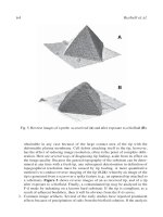
obtainable in any case because of the large contact area of the tip with the
deformable plasma membrane. Cell debris attaching itself to the tip, however,
has the effect of reducing image resolution, often to the point of complete oblit-
eration. Here are several ways of diagnosing tip fouling, aside from its effect on
the image quality. Because the general topography of the substrate can be deter-
mined at any time with a fresh tip, any subsequent deterioration in definition of
topographical resolution must be caused by tip fouling. A more quantitative
method is to conduct reverse imaging of the tip (8,26), whereby an image of the
tip is generated from a scan over a spiky feature (e.g., an upturned tip attached to
a substrate). Figure 5 shows reverse images of an as-received tip, and of a tip
after exposure to a biofluid. Finally, a contaminated tip may be analyzed in the
F-d mode by indenting on a known hard substrate. If the tip is compliant, as a
result of adherent biodebris, then it will be obvious from the F-d curves.
3. Common image artifacts. Several of the early studies have reported prominent
effects because of precipitation of salts from the biofluid solution. If the analysis
Fig. 5. Reverse images of a probe as-received (A) and after exposure to a biofluid (B).
Analysis of Human Fibroblasts by AFM 65
is conducted in an open cell, and the cell is subject to evaporative losses, then the
solution will become supersaturated in salts. Consequently, crystalline precipi-
tates will form within the field of view. Moreover, the biofluid will no longer be
compatible with cell viability. Frequent replacement of the biofluid will substan-
tially eliminate that problem.
Tip-broadening and other tip-related artifacts will occur when the actual
topography of the object being imaged is defined by radii of curvature less than
or comparable to the radius of curvature of the tip, and/or when there are gradients
exceeding that corresponding to the aspect ratio of the tip. For instance, images
of tobacco mosaic virus (TMV) attached to a flat substrate obtained by AFM
reveal the correct height of approx 18 nm, but the apparent lateral width will be
in the range 60–100 nm as a result of the tip-shape convolution (27). Because the
radius of the cylindrical TMV is known and is comparable to that of the apex of
the tip, the apparent width of the object, W, in is given by the following:
W = 2[(R
TMV
+ R
Tip
)
2
– (R
Tip
– R
TMV
)
2
]
1/2
When cytoskeletal structure is being imaged, the situation is somewhat more com-
plicated by filamentary objects located some distance above the substrate. The
aspect ratio then comes into play because the deformable membrane allows the
tip to indent the cell on either side of the filamentary object. The apparent width
will now depend on the height, h, of the object above the point of greatest inden-
tation by the tip on either side of the object. The relevant expression is now as
follows:
W ≈ 2[hA
r
–1
+ (r
tip
+ r
obj
)cos φ]
where the radii of the tip and object are r
tip
and r
obj
, respectively; A
r
, is the aspect
ratio of the tip, and the angle is defined by φ = tan
–1
A
r
–1
.
Finally, other grosser artifacts will occur when the dynamic range of the z
stage is exceeded; the image then becomes entirely featureless. A similar effect
occurs when the z-height corrugations of the object exceed the height of the tip,
and the surface of the lever defines the point of contact. The interaction is no
longer localized, and the details of the image become washed out. Likewise, F-d
analysis will now produce erroneous data since the spring constant will depend
on an unknown and changing point of contact and the contact area will also be
much greater leading to erroneous conclusions about indentation and adhesion.
Acknowledgments
Some of the work described above was funded in part by the Australian
Research Council.
References
1. Gould, S. A. C., Drake, B., Prater, C. B., Weisenhorn, A. L., Manne, S., Hansma,
H. G., et al. (1990) From atoms to integrated-circuit chips, blood-cells, and bacte-
ria with the atomic force microscope. J. Vac. Sci. Technol. A 8, 369–373.
66 Bushell et al.
2. Henderson, E., Haydon, P. G., and Sakaguchi, D. S. (1992) Actin filament
dynamics in living glial cells imaged by atomic force microscopy. Science 257,
1944–1946.
3. Hoh, J. H. and Hansma, P. K. (1992) Atomic force microscopy for high-resolu-
tion imaging in cell biology. Trends Cell Biol. 2, 208–212.
4. Hong, X. and Lei, Y. (1999) Atomic force microscopy of living cells: progress,
problems and prospects. Methods Cell Sci. 21, 1–17.
5. Bushell, G. R., Cahill, C., Clarke, F. M, Gibson, C. T., Myhra, S., and Watson, G.
S. (1999) Imaging and force-distance analysis of human fibroblasts in vitro by
atomic force microscopy. Cytometry 36, 254–264.
6. Pietrasanta, L. I., Schaper, A., and Jovin, T. M. (1994) Imaging subcellular struc-
tures of rat mammary carcinoma cells by scanning force microscopy. J. Cell Sci.
107, 2427–2437.
7. Gibson, C. T., Watson, G. S., and Myhra, S. (1996) Determination of the
spring constants of probes for force microscopy/spectroscopy. Nano-
technology 7, 259–262.
8. Gibson, C. T., Watson, G. S., and Myhra, S. (1997) Scanning force microscopy -
calibrative procedures for ‘best practice’. Scanning 19, 564–581.
9. Putman, C. A. J., van der Werf, K. O., de Grooth, B. G., van Hulst, N. F., and
Greve, J. (1994) Viscoelsticity of living cells allows high resolution imaging by
tapping mode atomic force microscopy. Biophys. J. 67,1749–1753.
10. Le Grimellec, C., Lesniewska, E., Giocondi, M C., Finot, E., and Goudonnet, J
P. (1997) Simultaneous imaging of the surface and submembraneous cytoskel-
eton hi living cells by tapping mode atomic force microscopy. Acad. Sci. Biophys.
320, 637–643.
11. Vie, V., Giocondi, M C., Lesniewska, E., Finot, E., Goudonnet, J P., and Le
Grimellec, C. (2000) Tapping-mode atomic force microscopy on intact cells:
optimal adjustment of tapping conditions by using the dflection signal. Ultrami-
croscopy 82, 279–288.
12. Schoenenberger, C A., and Hoh, J. H. (1994) Slow cellular dynamics in MDCK
and R5 cells monitored by time-lapse atomic force microscopy. Biophys. J. 67,
929–936.
13. Braet, F., Saynaeve, C., de Zanger, R., and Wisse, E. (1998) Imaging surface and
submembraneous structures with the atomic force microscope: a study on living
cancer cells, fibroblasts and macrophages. J. Microsc. 190, 328–338.
14. Rotsch, C. and Radmacher, M. (2000) Drug-induced changes of cytoskeletal struc-
ture and mechanics in fibroblasts: an atomic force microscopy study. Biophys. J.
78, 520–535.
15. Shroff, S. G., Saner, D. R., and Lai, R. (1995) Dynamic micromechanical proper-
ties of cultured rat atrial myocytes measured by atomic force microscopy. Am. J.
Physiol. 269, C286–C292.
16. Domke, J., Parak, W. J., George, M., Gaub, H. E., and Radmacher, M. (1999)
Mapping the mechanical pulse of single cardiomyocytes with the atomic force
microscope. Eur. Biophys. J. 28,179–186.
Analysis of Human Fibroblasts by AFM 67
17. Crossley, J. A. A., Gibson, C. T., Mapledoram, L. D., Huson, M. G., Myhra, S.,
Pham, D. K., et al. (2000) Atomic force microscopy analysis of wool fibre sur-
faces in air and under water. Micron 31, 659–667.
18. Blach, J., Loughlin, W., Watson, G., and Myhra, S. (2001) Surface characteriza-
tion of human hair by atomic force microscopy in the imaging and F-d modes. Int.
J. Cosm. Sci. 23,165–174.
19. Wu, H. W., Kuhn, T., and Moy, V. T. (1998) Mechanical properties of L929 cells
measured by atomic force microscopy: effects of anticytoskeletal drugs and mem-
brane crosslinking. Scanning 20, 389–397.
20. Kuznetsov, Y. G., Malkin, A. J., and McPherson, A. (1997) Atomic force micros-
copy studies of living cells: Visualization of motility, division, aggregation, trans-
formation and apoptosis. J. Struct. Biol. 120,180–191.
21. Wu, H. W., Kuhn, T., and Moy, V. T. (1998) Mechanical properties of L929 cells
measured by atomic force microscopy: effects of anticytoskeletal drugs and mem-
brane crosslinking. Scanning 20, 389–397.
22. Rotsch, C., Jacobson, K., and Radmacher, M. (1999) Dimensional and mechani-
cal dynamics of active and stable edges in motile fibroblasts investigated by using
atomic force microscopy. Proc. Natl. Acad. Sci. USA 96, 921–926.
23. Ricci, D., Tedesco, M., and Grattarola, M. (1997) Mechanical and morphological
properties of living 3T6 cells probed via scanning force microscopy. Microsc.
Res. Tech. 36, 165–171.
24. Haga, H., Sasaki, S., Kawabata, K., Ito, E., Ushiki, T., and Sambongi, T. (2000)
Elasticity mapping of living fibroblasts by AFM and immunofluorescence obser-
vation of the cytoskeleton. Ultramicroscopy 82, 253–258.
25. Haga, H., Nagayama., M., Kawabata, K., Ito, E., Ushiki, T., and Sambongi, T.
(2000) Time-lapse viscoelastic imaging of living fibroblasts using force modu-
lation in AFM. J. Electron Microsc. 49, 473–481.
26. Hellemans, L., Waeyaert, K., and Hennau, F. (1991) Can atomic force micros-
copy tips be inspected by atomic force microscopy? J. Vac. Sci. Technol. B9,
1309–1312.
27. Bushell, G. R., Watson, G. S., Holt, S.A., and Myhra, S. (1995) Imaging and
nano-dissection of tobacco mosaic virus by atomic force microscopy. J. Microsc.
180,174–181.
68 Bushell et al.
Corneal Tissue Observed by Means of AFM 69
69
6
Corneal Tissue Observed by Atomic Force Microscopy
Stylliani Lydataki, Miltiadis K. Tsilimbaris, Eric S. Lesniewska,
Alain Bron, and Iannis G. Pallikaris
1. Introduction
The cornea is the transparent avascular part of the anterior segment of the
eye and consists of a stratified nonkeratinizing squamous epithelium, a stromal
dense connective tissue layer, and an endothelium facing the anterior chamber.
The cornea contributes largely to the intraocular refraction of the light. Dam-
age can impair its tissue transparency and lead to loss of vision. Significant
diseases, such as corneal dystrophies, keratoconus, and refractive errors, are
related to the structure and integrity of the cornea.
In conventional scanning electron microscopy studies, the corneal surface
appears like a mosaic consisting of three types of cells, as it can be deduced from
their electron reflex and size (1–4). The apical membrane of these cells is covered
by the tear film. The inner corneal surface, facing the anterior chamber of the
eye, is the apical membrane of the endothelium, which forms a monolayer of
polygonal cells responsible for maintaining the state of relative deturgescence of
the stroma through active transport (5–10). The stromal layer consists of regu-
larly arranged dense connective tissue constituting 90% of the corneal thickness.
It comprises sheets of lamellae of highly ordered collagen fibrils, embedded in a
matrix of proteoglycans, and keratocytes. The former are interspersed between
the lamellae, forming an interlinking network throughout the cornea (11–13).
AFM has been recently introduced with success in the research of corneal
surfaces and components (11,14–16). Compared with other forms of micros-
copy used in corneal study, AFM offers several advantages: it can reach very
high magnifications with high resolution, it requires minimal tissue prepara-
tion, and it is able to image samples in aqueous environments, thus permitting
images to be obtained under conditions that resemble the tissue’s native envi-
ronment. Additional advantages include the possibility of dynamic in vivo
From:
Methods in Molecular Biology, vol. 242: Atomic Force Microscopy: Biomedical Methods and Applications
Edited by: P. C. Braga and D. Ricci © Humana Press Inc., Totowa, NJ
70 Lydataki et al.
study of biological processes and the capability of characterizing the
nanomechanical properties of relatively smooth surfaces. Limitations of the
method include the relatively small scan sizes and scan speeds and difficulties
in imaging very soft biological samples. Because of such limitations, the AFM
is currently used either as an investigational tool or as an adjuvant to other
microscopic techniques. In long term, however, it has the potential to evolve in
a unique multipotential instrument for the study of the morphology and
mechanical properties of various biological tissues (17).
This chapter describes the methodology used to study the surface of the
cornea in albino New Zealand rabbits and in humans. We describe the proce-
dures necessary in rabbits to study the normal epithelial and endothelial sur-
faces as well as the corneal stroma after mechanical and excimer laser ablation.
Samples were imaged in balanced salt solution (BSS) both fresh and after fixa-
tion in glutaraldehyde. We studied in humans the endothelial surface of two
corneal buttons received after corneal transplantation for endothelial dystro-
phy. The tissue was imaged in BSS after fixation in glutaraldehyde.
2. Materials
2.1. Tissue Collection and Preparation
1. Rabbit corneas: New Zealand albino rabbits with 3–4 kg body weight.
2. Human corneas: Transplant recipient corneal button.
3. Anesthesia solution: 10 mg/kg xylazine hydrochloride + 10 mg/kg ketamine
hydrochloride.
4. Proparacaine drops.
5. Operating microscope.
6. Surgical blades.
7. Excimer laser.
8. Surgical instruments for enucleation and corneal dissection.
9. Precision wipe paper.
10. Rinsing and observation solution (Alcon Laboratories, Fort Worth, TX).
11. Solutions for enzymatic preparation: 30 mU/mL neuraminidase in phosphate
buffer solution (Sigma Chemical Co., St. Louis, MO); 30 mU/mL hyaluronidase
in phosphate buffer solution (Sigma).
12. Fixative solution: glutaraldehyde, 2.5% buffered solution, pH 7.3, at 4°C.
13. Buffer solution for fixative preparation: 0.2 M stock solution of sodium cacody-
late, pH 7.3, kept at 4°C.
14. Euthanasia solution: sodium pentobarbital.
2.2. Microscopy Equipment
1. AFM (Nanoscope IIIa, Digital Instruments, Veeco Inst., Santa Barbara, CA),
including an optical viewing system and image analysis software.
2. Piezo-electric scanners, 12–150 µm.
Corneal Tissue Observed by Means of AFM 71
3. V-shaped silicon nitride tips with a spring constant of 10 mN/m (Microlever;
Park Scientific Instruments, Sunnyval, CA).
4. Magnetic stainless-steel punches.
5. Epoxy glue.
6. Fine forceps for tissue transfer and manipulation.
3. Methods
3.1. Tissue Collection (
see
Notes 1–5)
3.1.1. Rabbit Cornea
3.1.1.1. ANESTHESIA
The animals are anesthetized with a subcutaneous injection of xylazine and
ketamine. Additional topical anesthesia with proparacaine drops is used to
anesthetize the cornea.
3.1.1.2. STROMAL ABLATION
The anesthetized animal is placed under the operating microscope. Mechani-
cal ablation is performed using a sharp surgical blade, and the anterior one
third of the cornea is dissected taking care not to penetrate the cornea. Excimer
laser ablation is performed following a standard protocol for myopia correc-
tion; a myopic correction of three diopters is aimed.
3.1.1.3. EUTHANASIA
Animals are euthanized by an injection of sodium pentobarbital overdose
delivered via a peripheral ear vein.
3.1.1.4. ENUCLEATION
The eye globes are carefully enucleated as soon as possible after death. Spe-
cial care is taken not to contaminate the corneal surface with blood and not to
touch or stress the tissue during manipulation. Eyes that will be imaged fresh
are placed in BSS solution. For eyes that are going to be examined fixed, the
fixation process described in the next paragraph is followed.
3.1.2. Human Corneas
The recipient corneal buttons from patients undergoing corneal transplanta-
tion are collected.
3.2. Fixation Process
3.2.1. Rabbit Eyes
Immediately after enucleation, the eye globes are placed into fixative solu-
tion. After 30 min and while the eye globe is still in the solution, a hole is
72 Lydataki et al.
opened 6 mm behind the limbus to allow penetration of the fixative solution in
the interior of the eye. The fixative solution is replaced with freshly prepared
solution. The eyes are kept overnight in the solution at 4°C before AFM obser-
vation.
3.2.2. Human Corneal Buttons
Immediately after trephination, the recipient button is placed into fixative
solution. The eyes are kept overnight in the solution at 4°C before AFM obser-
vation.
3.3. Preparation of Corneal Specimens
Handle all cornea specimens withfine instruments under microscopic obser-
vation, paying attention not to distort the tissue during manipulations such as
cutting, transportation, and gluing
3.3.1. Rabbit Corneas
This step is performed immediately after enucleation in eyes that are going
to be imaged fresh. Fixed eyes are processed after completion of the fixation.
The anterior part of the eye is cut away and the cornea is freed from the under-
lying iris, cilliary body, and lens. The tissue is trimmed near the sclerocorneal
limbus and it is dissected in two semicircular pieces.
Corneal specimens are transferred to magnetic stainless-steel punches and
are fixed with epoxy glue. Specimens are maintained with the surface that is
going to be examined upwards. Before transfer, the excess of solution is
absorbed from the seating side by using a precision wipe paper. After transfer to
the magnetic punches all specimens are covered with BSS solution and placed
under the micoscope. For corneas that will be observed after enzymatic treat-
ment the process described below is followed prior to transfer to the punches.
3.3.2. Human Corneas
The corneal button is dissected in two semicircular pieces. Corneal speci-
mens are transferred to magnetic stainless-steel punches and are fixed with
epoxy glue. Specimens are maintained with the surface that is going to be
examined upwards. After transfer to the magnetic punches, all specimens are
covered with BSS solution and placed under the microscope.
3.3.3. Enzymatic Preparation
The cornea freed from the underlying iris, cilliary body, and lens is immersed
in neuraminidase or hyaluronidase enzymatic solution with the surface to be
examined directed upwards. The dishes containing the enzymatic solutions are
closed and kept at 37°C for 30 min. After the completion of this time, they are
Corneal Tissue Observed by Means of AFM 73
removed from the solution and rinsed gently with BSS for 5 min to remove the
excess of enzyme and the enzymatic digestion products. After that the speci-
mens are transferred to magnetic punches.
3.4. AFM Imaging (
see
Notes 6–15)
3.4.1. Image Aquisition
1. The area of interest is chosen using the optical microscope attached to the view-
ing window of the AFM. The central area at a distance of some millimeters from
the specimen’s edges is considered the area most appropriate for observation.
2. Imaging starts using large scanning areas, when possible. Large scanning areas
provide information about the general topography of the sample and allow for the
selection of flat regions without defects for small-scale imaging. For imaging of
areas from 20–100 µm (Fig. 1) a 100-µm scanner is used. For smaller areas ranging
from 10–0.2 µm, high resolution can be achieved with a 12-mm scanner (Fig. 2).
3. To obtain good images, the force curve needs to be corrected repeatedly. In fresh
tissue the adhesion of the surface glycocalyx sugars to the microscope tip, results
in fuzzy images. In these sample it is often difficult to achieve a good forces-vs-
distance curve and several tries are necessary until satisfactory images are
acquired (Fig. 3A). Imaging of fixed tissue is considerably easier because the
surface glycocalyx is removed during the fixation process (Fig. 3B).
4. The scan rate ranges between 0.5 and 10 Hz, depending on the scan size. Small
frequencies are used to scan large areas (Fig. 4) and vice versa.
5. Imaging forces of not more than 100 pN are used. High forces are applied only as
a means to mechanically remove the surface layer that adheres to the tip.
6. Images are obtained with a resolution 512 × 512 pixels of trace and retrace col-
lecting data. Three types of images can be obtained during the contact mode
imaging:
.a.In height images the color-coded contrast refers to the spatial variation of the
Z-height of the tip (Figs. 3 and 4).
b. In deflection images the contrast differences of the surface refer to the spatial
variation of the strength of the probe–specimen interaction (Fig. 5).
c. In lateral force microscopy or friction images, information concerning the
friction on the surface f the specimen during the movement of the tip is dis-
placed. However, interpretation and analysis of the later images of the cornea
remains difficult.
3.4.2. Image Analysis
1. For a better presentation, height images are processed using a plane-fit adjust-
ment, when the sample surface is not perpendicular to the scanner’s z-axis.
2. To evaluate the surface structure, sections on the height images are used that
present the profile of the surface. These sections are indispensable when features
like protrusions, particles, holes, fibrils, and so on have to be measured. The
sections are performed on raw data images. Zooming is necessary when small
features of large-scanning images have to be measured.
74 Lydataki et al.
3. Quantitative data are acquired after the measurement of several morphological
characteristics. The meta-analysis tools provided by the system’s software facili-
tate for the calculation of statistical and topographic parameters. These include
the ratio of the length along the longer axis over the height of measured struc-
tures as well as the measurement of surface roughness. Such quantitative analy-
sis gives more precise information about the morphology of the surface.
Fig. 1. Low-force contact-mode AFM image. Human corneal endothelium from a
patient with corneal endothelial dystrophy who underwent corneal transplantation. The
recipient corneal button was studied with AFM. 20-µm scan range; 1.5-Hz scan rate;
scanning force <100 pN.
Fig. 2. (opposite) Low-force contact-mode AFM images. (A) Height image of fixed
rabbit corneal endothelium showing a detail of the intercellular contact of two epithe-
lial cells and the micro-projections on their surface. 5-µm scan range; 2-Hz scan rate;
scanning force <100 pN. (B) Height image of fixed rabbit corneal stroma after
mechanical dissection. Collagen fibrils appear randomly arranged. In some of them the
periodicity is apparent. 10-µm scan range; 2-Hz scan rate; scanning force <100 pN.
Corneal Tissue Observed by Means of AFM 75
76 Lydataki et al.
4. Roughness statistics are performed on height images 5 × 5 µm. Mean roughness
(Ra) and root mean square (RMS), or R(q), are calculated. R(q) is the standard
deviation of the Z values in a given area whereasRa is the mean roughness value
of the surface relative to the center plane.
4. Notes
To be able to extract information from AFM imaging it is important to mini-
mize the risk of artifacts before or during the imaging.
Fig. 3. (A) Force-vs-distance curve recorded on the fresh corneal surfaces. The
cantilever’s deflection in the vertical axis is converted into force using the relationship
F = k
cl
, where k
cl
is the spring constant of the free cantilever. (B) Force-vs-distance
curve recorded on the fixed corneal surfaces. Note the difference between fixed and
fresh specimen curves.
Fig. 4. (opposite) Low-force contact-mode AFM images. (A) Height image of fixed
rabbit cornea showing one endothelial cell. 25-µm scan range; 1.5-Hz scan rate; scan-
ning force <100 pN. (B) Height image of fixed corneal endothelium showing a very
fine structure of a few nanometers on the surface. 200-nm scan range; 10 Hz-scan rate;
scanning force <100 pN.
Corneal Tissue Observed by Means of AFM 77
78 Lydataki et al.
1. Before imaging, fresh tissue poses considerable imaging difficulties. The inter-
action of glycoaminoglycans chains of the glycocalyx layer with the microscope
tip makes the imaging of the fresh tissue difficult (Fig. 6). When the imaging of
a specimen is important not to fail, consider fixation. The removal of the
glycocalyx that happens during fixation makes the imaging easier.
2. Enzymatic treatment represents another way to improve image acquisition from
fresh tissue. The enzymatic process increases the unevenness of the sample sur-
face thus increasing contrast. In addition, information concerning the sample’s
molecular composition can be revealed and help in the interpretation of the effect
of the enzyme on the surface morphology (Fig. 7).
3. When imaging of the outer corneal surface is intended, it is important to handle
the tissue very carefully during preparation. Contamination and distortion of the
superficial corneal layers can happen very easily and will alter the surface mor-
phology. When the tissue is going to be imaged fixed, the installation of a few
fixative drops on the corneal surface while the animal is in deep anesthesia just
Fig. 5. Deflection AFM image. Fixed rabbit corneal endothelial surface. The con-
tour of endothelial cells its easily detected. Because of their height contrast, these
images are suitable for counting the features on the surface. 50-µm scan range; 0.5-Hz
scan rate; scanning force <100 pN.
Corneal Tissue Observed by Means of AFM 79
prior to euthanasia ensures preservation of the superficial corneal layers in the
best possible condition.
4. Gentle manipulation of the tissue in general is very important. Prepare all the
instruments and materials in advance. It is essential to work under an operating
microscope or a stereoscope especially when cutting the samples to be imaged.
The working place, the instruments, and the solutions need to be very clean.
Prior to imaging, inspect the sample’s surface and ensure it is not defective or
contaminated.
5. Time optimization: tissue preparation, cutting, and gluing on the pounches must
be completed as quickly as possible to avoid tissue drying.
6. BSS represents our preferred medium for observation. This solution was selected
because it contains all the essential ions necessary for maintenance of the rabbit
and human corneal integrity (10,18,19).
7. Allow 15–30 min after the installation of the sample under the microscope for the
system to reach a thermal equilibrium. This will elliminate thermal drifting.
Fig. 6. Low-force contact-mode AFM image. Height image of fresh epithelium. 22-µm
scan range; 1.5-Hz scan rate; scanning force <100 pN. A large part of the surface
appears fuzzy.
80 Lydataki et al.
8. Adjust the level of the set-point force by using the force-vs-distance curve (Fig.
3). This determines the force that the tip applies to the sample. A set–point level
close to the jump-out point ensures an operation with minimal force.
9. When a soft cantilever is used, the applied force should be maintained in the sub-
nano-newton level. Higher forces produce significant surface alterations. This
effect is more pronounced in fresh tissue.
10. Duration of sample observation: in fixed tissue, the duration of observation of a
specimen can be extended to 2.5 h without obvious morphological alterations.
Observation of fresh tissue in BSS should not exceed 1–1.5 h. If the process is
prolonged over this time the tissue hydration changes and edema occurs.
11. When there is any doubt concerning the tip’s quality, check it and if necessary
replace it.
Fig. 7. Low-force contact-mode AFM image. Fresh rabbit corneal endothelium
treated with neuraminidase. A microgranular structure can be seen on the surface to-
gether with elevated aggregates of different sizes. 4-µm scan range; 3-Hz scan rate;
scanning force <100 pN.
Corneal Tissue Observed by Means of AFM 81
12. When the specimen’s surface is rough, it may be necessary to alternate various
imaging conditions (scan angle, scan frequency, etc.) to obtain an image of
acceptable quality.
13. If the surface does not permit imaging using a large scanning area it is preferable
to reduce the scanning area. This way you will save time and you will reduce the
risk of damaging the sample and/or the tip.
14. Zooming to a smaller scanning area should be accompanied by an increase in the
scanning speed.
15. Imaging is usually performed in contact mode. Taping mode gives height-images
with a quality inferior to that of contact mode. The phase and amplitude images
in tapping mode are usually of very good quality (Fig. 8). For the time being,
however, the interpretation of these images is not easy.
Fig. 8. Low-force tapping-mode AFM. Amplitude image of fixed rabbit cornea
showing the corneal endothelial cell interdigitations. 5-µm scan range; scanning force
<100 pN; 0.500-nm amplitude.
82 Lydataki et al.
References
1. Doughty, M. J. (1990) Morphometric analysis of the surface cells of rabbit cor-
neal epithelium by scanning electron microscopy. Am. J. Anat. 189, 316–328.
2. Doughty, M. J. (1990) On the evaluation of the corneal epithelial surface by scan-
ning electron microscopy. Optom. Vis. Sci. 67, 735–756.
3. Hazlett, L. D. (1993) Corneal and ocular surface histochemistry. Prog. Histochem.
Cytochem. 25, 1–60.
4. Hoffmann, F. (1972) The surface of epithelial cells of the cornea under the scan-
ning electron microscope. Ophthal. Res. 3, 207–214.
5. Hager, H., Hoffmann, F., and Dumitrescu, L. (1975) Scanning electron micros-
copy in ophthalmology. Ann. Ophthalmol. 7, 1361–1371.
6. Doughty, M. J., Bergmanson, J. P., and Blocker, Y. (1997) Shrinkage and distor-
tion of the rabbit corneal endothelial cell mosaic caused by a high osmolality
glutaraldehyde-formaldehyde fixative compared to glutaraldehyde. Tissue Cell
29, 533–547.
7. Doughty, M. J. (1994) The cornea and corneal endothelium in the aged rabbit.
Optom. Vis. Sci. 71, 809–818.
8. Hirsch, M., Renard, G., Faure, J P., and Pouliquen, Y. (1997) Study of the ultra-
structure of the rabbit corneal endothelium by freeze-fracture technique: apical
and lateral junctions. Exp. Eye Res. 25, 277–288.
9. Lea, P. J., Hollenberg, M. J., Menon, I. A., Temkin, R. J., Persad, S. D., and Basu,
P. K. (1989) High resolution scanning electron microscopy of rabbit corneal en-
dothelium to show effects of UV-visible irradiation in the presence of chlorprom-
azine. Lens Eye Toxic Res. 6, 119–133.
10. Edelhauser, H. F., Hanneken, A. M., Pederson, H. J., and Van Horn, D. L. (1981)
Osmotic tolerance of rabbit and human corneal endothelium. Arch. Ophthalmol.
99, 1281–1287.
11. Yamamoto, S., Hashiume, H., Hitomi, J., et al. (2000) The subfibrillar arrange-
ment of corneal and scleral collagen fibrils as revealed by scanning electron and
atomic force microscopy. Arch. Histol. Cytol. 63, 127–135.
12. Bairaktaris, G., Lewis, D., Fullwood, N. J., et al. (1998) An ultrastructural inves-
tigation into proteoglycan distribution in human corneas. Cornea 17, 396–402.
13. Borcherding, M. S., Blacik, L. J., Sittig, R. A., Bizzell, J. W., Breen, M., and
Weinstein, H. G. (1975) Proteoglycans and collagen fibre organization in human
corneoscleral tissue. Exp. Eye Res. 21, 59–70.
14. Fullwood, N. J., Hammiche, A., Pollock, H. M., Hourston, D. J., and Song, A. M. (1995)
Atomic force microscopy of the cornea and sclera. Curr. Eye Res. 14, 529–535.
15. Tsilimbaris, M. K., Lesniewska, E., Lydataki, S., Le Grimellec, C., Goudonnet, J.
P., and Pallikaris, I. G. (2000) The use of atomic force microscopy for the obser-
vation of corneal epithelium surface. Invest. Ophthalmol. Vis. Sci. 41, 680–686.
16. Meller, D., Peters, K., and Meller, K. (1997) Human cornea and sclera studied by
atomic force microscopy. Cell Tissue Res. 228, 111–118.
Corneal Tissue Observed by Means of AFM 83
17. Binning, G., Quate, C. F., and Gerber, C. (1986) Atomic force microscope. Phys.
Rev. Lett. 56, 930–933.
18. Doughty, M. J. (1992) Quantitative evaluation of the effects of a bicarbonate and
glucose- free balanced salt solution on rabbit corneal endothelium in vitro. Optom
Vis. Sci. 69, 846–857.
19. Doughty, M. J., Newlander, K., and Olejnik, O. (1993) Effect of bicarbonate-free
balanced salt solutions on fluid pump and endothelial morphology of rabbit cor-
neas in-vitro. J. Pharm. Pharmacol. 45, 102–109.