Báo cáo sinh học: " Happy together: genomic insights into the unique Nanoarchaeum/Ignicoccus association" pdf
Bạn đang xem bản rút gọn của tài liệu. Xem và tải ngay bản đầy đủ của tài liệu tại đây (382.02 KB, 4 trang )
Minireview
HHaappppyy ttooggeetthheerr:: ggeennoommiicc iinnssiigghhttss iinnttoo tthhee uunniiqquuee
NNaannooaarrcchhaaeeuumm//IIggnniiccooccccuuss
aassssoocciiaattiioonn
Patrick Forterre*
†
, Simonetta Gribaldo* and Céline Brochier-Armanet
‡
Addresses: *Institut Pasteur, 25 rue du Docteur Roux, 75015 Paris, France.
†
Université Paris-Sud, Institut de Génétique et Microbiologie,
CNRS UMR8621, 91405 Orsay Cedex, France.
‡
Université de Provence, Aix-Marseille I, CNRS UPR9043, Laboratoire de Chimie Bactérienne,
IFR88, 31 chemin Joseph Aiguier, 13402, Marseille, Cedex 20, France.
Correspondence: Patrick Forterre. Email:
AA uunniiqquuee aassssoocciiaattiioonn iinn tthhee aarrcchhaaeeaall wwoorrlldd
The discovery of Nanoarchaeum equitans in a hydrothermal
vent off the coast of Iceland by the group of Karl Stetter in
2002 has been one of the most exciting findings of the past
decade in microbiology [1,2]. This tiny regular coccus
(400 nm = 1% of the volume of Escherichia coli) lives at the
surface (equitans meaning ‘riding’) of its host, the crenarch-
aeon Ignicoccus hospitalis (hospitalis referring to the ability to
serve as a host for N. equitans), which belongs to the order
Desulfurococcales within the Crenarchaeota (Figure 1).
Members of the genus Ignicoccus are the only obligatory
anaerobic chemolithoautotrophic sulfur reducers within the
Desulfurococcales, coupling elemental sulfur respiration
and carbon dioxide fixation through a novel and unique
pathway called the dicarboxylate/4-hydroxybutyrate cycle
and using molecular hydrogen as electron donor ([3] and
references therein).
Ignicoccus species exhibit irregular coccus morphology and
are the only known archaeal cells to be surrounded by two
membranes ([3,4] and references therein). The cytoplasmic
membrane is separated from an outer membrane, which
has a distinct lipid composition and contains pores of
unique type, by a periplasmic space with a variable width of
between 20 and 500 nm. The association between N. equitans
and I. hospitalis is particularly interesting because it is the
first (and so far the only) known example of a parasitic/
symbiotic partnership involving two archaea, and moreover
two hyperthermophilic organisms [1]. N. equitans cannot be
cultivated in the absence of I. hospitalis, whereas the latter
thrives well without its putative symbiont [1]. Although no
benefits for the host can be detected in co-cultures, N.
equitans and I. hospitalis are pioneering colonizers in deep-
sea hydrothermal vents [5], and their association has been
proposed to help these chemolithotrophs compete with
heterotrophs in sulfide-rich environments. Often, the
genome of only one of the two partners in a symbiotic
association has been sequenced. That of N. equitans has
already been sequenced [2] and, fortunately, the genome of
I. hospitalis has now been published in Genome Biology by
Podar et al. [3], allowing for the first time a comprehensive
genomic analysis of this unique archaeal association.
AAbbssttrraacctt
The complete genome sequence of the crenarchaeon
Ignicoccus hospitalis
published recently
in
Genome Biology
provides a great leap forward in the dissection of its unique association
with another hyperthermophilic archaeon,
Nanoarchaeum equitans
.
Journal of Biology
2009,
88::
7
Published: 23 January 2009
Journal of Biology
2009,
88::
7 (doi:10.1186/jbiol110)
The electronic version of this article is the complete one and can be
found online at />© 2009 BioMed Central Ltd
AA hhoosstt wwiitthh aa hhiigghhllyy rreedduucceedd ggeennoommee
The most important finding of Podar et al. is that of few but
striking similarities between the genomes of I. hospitalis and
N. equitans. First, both have highly reduced genomes:
N. equitans harbors a compact genome of 490 kb encoding
552 genes [2], which represents the smallest known genome
for an exosymbiont, whereas the genome of I. hospitalis is
around 1.3 Mb, the smallest among free-living organisms
[3]. As pointed out by Podar et al., even the combined gene
complement of both genomes is significantly smaller than
that of the average free-living bacteria or archaea [3]. The
authors put forward the hypothesis that, as with N. equitans
[6,7], the small size of the genome of I. hospitalis is a derived
trait, resulting from a massive reduction that occurred after
the divergence of this crenarchaeon from other members of
the Desulfurococcales [3]. They estimate that around 500
genes corresponding to archaeal-specific clusters of
orthologous genes (arCOGs) [7], including 19 genes
otherwise common to all crenarchaeal genomes, have been
lost in the I. hospitalis genome [3]. The authors put forward
the hypothesis that these losses may be linked to the
adaptation of Ignicoccus to a strict anaerobic and
autotrophic lifestyle compared with its relatives.
Moreover, as in the case of N. equitans, the genome of
I. hospitalis shows little operon-structure conservation, very
few paralogs and no transposable elements, indicating that
both genomes have been streamlined by extensive
chromosomal rearrangements. This process has gone further
in the smaller N. equitans genome [2], and it has been
proposed that these features are primitive, leading to the
hypothesis that this archaeon is a living fossil [8]. To us, the
fact that N. equitans and I. hospitalis have these same features
seems to suggest instead that both genomes evolved through
7.2
Journal of Biology
2009, Volume 8, Article 7 Forterre
et al.
/>Journal of Biology
2009,
88::
7
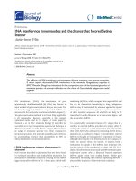
FFiigguurree 11
((aa))
Schematic diagram of archaeal phylogeny showing the evolutionary relationships between archaeal lineages for which completely sequenced
genomes are available (updated from [12]). The positions of
Ignicoccus hospitalis
and
Nanoarchaeum equitans
are indicated by arrows. The position
of
N. equitans
has been controversial [2,8], but the position suggested here has been recently supported by a specific synapomorphy (a derived
character state shared by two or more groups) [13] and signature genes [14].
((bb))
Electron micrograph showing a cell of
N. equitans
attached to a
cell of
I. hospitalis
. The scale bar is 100 nm. Courtesy of Dr Rachel Reinhard, Regensburg University. OM: outer membrane.
Thaumarchaeota
Thermoproteales
Crenarchaeota
Euryarchaeota
Sulfolobales
Desulfurococcales
“Thaumarchaeota”
Ignicoccus hospitalis
Nanoarchaeum equitans
Korarchaeota
Nanoarchaeota
Halobacteriales
Methanosarcinales
Methanomicrobiales
Thermoplasmatales
Archaeoglobales
Methanobacteriales
Methanococcales
Methanopyrales
Thermococcales
“Korarchaeota”
“Nanoarchaeota”
(a) (b)
a similar reduction process, in agreement with the position
of N. equitans in archaeal phylogenies as an euryarchaeon
possibly related to the Thermococcales [6] (Figure 1).
EExxppaannddeedd ggeennee ffaammiilliieess
Interestingly, despite an evolutionary trend toward reduc-
tion, the genome of I. hospitalis exhibits expansions of
specific gene families, notably those coding for proteins
harboring domains involved in the formation of macro-
molecular complexes, such as WD40 repeats, CBS and V4R
domains [3]. Moreover, the genome of I. hospitalis has the
lowest arCOG coverage among the Crenarchaeota: an
important fraction of its open reading frames (approxi-
mately 20%) cannot be affiliated to any arCOG [3].
The experimental characterization of these novel proteins
will surely help to decipher at the molecular level the
unique mechanisms responsible for the association between
I. hospitalis and N. equitans. The expanded family of proteins
with V4R domains will be especially interesting to study,
because these proteins are homologs to the Bet3 subunit of
the TRAPPI vesicle-tethering complex that is conserved in all
eukaryotes [9]. Microscopy observations have consistently
shown that both round and elongated membrane-coated
vesicles are released from the cytoplasmic membrane in the
periplasm of I. hospitalis and sometimes appear to come
into close proximity to, and fuse with, the outer membrane
([4] and references therein). This has led to the hypothesis
that these vesicles might be involved in transport of meta-
bolites from the host to N. equitans. It will be important to
determine whether proteins with VR4 domains indeed
participate in the vesicle-trafficking system observed in
I. hospitalis and if these vesicules are really involved in the
interaction with N. equitans. Alternative hypotheses suggest
that transport of metabolites or substrates between the two
cells occurs through unusual structures observed at the
contact point between them ([10] and references therein).
These structures could involve some of the I. hospitalis-
specific proteins revealed by the genome analysis.
HHoorriizzoonnttaall ggeennee ttrraannssffeerr iinn aa mmiinniimmaalliisstt ssyysstteemm
Using phylogenetic methods, Podar et al. [3] have identified
a number of genes that were probably acquired by hori-
zontal gene transfer (HGT), either from bacteria (4%) or
from Euryarchaeota (6%). Although limited in extent, some
of these HGT events might have been important in
permitting the combination of a streamlined genome with
efficient metabolic strategies. For instance, I. hospitalis may
use a transporter acquired from a euryarchaeon to import
ammonium for nitrogen assimilation, a wise strategy in a
highly reduced environment [9]. Similarly, I. hospitalis may
use a sulfur/polysulfide reductase complex of bacterial
origin in addition to the one normally found in Cren-
archaeota [3].
Podar et al. [3] present an extensive description of I. hospitalis
genes involved in genetic information processing, transport,
central metabolism, respiration and energy metabolism. A
major question is whether some of the genes for metabo-
lites and energy production originally present in the
N. equitans genome were transferred to the genome of
I. hospitalis and the gene products are now being imported
back into N. equitans. HGT from N. equitans to I. hospitalis
and vice versa has apparently occurred, but on a very limited
scale (only 13 such genes were identified), indicating that
N. equitans should be able to import and use metabolites
and energy (ATP) from I. hospitalis, as previously experi-
mentally demonstrated in the case of lipids and amino
acids [10].
WWhhaatt nneexxtt??
The discovery of the unique N. equitans/I. hospitalis system
has significantly increased our knowledge of the archaeal
domain in terms of its diversity (the discovery of a new
main lineage), ecology (association between two archaea)
and genomics (highly reduced archaeal genomes). The
sequencing of the genomes of both partners now provides
valuable data for elucidating the nature of this association.
Important biological questions remain to be answered,
however: for instance, we would like to know how the cell
cycles of N. equitans and I. hospitalis are coordinated. Another
important question concerns the wide diversity of associa-
tions involving a nanoarchaeal partner. Up to now, the
association between I. hospitalis and N. equitans has been
described as highly specific because attempts to infect other
species of Ignicoccus or other hyperthermophilic archaea
with N. equitans have failed. However, in the past few years,
molecular ecological studies have identified nanoarchaeal
sequences in hot marine and terrestrial environments from
geographically distant regions, suggesting that nanoarchaeota
are widely distributed around the world. More surprisingly,
nanoarchaeota have recently been reported from hyper-
saline mesophilic environments [11]. It will be extremely
interesting to determine if these sequences correspond to
free-living nanoarchaea or if they are symbiotic/parasitic
cells that have established associations similar to that
between N. equitans and I. hospitalis. In the latter case, the
presence of nanoarchaea in halophilic and mesophilic
environments might involve hosts other than I. hospitalis or
Desulfurococcales, because no mesophiles are currently
known in these lineages. The characterization of these
uncultivated nanoarchaea as well as that of their hosts will
surely bring answers to these questions.
/>Journal of Biology
2009, Volume 8, Article 7 Forterre
et al.
7.3
Journal of Biology
2009,
88::
7
AAcckknnoowwlleeddggmmeennttss
We thank Dr Rachel Reinhard for useful comments and the micrograph
in Figure 1b.
RReeffeerreenncceess
1. Huber H, Hohn MJ, Rachel R, Fuchs T, Wimmer VC, Stetter KO:
AA nneeww pphhyylluumm ooff AArrcchhaaeeaa rreepprreesseenntteedd bbyy aa nnaannoossiizzeedd hhyyppeerrtthheerr
mmoopphhiilliicc ssyymmbbiioonntt
Nature
2002,
441177::
63-67.
2. Waters E, Hohn MJ, Ahel I, Graham DE, Adams MD, Barnstead M,
Beeson KY, Bibbs L, Bolanos R, Keller M, Kretz K, Lin X, Mathur
E, Ni J, Podar M, Richardson T, Sutton GG, Simon M, Soll D,
Stetter KO, Short JM, Noordewier M:
TThhee ggeennoommee ooff
NNaann
ooaarrcchhaaeeuumm eeqquuiittaannss
:: iinnssiigghhttss iinnttoo eeaarrllyy aarrcchhaaeeaall eevvoolluuttiioonn aanndd
ddeerriivveedd ppaarraassiittiissmm
Proc Natl Acad Sci USA
2003,
110000::
12984-
12988.
3. Podar M, Anderson I, Makarova KS, Elkins JG, Ivanova N, Wall
MA, Lykidis A, Mavromatis K, Sun H, Hudson ME, Chen W, Deciu
C, Hutchison D, Eads JR, Anderson A, Fernandes F, Szeto E,
Lapidus A, Kyrpides NC, Saier MH Jr, Richardson PM, Rachel R,
Huber H, Eisen JA, Koonin EV, Keller M, Stetter KO:
AA ggeennoommiicc
aannaallyyssiiss ooff tthhee aarrcchhaaeeaall ssyysstteemm
IIggnniiccooccccuuss hhoossppiittaalliiss
NNaann
ooaarrcchhaaeeuumm eeqquuiittaannss
Genome Biol
2008,
99::
R158.
4. Junglas B, Briegel A, Burghardt T, Walther P, Wirth R, Huber H,
Rachel R:
IIggnniiccooccccuuss hhoossppiittaalliiss
aanndd
NNaannooaarrcchhaaeeuumm eeqquuiittaannss
:: uullttrraa
ssttrruuccttuurree,, cceellll cceellll iinntteerraaccttiioonn,, aanndd 33DD rreeccoonnssttrruuccttiioonn ffrroomm sseerriiaall
sseeccttiioonnss ooff ffrreeeezzee ssuubbssttiittuutteedd cceellllss aanndd bbyy eelleeccttrroonn ccrryyoottoommooggrraa
pphhyy
Arch Microbiol
2008,
119900::
395-408.
5. McCliment EA, Voglesonger KM, O’Day PA, Dunn EE, Holloway
JR, Cary SC:
CCoolloonniizzaattiioonn ooff nnaasscceenntt,, ddeeeepp sseeaa hhyyddrrootthheerrmmaall vveennttss
bbyy aa nnoovveell AArrcchhaaeeaall aanndd NNaannooaarrcchhaaeeaall aasssseemmbbllaaggee
Environ Micro-
biol
2006,
88::
114-125.
6. Brochier C, Gribaldo S, Zivanovic Y, Confalonieri F, Forterre P:
NNaannooaarrcchhaaeeaa:: rreepprreesseennttaattiivveess ooff aa nnoovveell aarrcchhaaeeaall pphhyylluumm oorr aa ffaasstt
eevvoollvviinngg eeuurryyaarrcchhaaeeaall lliinneeaaggee rreellaatteedd ttoo TThheerrmmooccooccccaalleess??
Genome
Biol
2005,
66::
R42.
7. Makarova KS, Wolf YI, Sorokin AV, Koonin EV:
CClluusstteerrss ooff oorrtthhoo
llooggoouuss ggeenneess ffoorr 4411 aarrcchhaaeeaall ggeennoommeess aanndd iimmpplliiccaattiioonnss ffoorr eevvoolluu
ttiioonnaarryy ggeennoommiiccss ooff aarrcchhaaeeaa
Biol Direct
2007,
22::
33.
8. Di Giulio M:
NNaannooaarrcchhaaeeuumm eeqquuiittaannss
iiss aa lliivviinngg ffoossssiill
J Theor Biol
2006,
224422::
257-260.
9. Podar M, Wall MA, Makarova KS, Koonin EV:
TThhee pprrookkaarryyoottiicc
VV44RR ddoommaaiinn iiss tthhee lliikkeellyy aanncceessttoorr ooff aa kkeeyy ccoommppoonneenntt ooff tthhee
eeuukkaarryyoottiicc vveessiiccllee ttrraannssppoorrtt ssyysstteemm
Biol Direct
2008,
33::
2.
10. Jahn U, Gallenberger M, Paper W, Junglas B, Eisenreich W, Stetter
KO, Rachel R, Huber H:
NNaannooaarrcchhaaeeuumm eeqquuiittaannss
aanndd
IIggnniiccooccccuuss
hhoossppiittaalliiss
:: nneeww iinnssiigghhttss iinnttoo aa uunniiqquuee,, iinnttiimmaattee aassssoocciiaattiioonn ooff ttwwoo
aarrcchhaaeeaa
J Bacteriol
2008,
119900::
1743-1750.
11. Casanueva A, Galada N, Baker GC, Grant WD, Heaphy S, Jones B,
Yanhe M, Ventosa A, Blamey J, Cowan DA:
NNaannooaarrcchhaaeeaall 1166SS
rrRRNNAA ggeennee sseeqquueenncceess aarree wwiiddeellyy ddiissppeerrsseedd iinn hhyyppeerrtthheerrmmoopphhiilliicc
aanndd mmeessoopphhiilliicc hhaalloopphhiilliicc eennvviirroonnmmeennttss
Extremophiles
2008,
1122::
651-656.
12. Brochier-Armanet C, Boussau B, Gribaldo S, Forterre P:
MMeessoopphhiilliicc CCrreennaarrcchhaaeeoottaa:: pprrooppoossaall ffoorr aa tthhiirrdd aarrcchhaaeeaall pphhyylluumm,,
tthhee TThhaauummaarrcchhaaeeoottaa
Nat Rev Microbiol
2008,
66::
245-252.
13. Urbonavicius J, Auxilien S, Walbott H, Trachana K, Golinelli-
Pimpaneau B, Brochier-Armanet C, Grosjean H:
AAccqquuiissiittiioonn ooff aa
bbaacctteerriiaall RRuummAA ttyyppee ttRRNNAA((uurraacciill 5544,,CC55)) mmeetthhyyllttrraannssffeerraassee bbyy
AArrcchhaaeeaa tthhrroouugghh aann aanncciieenntt hhoorriizzoonnttaall ggeennee ttrraannssffeerr
Mol Micro-
biol
2008,
6677::
323-335.
14. Dutilh BE, Snel B, Ettema TJ, Huynen MA:
SSiiggnnaattuurree ggeenneess aass aa
pphhyyllooggeennoommiicc ttooooll
Mol Biol Evol
2008,
2255::
1659-1667.
7.4
Journal of Biology
2009, Volume 8, Article 7 Forterre
et al.
/>Journal of Biology
2009,
88::
7