Báo cáo sinh học: "Generalized immune activation as a direct result of activated CD4+ T cell killing" doc
Bạn đang xem bản rút gọn của tài liệu. Xem và tải ngay bản đầy đủ của tài liệu tại đây (695.35 KB, 18 trang )
Open Access
Research
Generalized immune activation as a direct result of activated CD4+ T cell
killing
Rute Marques*, Adam Williams†¶, Urszula Eksmond*, Andy Wullaert‡,
Nigel Killeen§, Manolis Pasparakis‡, Dimitris Kioussis†
and George Kassiotis*
Addresses: *Division of Immunoregulation and †Division of Molecular Immunology, MRC National Institute for Medical Research,
The Ridgeway, London NW7 1AA, UK. ‡Institute for Genetics, University of Cologne, Zülpicher Strasse 47, 50674 Cologne, Germany.
§Department of Microbiology and Immunology, University of California, San Francisco, CA 94143, USA. ¶Current address: Department of
Immunobiology, Yale University School of Medicine, New Haven, CT 06520, USA.
Correspondence: George Kassiotis. Email:
Published: 27 November 2009
Received: 15 September 2009
Revised: 6 October 2009
Accepted: 7 October 2009
Journal of Biology 2009, 8:93
The electronic version of this article is the complete one and can be
found online at />
© 2009 Marques et al.; licensee BioMed Central Ltd.
This is an Open Access article distributed under the terms of the Creative Commons Attribution License ( />which permits unrestricted use, distribution, and reproduction in any medium, provided the original work is properly cited.
Abstract
Background: In addition to progressive CD4+ T cell immune deficiency, HIV infection is
characterized by generalized immune activation, thought to arise from increased microbial
exposure resulting from diminishing immunity.
Results: Here we report that, in a virus-free mouse model, conditional ablation of activated
CD4+ T cells, the targets of immunodeficiency viruses, accelerates their turnover and
produces CD4+ T cell immune deficiency. More importantly, activated CD4+ T cell killing also
results in generalized immune activation, which is attributable to regulatory CD4+ T cell
insufficiency and preventable by regulatory CD4+ T cell reconstitution. Immune activation in
this model develops independently of microbial exposure. Furthermore, microbial translocation in mice with conditional disruption of intestinal epithelial integrity affects myeloid but
not T cell homeostasis.
Conclusions: Although neither ablation of activated CD4+ T cells nor disruption of intestinal
epithelial integrity in mice fully reproduces every aspect of HIV-associated immune dysfunction in humans, ablation of activated CD4+ T cells, but not disruption of intestinal
epithelial integrity, approximates the two key immune alterations in HIV infection: CD4+
T cell immune deficiency and generalized immune activation. We therefore propose activated
CD4+ T cell killing as a common etiology for both immune deficiency and activation in HIV
infection.
See minireview />
Journal of Biology 2009, 8:93
93.2 Journal of Biology 2009,
Volume 8, Article 93
Marques et al.
Background
T lymphocyte numbers in the human body are kept
constant by homeostatic mechanisms balancing cell gain
and loss. These mechanisms eventually fail in HIV infection,
which is characterized by progressive immune deficiency,
because of loss of CD4+ T cell function [1]. HIV infection is
also associated with increased T cell turnover and activation,
which extends to uninfected cells, resulting in a state of
chronic generalized immune activation [2-5]. Indeed, the
level of activation and turnover in CD8+ T cells, which are
not infected by HIV, can be higher than in CD4+ T cells, and
this is a powerful predictor of disease progression [2,4,5].
Early views of generalized immune activation as a compensatory mechanism to achieve T cell homeostasis after virusmediated CD4+ T cell destruction [6-8] have been replaced
by alternative models in which immune activation is the
cause, rather than the consequence, of CD4+ T cell loss. In
the latter models, immune activation is considered to be
directly responsible for increased proliferation and death of
both CD4+ and CD8+ T cells [9-11]. There is a strong
positive correlation between T cell immune activation and
CD4+ T cell loss in HIV infection [12]. However, as the
precise origin of generalized immune activation is still not
fully understood, the direction of causality between CD4+
T cell loss and immune activation remains unclear.
Immunodeficiency viruses are highly selective for activated/
memory CD4+ T cells owing to the restricted expression
solely in these cells of CCR5, the co-receptor for HIV and
simian immunodeficiency virus (SIV) [13,14], or CD134
(also called OX40 or Tumor necrosis factor receptor superfamily 4, TNFRSF4), the cellular receptor for feline
immunodeficiency virus (FIV) [15]. This fraction of CD4+
T cells is characterized by substantial heterogeneity and
consists of T cells with distinct homeostatic behavior and
functional role. The two major and best characterized
subsets are antigen-experienced memory CD4+ T cells and
regulatory T (Treg) cells. Similarly to naïve CD4+ T cells,
Treg cells, which are equipped with immune-suppressive
capacity, are generated in the thymus [16,17]. Newly
generated Treg cells have a pre-activated phenotype and a
considerable fraction also show higher turnover rates than
naïve CD4+ T cells in the periphery [18,19]. Peripheral Treg
cell numbers are also regulated homeostatically. However,
the requirements for peripheral maintenance of the Treg cell
pool may differ from those for other CD4+ T cell subsets,
and precise knowledge of the relative contribution of
thymic or peripheral generation to maintenance of Treg cell
numbers remains incomplete [16,17]. Memory CD4+ T cells
are generated following the response of naïve CD4+ T cells
to infection or immunization in the periphery and mediate
immunity to re-infection. However, in contrast to the naïve
CD4+ T cell pool, maintenance of which relies to a large
/>
extent on continuous thymic production, the memory CD4+
T cell pool has considerable self-renewal capacity, regulated
independently from the naïve CD4+ T cell compartment,
and can be maintained long-term in the absence of thymic
function [20,21]. Although at the population level memory
CD4+ T cells are much longer lived than naïve CD4+ T cells,
at the individual-cell level memory CD4+ T cells show a
considerably higher turnover rate than relatively quiescent
naïve CD4+ T cells [20,22]. The high turnover rate within
the memory CD4+ T cell pool is thought to be driven, to a
variable degree, by antigen and homeostatic cytokines [20].
Although memory CD4+ T cells are the most frequent
targets for HIV replication, they do not necessarily suffer the
biggest loss during the chronic phase of infection. Indeed,
the proportion of activated CCR5+CD4+ T cells during HIV
or SIV infection correlates strongly with the degree of pathogenesis. In contrast to their loss during progressive HIV-1
infection, CCR5+CD4+ T cells are preserved in individuals
who spontaneously control HIV-1 infection [23] and are
even increased during the less pathogenic HIV-2 infection
[24]. Similarly, CCR5+CD4+ T cells are quickly lost during
rapidly progressing SIV infection of Indian-origin rhesus
macaques, but are increased in frequency during SIV infection of Chinese-origin macaques, characterized by much
slower progression to disease [25]. The paradoxical increase
in the proportion of CCR5+CD4+ T cells during less pathogenic HIV and SIV infection is thought to result from robust
replenishment of lost CD4+ T cells as part of the physiological homeostatic process, and it may also be partly fueled
by immune activation [11].
We have applied a reductionist approach to study the effect
of depletion of activated CD4+ T cells, the targets of
immunodeficiency viruses, in a virus-free mouse model. We
show here that conditional ablation of activated CD4+ T
cells greatly accelerates their turnover, with minimal apparent
effect on their numbers, and results in CD4+ T cell immune
deficiency. More importantly, activated CD4+ T cell killing
in this model also results in generalized immune activation,
independently of viral infection, reactivity to apoptotic T
cells and microbial exposure. In contrast, we further show
that generalized immune activation following activated
CD4+ T cell killing is due to an insufficiency of Treg cells.
Results
Conditional deletion of activated CD4+ T cells
To examine the consequences of activated CD4+ T cell
deletion for immune homeostasis, we generated a genetic
mouse model in which activated CD4+ T cells were killed in
the absence of retroviral infection. Activated CD4+ T cells
were targeted by conditional gene activation mediated by
Journal of Biology 2009, 8:93
Journal of Biology 2009,
/>
YFP+ cells (%)
40
40
0
+
d1
60
40
20
0
YFP
Memory
Naive
Total
Memory
Reg
(h)
CD134+
YFP+
80
Annexin V+ cells (%)
(g)
d0
Naïve
Total
0
20
Naïve Memory Reg
60
0
Marker cells (%)
40
60
0
YFP/+
YFP/DTA
60
SP
LN
20
CD134
YFP+ cells (%)
80
CD8+
80
20
(f)
100
SP
LN
20
YFP
(e)
YFP+ cells (%)
40
(d) 100
CD4+
80
Other
7
60
CD19
LN
SP
LN
80
CD8
Side scatter
4
(c) 100
100
CD4
(b)
SP
Lineage marker+ cells
(% of YFP+ cells)
(a)
Marques et al. 93.3
Volume 8, Article 93
WT
DTA
50
40
30
20
10
WT
DTA
0
0
24
48
72
Hours after activation
0
24
48
72
Hours after activation
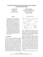
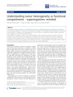
Figure 1
Specific targeting of memory and regulatory CD4+ T cells by Tnfrsf4-driven Cre expression. (a) Activated YFP expression in a subset of splenic (SP)
and lymph node cells (LN) isolated from Tnfrsf4Cre/+ R26Yfp/+ mice. Numbers within dot plots denote the percentage of YFP+ cells. (b) Percentage
(mean ± SEM, n = 6-9) of CD4+, CD8+ or CD19+ cells or cells negative for all three markers (other) in gated YFP+ cells from the spleen and lymph
nodes of Tnfrsf4Cre/+ R26Yfp/+ mice. (c,d) Percentage (mean ± SEM, n = 6-9) of YFP+ cells in (c) total, naïve (CD44loCD25-), memory (CD44hiCD25-)
and regulatory (reg; CD25+) CD4+ T cells and in (d) total, naïve (CD44loCD25-) and memory (CD44hiCD25-) CD8+ T cells, both from the spleen
and lymph nodes of Tnfrsf4Cre/+ R26Yfp/+ mice. (e) Percentage (mean ± SEM, n = 4-8) of YFP+ cells in naïve, memory and regulatory CD4+ T cells
from Tnfrsf4Cre/+ R26Yfp/+ (YFP/+) and Tnfrsf4Cre/+ R26Yfp/Dta (YFP/DTA) mice. (f) Flow cytometric example of YFP and CD134 induction 1 day (d1)
after in vitro stimulation of sorted naïve YFP- CD4+ T cells (d0) from Tnfrsf4Cre/+ R26Yfp/+ mice. (g) Percentage of YFP+ and CD134+ cells (mean ±
SEM, n = 4-6) in CD4+ T cells stimulated as in (f). (h) Percentage of annexin V+ cells following in vitro activation of sorted naïve CD4+ T cells from
Tnfrsf4Cre/+ R26Dta/+ (DTA) or control Tnfrsf4Cre/+ R26+/+ (WT) mice.
Tnfrsf4-driven Cre recombinase expression [26]. Specificity
for CD4+ T cells was confirmed by activation of a yellow
fluorescent protein (YFP) reporter gene in the Gt(ROSA)26Sor
(R26) locus [27], which revealed that about 95% of YFP+
cells were CD4+ T cells (Figure 1a,b). YFP expression in
Tnfrsf4Cre/+ R26Yfp/+ mice marked 55% and 80% of memory
and regulatory CD4+ T cells, respectively (Figure 1c). In
contrast, the vast majority of naïve CD4+ T cells (92%) and
naïve and memory CD8+ T cells (99% and 97%, respectively) were YFP- (Figure 1c,d). Conditional deletion of
CD134-expressing CD4+ T cells was achieved by Cremediated activation of a gene encoding diphtheria toxin
fragment A (DTA) independently targeted into the R26
locus (Additional data file 1).
The efficiency of DTA-mediated T cell deletion was assessed
in Tnfrsf4Cre/+ R26Yfp/Dta heterozygous mice. In comparison
with Tnfrsf4Cre/+ R26Yfp/+ mice, the proportion of YFP+
memory and regulatory CD4+ T cells in Tnfrsf4Cre/+ R26Yfp/Dta
mice was reduced by more than half (Figure 1e), suggesting
that more than 50% of the cells that were tagged with YFP
in the absence of DTA expression were killed on DTA
activation. However, this analysis ignored the dynamic
nature of T cell death and replacement. The relative
Journal of Biology 2009, 8:93
0.0
45
MIG
IL-1β
IP-10
(e)
3
2
30
9
1
9
15
26
17
0.0002
2
1
0
WT DTA
WT
DTA
Absolute cell number (x10-6)
Mphi
T cells
B cells
0
IFN-γ
(d)
CD4:CD8 ratio
0.0001
0.0001
0.04
0.04
0.0002
0.0002
0.02
0.02
0.01
0.01
0.01
0.01
0.002
0.002
0.2
MCP-1
1cm
1 cm
0.4
WT
DTA
MIP-1α
SP
DTA
Total cells
0.6
IL-12
WT
iLN
aLN
bLN
cLN
mLN
/>
(c)
(b)
Serum cytokine levels (ng/ml)
(a)
Marques et al.
Volume 8, Article 93
Absolute cell number (x10-7)
93.4 Journal of Biology 2009,
CD4+
CD8+
Memory
Naïve
Reg.
Reg
Memory
Naïve
60
5
8
40
3
8
14
6
20
41
36
26
27
0
WT DTA
WT DTA
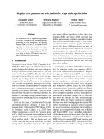
Figure 2
Immunological consequences of DTA-mediated deletion of CD134+CD4+ T cells. (a) Size of inguinal (iLN), axillary (aLN), brachial (bLN), cervical
(cLN), mesenteric (mLN) lymph nodes and spleen (SP) from Tnfrsf4Cre/+ R26Dta/+ (DTA) and littermate control Tnfrsf4Cre/+ R26+/+ (WT) mice.
(b) Serum levels (mean ± SEM, n = 5-7) of MCP-1, IL-12 (p40), IFN-γ, MIP-1α, IP-10 (CXCL10), IL-1β and MIG (CXCL9) in the same mice. (c) Total
numbers of B cells, T cells and macrophages (Mphi). P = 0.02 and P = 0.03 for total cells and B cells, respectively. (d) CD4:CD8 ratio. (e) Total
numbers (mean, n = 9-12) of naïve, memory and regulatory (reg) CD4+ T cells and naïve and memory CD8+ T cells. P = 0.0008 for regulatory CD4+
T cells; P = 0.04 for total CD8+ T cells; P = 0.0003 for memory CD8+ T cells. Numbers within bars in (c,e) denote the absolute number, ×10-7 and
×10-6, respectively, of each cell type.
presence of activated CD4+ T cells and proportion of YFP+
T cells in Tnfrsf4Cre/+ R26Yfp/Dta mice reflected equilibrium
between DTA-mediated killing, which would reduce, and
homeostatic replacement, which would increase, the
number of YFP+ activated CD4+ T cells, in addition to the
relative kinetics of YFP and DTA induction following T cell
activation. In vitro activated purified CD134-YFP- naïve
CD4+ T cells from Tnfrsf4Cre/+ R26Yfp/+ mice began to express
YFP by the first day of culture, with a delay of about 1 day
relative to CD134 induction (Figure 1f,g). However, the
effect of DTA activation on survival of in vitro activated
naïve CD4+ T cells from Tnfrsf4Cre/+ R26Dta/+ mice was not
evident until the second day of culture (Figure 1h).
Consequences of activated CD4+ T cell killing for CD4+ T
cell homeostasis
To determine whether activated CD4+ T cell deletion had any
impact on lymphocyte population dynamics, we analyzed
lymphoid organ cellularity and composition. We observed
significant systemic lymph node enlargement in Tnfrsf4Cre/+
R26Dta/+ mice compared with control Tnfrsf4Cre/+ R26+/+ mice
(Figure 2a), which was also associated with elevated serum
levels of several proinflammatory mediators (Figure 2b).
Spleen size was not appreciably affected (Figure 2a). We thus
calculated the total size of lymphocyte and myeloid
populations as the sum of the cellular contents of the spleen
and of inguinal, axillary, brachial, mesenteric and superficial
cervical lymph nodes. B cells, but not T cells or myeloid cells,
were significantly more numerous in Tnfrsf4Cre/+ R26Dta/+
mice than in control Tnfrsf4Cre/+ R26+/+ mice (Figure 2c).
Deletion of activated CD4+ T cells resulted in a substantial
systemic drop in the CD4:CD8 ratio (Figure 2d). Remarkably, compared with control mice, total CD4+ T cell
numbers in Tnfrsf4Cre/+ R26Dta/+ mice were only marginally
reduced and remained stable throughout a 6-month
observation period (Figure 2e). To assess whether activated
CD4+ T cell numbers were selectively reduced in Tnfrsf4Cre/+
R26Dta/+ mice, we determined the composition of the CD4+
T cell pool. Numbers of naïve CD4+ T cells and, notably, of
memory CD4+ T cells were similar between Tnfrsf4Cre/+
R26Dta/+ and control Tnfrsf4Cre/+ R26+/+ mice (Figure 2e),
whereas numbers of regulatory CD4+ T cells were reduced
by about 40% in Tnfrsf4Cre/+ R26Dta/+ mice (Figure 2e). In
contrast to CD4+ T cells, total numbers of CD8+ T cells were
elevated in Tnfrsf4Cre/+ R26Dta/+ mice compared with
Tnfrsf4Cre/+ R26+/+ mice, resulting from a systemic expansion
exclusively of memory CD8+ T cells (Figure 2e), which was
primarily responsible for the systemic reduction in the
CD4:CD8 ratio.
Preservation of CD4+ T cell numbers despite killing of
activated CD4+ T cells in Tnfrsf4Cre/+ R26Dta/+ mice suggested
increased replenishment, which would be associated with
functional and phenotypic activation. Phenotypic differences between naïve or memory CD4+ T cells in Tnfrsf4Cre/+
R26Dta/+ and those in control Tnfrsf4Cre/+ R26+/+ mice were
largely unremarkable, with modest increases in expression
of cytokines and of the activation markers CD43 and
CD49b in memory CD4+ T cells isolated from Tnfrsf4Cre/+
R26Dta/+ mice (Figure 3a,b). In contrast to regulatory CD4+
Journal of Biology 2009, 8:93
Journal of Biology 2009,
/>
CD44
57
33
22
68
49
18
31
13
41
74
63
72
CD62L
CD43
CD49b
CD103
0
3
0
47
22
31
36
3
TNF-α
1
Marques et al. 93.5
2
TNF-α
Cell number
1
90 59
FoxP3
6
92 38
DTA
9
8 67
WT
2
TNF-α
CD25
(c)
DTA
1
1
WT
6
15 65
DTA
Cell number
CD25
(b)
2 10
WT
Cell number
CD25
(a)
Volume 8, Article 93
0
9
1
IFN-γ
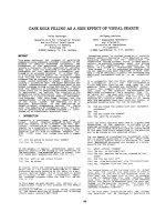
Figure 3
Effect of CD134+CD4+ T cell killing on the phonotype of CD4+ T cells. (a) Naïve, (b) memory and (c) regulatory CD4+ T cell expression of FoxP3
and activation markers, and production of cytokines following in vitro re-stimulation. Numbers within the plots represent the percentage of CD4+
T cells that were positive for each marker. Plots are representative of 4-7 mice per group.
T cells from control mice, those from Tnfrsf4Cre/+ R26Dta/+
mice showed a highly activated phenotype, characterized by
downregulation of CD62L and upregulation of CD44,
CD43, CD49b and CD103 (Figure 3c). Thus, DTA-mediated
destruction of activated CD4+ T cells had a significant effect
on regulatory CD4+ T cell numbers and activation state, but
little apparent effect on memory CD4+ T cells.
Memory CD4+ T cells, under physiological conditions,
display higher turnover rates, self-renewal potential and
activation profile than either naïve or regulatory CD4+ T cells
[1]. Indeed, naïve CD4+ T cells from either Tnfrsf4Cre/+ R26Dta/+
or control mice showed little evidence for cell division
assessed either by incorporation of bromodeoxyuridine
(BrdU) or staining with the Ki67 antibody (Figure 4a). In
contrast, population turnover rates were very high in memory
CD4+ T cells from both Tnfrsf4Cre/+ R26Dta/+ and control mice
(Figure 4b). Ki67 staining, but not BrdU incorporation, in
memory CD4+ T cells from Tnfrsf4Cre/+ R26Dta/+ mice was
elevated in comparison with that in memory CD4+ T cells
from control mice (Figure 4b). Moreover, regulatory CD4+ T
cells had a significantly higher turnover rate in Tnfrsf4Cre/+
R26Dta/+ mice than in control mice, which approached the
high turnover rate of memory CD4+ T cells (Figure 4c).
Journal of Biology 2009, 8:93
BrdU
23
10
44
MIXED
Mixed
16
4
26
6
CD44
(g)
YFP/+
YFP/DTA
100
75
50
25
0
(h)
YFP/+
YFP/DTA
40
2
4
6
8
Weeks post transfer
Reg
Memory
Naïve
75
30
20
10
0
0
2
4
6
5
0
8
Weeks post transfer
(i)
CD4+ T cells
2
6
50
52
WT host
23
25
3
6
0
0
10
Ki67
Absolute cell number (x10-6)
16
(f)
BrdU
CD4+ T cells in blood (%)
SINGLE
Single
Ki67
DTA
0.006
Reg
DTA
Ki67
DTA BM-origin
WT
30
0.025
15
Memory
WT
39
WT
DTA
20
Naïve
DTA 41
(d)
12
CD44
48
14
35
Loss of BrdU+ cells (% of subset)
DTA
WT
YFP/DTA
45
Regulatory CD4+ T cells
DTA
BrdU
B6 BM-origin
(c)
YFP/+
WT
3
DTA
CD25
WT
2
WT
2
/>
Memory CD4+ T cells
Cell number
Cell number
3
(e)
(b)
Naïve CD4+ T cells
YFP+ cells (% of CD4+ T cells)
(a)
Marques et al.
Volume 8, Article 93
CD44
93.6 Journal of Biology 2009,
DTA host
CFSE
Figure 4
Effect of DTA-mediated deletion of memory and regulatory CD4+ T cells on CD4+ T cell homeostasis. (a) Naïve, (b) memory and (c) regulatory
CD4+ T cell BrdU incorporation during a 6-day administration period, and expression of Ki67 nuclear antigen. Numbers within the plots denote the
percentage of positive CD4+ T cells and are representative of 4-6 mice per group. P = 0.011 for Ki67 staining in memory CD4+ T cells; P = 0.006 for
BrdU incorporation and P = 0.0004 for Ki67 staining in regulatory CD4+ T cells. (d) Loss of BrdU+ naïve, memory or regulatory CD4+ T cells 3 days
after cessation of a 6-day BrdU administration period. Values are the percentage (± SEM) of BrdU+ cells in each subset on day 3 after cessation of
BrdU administration minus the percentage of BrdU+ cells at the peak (day 6 of the administration period), and are representative of three mice per
group. (e) CD45.2+ Tnfrsf4Cre/+ R26Dta/+ (DTA) and CD45.1+ C57BL/6 (B6) bone marrow (BM) cells were injected separately (single) or mixed
together at 1:1 ratio (mixed) into non-irradiated Rag1-/- recipients and lymphoid organs were analyzed 12 weeks later. CD25 and CD44 expression
in gated DTA BM-origin or B6 BM-origin CD4+ T cells in these recipients is shown. Numbers within the plots denote the percentage of positive
CD4+ T cells and are representative of 4-8 mice per group analyzed in two independent experiments. (f-h) 1 × 10⁶ purified CD4+ T cells from
Tnfrsf4Cre/+ R26Yfp/+ (YFP/+) or Tnfrsf4Cre/+ R26Yfp/Dta (YFP/DTA) mice were adoptively transferred into Rag1-/- recipients and followed over time.
(f) Percentage of YFP+ cells in CD4+ T cells in the blood. (g) Percentage of CD4+ T cells in blood mononuclear cells. (h) Total numbers of naïve,
memory and regulatory (reg) CD4+ T cells in lymphoid organs at the end of the 7-week observation period. P < 0.0001 for memory CD4+ T cells;
P = 0.006 for regulatory CD4+ T cells. Values in (f-h) are the means (± SEM) of five mice per group. Numbers within bars in (h) denote the absolute
number, ×10-6, of each cell type. (i) 5 × 10⁶ purified naïve (CD44loCD25-) CD45.1+ CFSE-labeled wild-type CD4+ T cells were adoptively transferred
into Tnfrsf4Cre/+ R26Dta/+ (DTA host) and control Tnfrsf4Cre/+ R26+/+ (WT host) recipient mice. CFSE dilution and CD44 expression on gated
CD45.1+ donor CD4+ T cells isolated from the spleens and lymph nodes of recipients 6 days after transfer are shown. Numbers within the plots
denote the percentage of CFSE-CD44hi CD4+ T cells and are representative of three mice per group.
Comparable representation of BrdU+ memory CD4+ T cells
in both Tnfrsf4Cre/+ R26Dta/+ and control mice at the end of
the 6-day BrdU pulsing period (Figure 4b) seemed discordant with the expected elevated turnover of memory CD4+
T cells in Tnfrsf4Cre/+ R26Dta/+ mice. However, in memory
CD4+ T cells from Tnfrsf4Cre/+ R26Dta/+ mice, increased BrdU
incorporation would be masked by increased DTA-
mediated death of the proliferating cells during the pulsing
period. We therefore examined the fate of BrdU+ memory
CD4+ T cells 3 days after termination of BrdU administration (chase period). In contrast to the opposing action of
cell proliferation and death, which would increase or
decrease, respectively, the percentage of BrdU+ cells during
the pulsing period, cell proliferation, by dilution of BrdU
Journal of Biology 2009, 8:93
Journal of Biology 2009,
/>
label, and cell death would both decrease the percentage of
BrdU+ cells during the chase period. Indeed, almost twice as
many BrdU+ memory CD4+ T cells were lost during the
3-day chase period in Tnfrsf4Cre/+ R26Dta/+ mice as in control
mice (Figure 4d). Consistent with the Ki67 staining, the
difference in the loss of BrdU+ cells was even more pronounced in regulatory CD4+ T cells (Figure 4d). These data
together suggested that memory CD4+ T cells, and to a
higher degree regulatory CD4+ T cells, had significantly
elevated turnover rates in Tnfrsf4Cre/+ R26Dta/+ mice than in
control mice.
To further reveal the full extent of memory CD4+ T cell
killing in Tnfrsf4Cre/+ R26Dta/+ mice, we generated mixed
bone marrow chimeras. Compared with immunodeficient
mice reconstituted with wild-type bone marrow alone, those
reconstituted with Tnfrsf4Cre/+ R26Dta/+ bone marrow alone
showed a paradoxical increase in memory CD44+CD4+
T cells and a small reduction in regulatory CD25+CD4+
T cells (Figure 4e). In contrast, mice reconstituted with a
mixture of wild-type and Tnfrsf4Cre/+ R26Dta/+ bone marrow
had a severe reduction in memory and regulatory CD4+
T cell numbers of Tnfrsf4Cre/+ R26Dta/+ origin (Figure 4e).
Comparison of the Tnfrsf4Cre/+ R26Dta/+:wild-type ratio in
thymocyte and peripheral lymphocyte subsets confirmed a
significant selective loss of Tnfrsf4Cre/+ R26Dta/+-origin memory
and regulatory CD4+ T cells (Additional data file 2). Thus,
although both subsets were being killed with equal
efficiency by DTA activation in Tnfrsf4Cre/+ R26Dta/+ mice,
regulatory CD4+ T cells were incompletely replenished,
whereas the loss of memory CD4+ T cells was overcompensated.
Continuous replenishment of memory CD4+ T cells in
Tnfrsf4Cre/+ R26Dta/+ mice could be due to constant recruitment of new cells from either memory CD4+ T cells that did
not express CD134 or naïve CD4+ T cells, thymic production and peripheral numbers of which were minimally
affected in these mice. To examine the contribution of
thymic T cell production and of naïve CD4+ T cells to the
preservation of memory CD4+ T cell numbers in Tnfrsf4Cre/+
R26Dta/+ mice, we infused purified CD4+ T cells into
lymphopenic recipients (adoptive transfer). Rag1-/- mice,
which are lymphopenic due to genetic deficiency in the
V(D)J recombination activation gene Rag1 that precludes
generation of lymphocytes, were chosen as recipients.
Adoptive transfer of Tnfrsf4Cre/+ R26Yfp/+ CD4+ T cells
revealed that T cell proliferation in lymphopenic Rag1-/recipients was sufficient to drive full activation, as nearly all
of the transferred CD4+ T cells expressed the YFP reporter
within 3 weeks of transfer (Figure 4f). In contrast, the
proportion of YFP+ cells in transferred Tnfrsf4Cre/+ R26Yfp/Dta
CD4+ T cells that could activate both YFP and DTA
Volume 8, Article 93
Marques et al. 93.7
remained low throughout the 7-week observation period
(Figure 4f), similar to the low percentage of YFP+ memory
CD4+ T cells found in donor Tnfrsf4Cre/+ R26Yfp/Dta mice
(Figure 1e). More importantly, in contrast to Tnfrsf4Cre/+
R26Yfp/+ CD4+ T cells, which progressively expanded over
time in Rag1-/- recipients (Figure 4g), numbers of transferred
Tnfrsf4Cre/+ R26Yfp/Dta CD4+ T cells remained low in the
blood throughout the observation period (Figure 4g) and in
the lymphoid organs of Rag1-/- recipients 7 weeks after
transfer (Figure 4h). Failure of Tnfrsf4Cre/+ R26Yfp/Dta memory
CD4+ T cells to accumulate in the setting of naïve CD4+
T cell deficiency demonstrated the requirement for recruitment of naïve CD4+ T cells in order to maintain the pool of
memory CD4+ T cells.
To directly visualize the recruitment of naïve CD4+ T cells
into the memory pool of Tnfrsf4Cre/+ R26Dta/+ mice, we
adoptively transferred purified carboxyfluorescein succinimidyl ester (CFSE)-labeled naïve (CD44loCD25-) CD4+
T cells from wild-type donor mice expressing the allotypic
marker CD45.1. As expected, naïve CD4+ T cells failed to
proliferate or activate within 6 days of transfer into control
Tnfrsf4Cre/+ R26+/+ mice (Figure 4i). In contrast, a substantial
proportion of the progeny of transferred naïve CD4+ T cells
had divided extensively and acquired CD44 expression
during the same time in Tnfrsf4Cre/+ R26Dta/+ mice
(Figure 4i). Together, these observations support a model in
which both memory and regulatory CD4+ T cells are being
killed with equal efficiency by expression of DTA in
Tnfrsf4Cre/+ R26Dta/+ mice. However, as long as production
and maintenance of naïve CD4+ T cells is unaffected, the
continual loss of memory but not regulatory CD4+ T cells
can be efficiently compensated for by continual recruitment
of naïve CD4+ T cells.
Effect of activated CD4+ T cell killing on immune
competence
To investigate whether the accelerated death and replenishment of memory CD4+ T cells compromised immune
competence, we evaluated CD4+ T cell function in
Tnfrsf4Cre/+ R26Dta/+ mice. In comparison with Tnfrsf4Cre/+
R26+/+ littermates and C57BL/6 (B6) control mice, which
showed a strong neutralizing antibody (nAb) response to
and effectively contained infection with Friend virus (FV), a
retrovirus that causes persistent infection in mice, the FVspecific nAb response was undetectable in four out of seven
Tnfrsf4Cre/+ R26Dta/+ mice and significantly delayed in the rest
(Figure 5a). Defective FV-specific nAb response also correlated with inability of Tnfrsf4Cre/+ R26Dta/+ mice to control FV
replication (Figure 5b). Furthermore, following acute influenza A virus (IAV) infection, titers of CD4+ T cell-dependent
virus-neutralizing antibodies (nAbs) in the serum of
Tnfrsf4Cre/+ R26Dta/+ mice were reduced to 48% and to 36%
Journal of Biology 2009, 8:93
(b)
3
B6
WT
DTA
(c)
Ter119
3
3
WT
0
DTA
5
2
1
0
7
14
21
28
35
/>
Glyco-Gag
(d)
6
40
B6
WT
DTA
Change in body weight (%)
(a)
Neutralizing antibody titer (log10)
Marques et al.
Volume 8, Article 93
Neutralizing antibody titer (x10-3)
93.8 Journal of Biology 2009,
4
2
0/4
0/2
0
4/4
-20
B6
WT
DTA
-/MHC II−/−
-40
0
6
12
18
Days after IAV infection
Days after FV infection
20
10
12
14
16
6/6
18
Weeks after P. murina infection
Figure 5
Effect of activated CD4+ T cell killing on immune competence. (a) Serum titers (mean ± SEM, n = 5-7) of neutralizing antibodies (nAb) in FV-infected
DTA, wild-type (WT) and B6 mice. P ≤ 0.018 and P ≤ 0.007 on days 21 and 28, respectively, between DTA mice and either WT or B6 mice.
(b) FV-encoded glyco-Gag on the surface of infected erythroid precursor (Ter119+) cells in the same mice. Glyco-Gag+Ter119+ cells were detected
in four out of seven DTA mice. (c) Serum titers (mean ± SEM, n = 6-9) of nAb in IAV-infected DTA, WT and B6 mice. P = 0.002 between DTA and
WT mice and P = 0.00003 between DTA and B6 mice on day 18. (d) Body weight changes in Pneumocystis murina-infected DTA, WT, B6 and MHC
II-deficient (MHC II-/-) mice. P ≤ 0.04 between DTA and WT or B6 mice on weeks 16 and 17 after infection for body weight changes. Numbers
within the plot denote the ratio of mice tested positive for P. murina at the end of the observation period (P = 0.004 between DTA and control mice).
of those in Tnfrsf4Cre/+ R26+/+ littermates and B6 mice,
respectively (Figure 5c). Titration of CD4+ T cells into T celldeficient mice revealed that this degree of reduction in IAVspecific nAb titers corresponded to about 80% loss of CD4+
T cells (Additional data file 3). Lastly, in contrast to
Tnfrsf4Cre/+ R26+/+ and B6 mice, which effectively cleared
experimental infection with Pneumocystis murina, Tnfrsf4Cre/+
R26Dta/+ mice remained persistently infected and failed to
thrive, with susceptibility intermediate between T cellreplete and MHC II-deficient mice, which lacked CD4+
T cells (Figure 5d). These results showed that, despite the
presence of normal numbers of CD4+ T cells, Tnfrsf4Cre/+
R26Dta/+ mice were CD4+ T cell immune deficient.
Consequences of activated CD4+ T cell killing for CD8+ T
cell homeostasis
CD8+ T cells in HIV infection are increased in numbers and
also have a higher turnover rate and activation state [2,4,5].
This could result from either a composition change from
quiescent naïve to activated memory CD8+ T cells and/or
further activation within the memory pool. Following in
vitro stimulation of total spleen and lymph node cells, the
expanded population of memory CD8+ T cells in Tnfrsf4Cre/+
R26Dta/+ mice showed a pattern of cytokine production
similar to that in control mice (Figure 6a), indicating that
they were functionally intact. Compared with the relatively
homogeneous memory CD8+ T cell pool in control mice,
memory CD8+ T cells in Tnfrsf4Cre/+ R26Dta/+ mice contained
higher numbers of recently activated/effector CD8+ T cells,
characterized by downregulation of CD62L and upregulation of CD43 expression (Figure 6a). To examine whether
or not the expansion of the memory CD8+ T cell pool in
Tnfrsf4Cre/+ R26Dta/+ mice was due to recruitment of a greater
fraction of naïve CD8+ T cells, we adoptively purified
CD45.1+ naïve (CD44lo) CD8+ T cells labeled with CFSE
from wild-type donor mice. Naïve CD8+ T cells remained
undivided, as evident from the retention of CFSE label
6 days after transfer into either Tnfrsf4Cre/+ R26Dta/+ or
control mice (Figure 6b). In contrast, naïve CD8+ T cells
divided extensively in lymphopenic Rag1-/- recipients
(Figure 6b). This finding suggested that naïve CD8+ T cells
were not constantly recruited into the memory pool of
Tnfrsf4Cre/+ R26Dta/+ mice. The proportion of proliferating
cells in memory CD8+ T cells, assessed by BrdU incorporation during a 6-day administration period, was similarly
high in Tnfrsf4Cre/+ R26Dta/+ and control mice (Figure 6c).
However, because of the difference in memory CD8+ T cell
pool size, absolute numbers of BrdU+ memory CD8+ T cells
were significantly higher in Tnfrsf4Cre/+ R26Dta/+ mice than in
control mice (Figure 6d). Thus, the expanded memory CD8+
T cell population in Tnfrsf4Cre/+ R26Dta/+ mice seemed to be
maintained by elevated turnover within the memory pool.
We next confirmed that expansion and activation of memory
CD8+ T cells in Tnfrsf4Cre/+ R26Dta/+ mice was a consequence of
accelerated turnover of activated CD4+ T cells. In mice
Journal of Biology 2009, 8:93
Journal of Biology 2009,
/>
(a)
(b)
CD8+
CD8+ CD44hi
30
WT
CD8+ CD44hi
48
22
CD8+ CD44hi
0
0
WT
WT
WT host
51
37
DTA
40
8
DTA
2
DTA host
Rag1-/- host
DTA
DTA
24
6
CD43
B6 BM-origin
CFSE
DTA BM-origin
(f)
Single
SINGLE
3
0.003
1
7
45
55
4
Mixed
MIXED
2
1
43
0
BrdU
1
37
CD4:CD8 ratio
23
(e)
WT
DTA
CD25
WT
CD62L
(d)
BrdU+ CD44hi CD8+ T cells (x10-6)
CD8+ CD44hi
98
17
IFN-γ
CD44
Cell number
Cell number
28
CD25
DTA
WT
Cell number
3
TNF-α
CD25
23
(c)
Marques et al. 93.9
Volume 8, Article 93
B6 BM
DTA BM
Mixed
MIXED
0.001
2
1
0
CD44
Figure 6
Effect of DTA-mediated deletion of CD134+CD4+ T cells on CD8+ T cell homeostasis. (a) TNF-α and IFN-γ production and CD62L, CD43 and
CD25 expression in gated memory CD44hiCD8+ T cells from Tnfrsf4Cre/+ R26Dta/+ (DTA) and littermate control Tnfrsf4Cre/+ R26+/+ (WT) mice
(n = 5-12). Cytokine production was assessed following 4-h in vitro stimulation of total spleen and lymph node cells. (b) CFSE dilution profiles of
purified naïve (CD44lo) CD45.1+ CFSE-labeled wild-type CD8+ T cells 6 days following adoptive transfer into Tnfrsf4Cre/+ R26Dta/+ (DTA host),
control Tnfrsf4Cre/+ R26+/+ (WT host) or lymphopenic Rag1-/- recipient mice (Rag1-/- host). Numbers within the plot represent the mean percentage
of CFSE- donor CD8+ T cells in three mice per group. (c) Percentage of BrdU+ cells in memory CD44hiCD8+ T cells and (d) absolute number (mean
± SEM, n = 4) of BrdU+ cells in total CD8+ T cells following a 6-day period of BrdU administration. (e) CD44 and CD25 expression in gated CD8+ T
cells of either DTA BM origin or B6 BM origin and (f) CD4:CD8 ratio (± SEM) in Rag1-/- recipients reconstituted with either DTA or CD45.1+ B6
bone marrow (single) or a 1:1 mixture of DTA and CD45.1+ B6 bone marrow (mixed) (n = 4-7).
reconstituted with a mixture of wild-type and Tnfrsf4Cre/+
R26Dta/+ bone marrow, in which CD4+ T cell homeostasis is
restored by wild-type CD4+ T cells (Figure 4e), memory CD8+
T cells of either wild-type or Tnfrsf4Cre/+ R26Dta/+ origin were
comparable, with no signs of activation (Figure 6e) or
competitive disadvantage (Additional data file 2), and
CD4:CD8 ratios were restored (Figure 6f). Furthermore,
memory CD8+ T cells in MHC II-deficient mice contained
higher percentages of CD62L- and CD43+ activated/effector
CD8+ T cells than wild-type mice, despite proportional
increases in numbers of both naïve and memory CD8+ T cells
(Additional data file 4). Thus, memory CD8+ T cell activation
and expansion in Tnfrsf4Cre/+ R26Dta/+ mice resulted from CD4+
T cell insufficiency, rather than a cell-autonomous effect.
Causes of immune activation following activated CD4+ T cell
depletion
Accelerated turnover of activated CD4+ T cells could trigger
generalized immune activation by several distinct mechanisms, including self-reactivity to apoptotic CD4+ T cells
[28], translocation of microbial products into the intestinal
mucosa as a result of local effector CD4+ T cell depletion,
Journal of Biology 2009, 8:93
Marques et al.
/>
Although we found no evidence for intestinal pathology in
Tnfrsf4Cre/+ R26Dta/+ mice, it could be that a small degree of
histologically undetectable bacterial translocation was
occurring. To answer this question, we examined the effect
0.002
40
20
(d)
Mphi
T cells
B cells
45
30
15
(c)
12
2
5
WT
46
6
12
9
20
0
WT
DTA
Total cells
(h)
Mphi
T cells
B cells
45
30
2
1
15
1
7
7
14
1
0
−
−
DTA
0.00003
2
19
0
40
Memory
Naïve
5
3
7
10
24
24
20
6
7
13
10
WT NEMO WT NEMO
(i)
3
Reg.
Reg
Memory
Nạve
60
0
0
CD4:CD8 ratio
40
(g)
Absolute cell number (x10-7)
Serum LBP (µg/ml)
60
2
CD8+
CD4+
F4/80
WT NEMO
(f)
WT
NEMO
NEMO
0
0
(e)
3
9
Absolute cell number (x10-6)
Serum LBP (µg/ml)
WT
NEMO
Total cells
CD11b
(b)
60
Absolute cell number (x10-7)
(a)
of microbial translocation on the immune system of Ikbkgfl/Y
Vil-Cre mice, in which intestinal epithelial cell-specific
deletion of NFκB essential modulator (NEMO, encoded by
Ikbkg) leads to epithelial cell apoptosis, translocation of
bacteria into the mucosa and myeloid differentiation
primary response gene 88 (MyD88)-dependent intestinal
inflammation and colitis [31]. Consistent with disruption
of the intestinal epithelial barrier, Ikbkgfl/Y Vil-Cre mice
showed significantly elevated serum levels of lipopolysaccharide-binding protein (LBP), a surrogate marker for the
systemic presence of bacterial lipopolysaccharide, in
comparison with control mice (Figure 7a). Numbers of
CD4+
Reg.
Reg
Memory
60
Naïve
60
40
CD8+
4
5
37
2
640
20
35
CD8+ CD44hi
−
Memory
Naïve
13
20
(j)
CD62L
and loss of regulatory CD4+ T cell activity [29,30]. Reactivity
to apoptosis-related self peptides could be excluded as the
cause of CD8+ T cell activation in Tnfrsf4Cre/+ R26Dta/+ mice
because activation of these cells was not observed in mixed
bone marrow chimeras in the presence of wild-type CD4+
T cells (Figure 6e), despite continuous apoptosis of
Tnfrsf4Cre/+ R26Dta/+ CD4+ T cells.
Absolute cell number (x10-6)
Volume 8, Article 93
CD4:CD8 ratio
93.10 Journal of Biology 2009,
2
DTA
5
20
24
13
0
DTA
−
Myd88-/-
DTA
Myd88-/-
−
DTA
CD43
Myd88-/-
Figure 7
Influence of microbial exposure on immune homeostasis. (a) Serum LBP levels (mean ± SEM, n = 6-9) in Ikbkgfl/Y Vil-Cre (NEMO) and littermate
control Ikbkgfl/Y (WT) mice. The number above the bars is the P-value. (b) Total numbers (mean, n = 8-9) of B cells, T cells and macrophages (Mphi)
in Ikbkgfl/Y Vil-Cre (NEMO) and littermate control Ikbkgfl/Y (WT) mice. P = 0.0007 for macrophages. (c) Flow cytometric example of CD11b+F4/80+
macrophage expansion in the spleen of NEMO mice, compared with WT mice. (d) CD4:CD8 ratio (± SEM) in the same mice. (e) Total numbers of
naïve, memory and regulatory (reg) CD4+ T cells and naïve and memory CD8+ T cells from NEMO and control WT mice. (f) Serum LBP levels
(mean ± SEM, n = 9-13) in Tnfrsf4Cre/+ R26Dta/+ (DTA) and control Tnfrsf4Cre/+ R26+/+ (WT) mice. (g) Total numbers (mean, n = 4-6) of B cells, T
cells and macrophages (Mphi), (h) CD4:CD8 ratio and (i) total numbers of naïve, memory and regulatory (reg) CD4+ T cells and naïve and memory
CD8+ T cells in Tnfrsf4Cre/+ R26Dta/+ MyD88-/- (DTA) and littermate control Tnfrsf4Cre/+ R26+/+ MyD88-/- (-) mice. P = 0.035 for total cells; P = 0.015
for B cells; P = 0.0003 for regulatory CD4+ T cells; P = 0.018 for total CD8+ T cells; and P = 0.0003 for memory CD8+ T cells. (j) CD62L and CD43
expression in gated memory CD44hiCD8+ T cells from the same mice. Numbers within bars denote the absolute number, ×10-7 in (b,g), and ×10-6 in
(e,i), of each cell type.
Journal of Biology 2009, 8:93
Journal of Biology 2009,
/>
B cells, CD4+ and CD8+ T cell subsets and the CD4:CD8
ratio were comparable between Ikbkgfl/Y Vil-Cre mice and
control Ikbkgfl/Y mice (Figure 7b-e) and sporadic lymphocyte
expansions at inflamed mesenteric lymph nodes [31] were
at the expense of splenic counterparts (Figure 7e; and data
not shown). Moreover, T cell turnover was not systemically
increased in Ikbkgfl/Y Vil-Cre mice compared with control
mice, and an increase in BrdU incorporation in memory
CD8+ T cells in mesenteric lymph nodes was balanced by a
proportional decrease in the spleen (Additional data file 5).
In contrast to lymphocytes, numbers of cells expressing the
myeloid markers CD11b+ and F4/80+ were dramatically
elevated in all secondary lymphoid organs of Ikbkgfl/Y Vil-Cre
mice and outnumbered B cells or T cells (Figure 7b-e). Thus,
microbial translocation in this mouse model was associated
with systemic expansion of myeloid cells, but was not
sufficient for T cell activation.
We next examined the contribution of microbial exposure
to the immune activation status of Tnfrsf4Cre/+ R26Dta/+ mice.
In agreement with the absence of obvious intestinal
pathology, serum levels of LBP in Tnfrsf4Cre/+ R26Dta/+ mice
were found to be similar to those in control mice (Figure 7f),
arguing against a requirement for microbial translocation
for immune activation in Tnfrsf4Cre/+ R26Dta/+ mice. To
formally exclude any potential contribution of microbial
exposure, we rendered Tnfrsf4Cre/+ R26Dta/+ mice deficient in
MyD88. In contrast to the essential role of MyD88 in
microbial recognition driving disease in Ikbkgfl/Y Vil-Cre
mice [31], MyD88 deficiency had no effect on immune
activation in Tnfrsf4Cre/+ R26Dta/+ mice, which still experienced lymph node enlargement with accumulation of
B cells, expansion of memory CD8+ T cells, particularly of
the activated CD62L-CD43+ phenotype, and a drop in the
CD4:CD8 ratio (Figure 7g-j). Thus, MyD88-dependent
microbial exposure was not necessary for immune
activation in Tnfrsf4Cre/+ R26Dta/+ mice.
A deficit in Treg cells has been linked by some studies to
immune activation and disease progression in HIV infection
[29,30]. We therefore examined whether immune activation
in Tnfrsf4Cre/+ R26Dta/+ mice resulted from Treg cell
insufficiency. Wild-type regulatory CD4+ T cells adoptively
transferred into Tnfrsf4Cre/+ R26Dta/+ mice, but not into CD4+
T cell-replete control mice, expanded efficiently, reaching
numbers comparable to the number of regulatory CD4+
T cells in wild-type mice, and maintained expression of
FoxP3, the master regulator of Treg cell differentiation
(Figure 8a,b), revealing a homeostatic deficit in the Treg cell
pool of Tnfrsf4Cre/+ R26Dta/+ mice. The adoptively transferred
regulatory CD4+ T cells had a significant effect on the
proportion of memory CD8+ T cells, particularly those with
an activated CD44hiCD62L- phenotype, in Tnfrsf4Cre/+
Volume 8, Article 93
Marques et al. 93.11
R26Dta/+ mice; the proportion returned to wild-type levels
over a period of 3 weeks (Figure 8c,d). Regulatory T cell
reconstitution was also associated with reduction of proinflammatory cytokine serum levels in Tnfrsf4Cre/+ R26Dta/+
mice to levels comparable to those of control mice
(Figure 8e). In addition to Treg cell insufficiency, Tnfrsf4Cre/+
R26Dta/+ mice were also characterized by impaired effector
CD4+ T cell function. To examine whether reduction of
immune activation in Tnfrsf4Cre/+ R26Dta/+ mice upon
transfer of wild-type CD4+ T cells was specific to Treg cells,
we performed similar adoptive transfer experiments using
total wild-type CD4+ T cells, consisting predominantly of
non-regulatory conventional CD4+ T cells, which gave rise
to effector CD4+ T cells. Although wild-type effector CD4+
T cells efficiently reconstituted Tnfrsf4Cre/+ R26Dta/+ mice, but
not control mice, they had no effect on the proportion of
memory CD8+ T cells or activated CD44hiCD62L-CD8+
T cells (Additional data file 6). Reconstitution of Tnfrsf4Cre/+
R26Dta/+ mice with wild-type effector CD4+ T cells also failed
to rescue the reduced host Treg cell numbers in Tnfrsf4Cre/+
R26Dta/+ mice (Additional data file 7). Thus, immune activation in Tnfrsf4Cre/+ R26Dta/+ mice was due to a deficit in
regulatory, but not effector, CD4+ T cell function.
Discussion
The complex and contrasting immune alterations in HIV
infection are currently thought to have distinct etiologies
[11,32]. Instead, our results suggest that the complexity of
immune dysfunction in infection with HIV may simply
reflect the functional heterogeneity of its targets. Conditional
virus-free killing of activated CD4+ T cells, which include
both memory and regulatory subsets, was directly
responsible for the development not only of immune
deficiency, but also of activation.
As a result of the expression pattern of their receptors/coreceptors [13-15], replication of immunodeficiency viruses
is largely restricted to the activated fraction of CD4+ T cells.
The finding that viruses such as HIV and FIV, which use
different combinations of receptors/co-receptors to infect
activated CD4+ T cells, cause similar disease [13-15]
indicates that specificity for target cells is more important
for disease development than the cellular receptor
conferring this specificity. Another important determinant
of the rate of disease progression is the age at which HIV
infection is acquired. Neonates generally develop symptomatic infection faster than adults, possibly because of the
relative immaturity of the neonatal immune system and
thus its inability to fully respond to the infection. Although
a difference in an antiviral immune response would not
influence the phenotype resulting from DTA-mediated
CD4+ T cell killing between neonate and adult mice, other
Journal of Biology 2009, 8:93
93.12 Journal of Biology 2009,
(a)
/>
(c)
6
(d)
(e)
hi
CD44hi
WT host
DTA host
4
0.60
90
45
CD44hi
WT host
hos
DTA host
ho
30
IFN-γ
WT host
DTA host
60
WT host
DTA host
0.40
0.013
0
0
2
4
6
8
Weeks after transfer
(b)
30
0
CD44hi CD62L−CD44hi CD62L
0
hi CD62L
CD44hi CD62L-
1
17
FoxP3
20
10
0.0047
0.20
0.00
MCP-1
0.033
0.50
11
CD45.1
0.2
10
5
DTA host
WT host
0.2
15
% of CD8+ T cells
2
Serum cytokine levels (ng/ml)
0.0053
% of CD8+ T cells in blood
Donor cells (% of CD4+ T cells)
Marques et al.
Volume 8, Article 93
4
0
0.25
0
0
2
4
6
0.00
−
8
+ Treg
−
+ Treg
Weeks after transfer
Figure 8
Restoration of immune homeostasis in Tnfrsf4Cre/+ R26Dta/+ mice by regulatory T cells. (a) Expansion of donor-type T cells in the blood of
Tnfrsf4Cre/+ R26Dta/+ (DTA host) and control Tnfrsf4Cre/+ R26+/+ (WT host) recipients of purified regulatory CD45.1+CD25+CD4+ T cells. (b)
Expansion and retention of FoxP3 expression in donor CD45.1+CD4+ T cells. Plots show gated CD4+ T cells from lymphoid organs of recipient mice
at the end of a 10-week observation period. (c) Percentage of CD44hi (top) and CD62L-CD44hi (bottom) CD8+ T cells in the blood of the same
recipients of regulatory CD25+CD4+ T cells. (d) Absolute numbers of CD44hi (top) and CD62L-CD44hi (bottom) CD8+ T cells in lymphoid organs of
either DTA and WT mice that did not receive regulatory CD25+CD4+ T cells (-) or DTA and WT mice 10 weeks after transfer of CD25+CD4+ T
cells (+ Treg). Values in (a-d) represent the mean (± SEM) of 7-10 mice per group pooled from three independent experiments. (e) Mean serum
levels (± SEM) of IFN-γ and MCP-1 in the same mice. Similar results were obtained for IL-12p40, MIP-1α and IL-1β.
factors may influence its severity. A limitation of the current
approach is that CD4+ T cell killing by CD134-driven DTA
activation starts as soon as activated T cells are generated
(within the first 3 days of birth in mice), which would thus
correspond only to neonatal HIV infection. It would be
important, once the tools become available, to compare the
effect of DTA-mediated CD4+ T cell killing in neonate and
adult mice.
During the chronic phase of HIV or SIV infection only a
small proportion of total CD4+ T cells thought to be
susceptible have been found to be infected at any one time
[33]. In contrast, studies with SIV in rhesus macaques have
revealed that up to 50% of all memory CD4+ T cells are
systemically killed during acute SIV infection [34,35].
Similarly, although approximately 50% of memory CD4+
T cells were cumulatively marked by selection-neutral YFP
expression in Tnfrsf4Cre/+ R26Yfp/+ mice, the potential for
DTA-mediated death upon CD4+ T cell activation in
Tnfrsf4Cre/+ R26Dta/+ mice had surprisingly little effect on
memory CD4+ T cell survival and homeostasis. The reasons
for this apparent ‘resistance’ to virus-mediated killing of
susceptible CD4+ T cell targets during the chronic phase of
HIV infection are not known, but may be related to the
naturally short lifespan of activated CD4+ T cells, even when
uninfected. The lifespan of HIV-infected CD4+ T cells (the
interval between virus entry and T cell death) has been
estimated to be about 48 hours [36], which is very similar
to the lifespan of activated CD4+ T cells in Tnfrsf4Cre/+
R26Dta/+ mice (there is about a 48 hour interval between
T cell activation and DTA-mediated death). It has been
postulated that HIV replication is mostly restricted to
Journal of Biology 2009, 8:93
Journal of Biology 2009,
/>
relatively short-lived cellular targets [11], and it is therefore
possible that the high natural turnover of activated CD4+
T cells masks virus-induced death. Alternatively, the apparent
‘resistance’ of activated CD4+ T cells during chronic HIV
infection may represent selection for true HIV resistance in
the CD4+ T cell population [37].
Despite being the major target of virus replication, the
proportion of CCR5+CD4+ T cells paradoxically increases
during less pathogenic HIV and SIV infection [11]. It may
thus be unsurprising that despite efficient killing of memory
CD4+ T cells, their numbers in Tnfrsf4Cre/+ R26Dta/+ mice are
preserved or even elevated under conditions of low thymic
output. Although it will be important to establish why
memory CD4+ T cell replenishment eventually fails in more
pathogenic HIV and SIV infection, our results also indicate
that there is still CD4+ T cell immunodeficiency even
though numbers of memory CD4+ T cells are not reduced at
the population level. Studies in HIV infection have
established that susceptibility to different infections is
related to the degree of reduction in CD4+ T cell counts in
the blood [38]. These findings could suggest that protection
against different infections requires a different number of
CD4+ T cells. Alternatively, susceptibility to different infections at different CD4+ T cell counts could indicate a
progressive decline in other arms of the adaptive immune
system, especially CD8+ T cells, a decline that correlates
with the decline in CD4+ T cells. The finding that Tnfrsf4Cre/+
R26Dta/+ mice show immunodeficiency in assays for CD4+
T cell-mediated protection suggests that apart from the total
number of memory CD4+ T cells, the lifespan of individual
clones and the clonal composition of the total memory
pool are also crucial for immune competence.
In addition to immunodeficiency, conditional deletion of
activated/memory CD4+ T cells by CD134-driven DTA
activation also leads to generalized immune activation,
which shares many features with HIV infection-associated
immune activation. Several distinct mechanisms have
recently been proposed to underlie immune activation in
HIV infection. A strong innate response to HIV components
is thought to contribute to generalized immune activation
and an attenuated innate response has been correlated with
the non-pathogenic nature of SIV infection in sooty
mangabeys [39]. Incomplete removal of apoptotic material
as a result of accelerated T cell death in HIV infection has
been proposed to induce self-reactive CD8+ T cells [28]. HIV
infection induces an early and extensive depletion of
effector CD4+ T cells at the intestinal mucosa, and it is
thought that diminishing local immunity permits
translocation of microbial products, which in turn causes
generalized immune activation [32]. Lastly, the regulatory
subset of CD4+ T cells is targeted by HIV, SIV and FIV
Volume 8, Article 93
Marques et al. 93.13
[40-43] and a relative deficit in this subset has been linked
by certain studies to immune activation and disease
progression [29,30,40,42,44,45].
Our results support a model in which generalized immune
activation originates primarily from a relative insufficiency
in Treg cells. The immune activation that develops in
Tnfrsf4Cre/+ R26Dta/+ mice is diminished upon restoration of
CD4+ T cell homeostasis by wild-type CD4+ T cells in bone
marrow chimeras, demonstrating that immune activation in
these mice results from dysregulated CD4+ T cell homeostasis. The two subsets of CD4+ T cells that are affected in
Tnfrsf4Cre/+ R26Dta/+ mice include memory/effector and
regulatory CD4+ T cells, whereas naïve CD4+ T cells are
unaffected, suggesting that immune activation is due to
insufficiency in either memory/effector or regulatory CD4+
T cells, or both (general activated CD4+ T cell lymphopenia). Treg cell numbers are reduced in Tnfrsf4Cre/+ R26Dta/+
mice, and the degree of relative Treg cell-specific lymphopenia in these mice is fully revealed by the expansion of
adoptively transferred wild-type Treg cells. Furthermore,
adoptive transfer of wild-type Treg cells reduces the immune
activation seen in untreated Tnfrsf4Cre/+ R26Dta/+ mice. In
contrast, adoptive transfer of total CD4+ T cells, consisting
largely of memory/effector CD4+ T cells, does not appreciably reduce immune activation in Tnfrsf4Cre/+ R26Dta/+
mice. Lastly, immune activation in Tnfrsf4Cre/+ R26Dta/+ mice
bears many similarities to the inflammatory disease that
develops in mice with genetic deficiency in Treg cells [46].
Together, these findings indicate a causal link between Treg
cell insufficiency and generalized immune activation in
Tnfrsf4Cre/+ R26Dta/+ mice.
It is now clear that Treg cells are targeted by HIV, SIV and
FIV [40-43]. However, their fate during infection remains
controversial. The presence of Treg cell activity can be
demonstrated during progression of HIV or SIV infection,
and several studies have suggested that Treg cells are
numerically increased and functionally activated during
infection [29,47-50]. In contrast, other studies have
indicated that Treg cells are lost during progression of HIV
and SIV infection and shown a correlation between this loss
and immune activation [29,30,40,42,44,45]. Given that
Treg cell homeostasis relies mainly on growth factors, such
as interleukin (IL)-2, produced by activated effector T cells,
loss of effector CD4+ T cells during pathogenic HIV or SIV
infection would be expected to contribute to the loss of Treg
cells, in addition to virus-mediated destruction. Loss of Treg
cells in our model seems to be mainly due to intrinsic
CD134-driven DTA activation, rather than loss of effector
CD4+ T cells. Firstly, expansion and maintenance of wildtype Treg cells adoptively transferred into Tnfrsf4Cre/+
R26Dta/+ mice is fully supported, indicating sufficient
Journal of Biology 2009, 8:93
93.14 Journal of Biology 2009,
Volume 8, Article 93
Marques et al.
provision of Treg cell growth factors in these mice.
Secondly, Treg cells of Tnfrsf4Cre/+ R26Dta/+ origin are severely
reduced in numbers in bone marrow chimeras between
Tnfrsf4Cre/+ R26Dta/+ and wild-type cells, despite the presence
of normal numbers of wild-type effector CD4+ T cells in
these chimeric mice. Lastly, reconstitution of effector T cells,
by adoptive transfer of wild-type CD4+ T cells into
Tnfrsf4Cre/+ R26Dta/+ mice, fails to restore the numbers of host
Treg cells, suggesting that the defect in their homeostasis is
intrinsic and not due to lack of effector CD4+ T cells.
Thus, although a role for Treg cells in HIV disease
progression is highlighted by all studies, it has remained
unclear whether HIV infection is facilitated by excessive or
insufficient Treg cell activity. Differences in methodology
used to quantify Treg cells notwithstanding, whether Treg
cell activity is increased or lost during progression of HIV
infection will also depend on the behavior of cells that need
to be regulated. Indeed, expansion and activation of Treg
cells is not at odds with insufficient regulation if expansion
and activation of effector CD4+ and CD8+ T cells is
disproportionally higher. Furthermore, studies in natural
hosts of SIV have suggested that preservation or functional
redundancy of regulatory subsets could be responsible for
the lack of immune activation and disease progression in
non-pathogenic SIV infection [42,51].
The immune alterations that arise from conditional
ablation of activated CD4+ T cells in the system used here
do not reproduce the entire spectrum of immune dysfunction that characterizes the various stages of HIV infection,
indicating a multifactorial origin. Nevertheless, the enlargement of the lymph nodes, elevated serum levels of proinflammatory cytokines and chemokines, hyperplasia of the
B cell compartment and increased T cell turnover and
activation in Tnfrsf4Cre/+ R26Dta/+ mice lend further support
to the idea that generalized immune activation may result
from Treg cell insufficiency. However, it also raises the
question of why immune activation in the setting of HIV
infection and in Tnfrsf4Cre/+ R26Dta/+ mice is not associated
with overt autoimmunity, as it is in Treg cell-deficient mice
[46]. Perhaps disastrous autoimmunity develops during
complete Treg cell deficiency, whereas HIV infection and
Tnfrsf4Cre/+ R26Dta/+ mice show only partial Treg cell loss.
Furthermore, most of the effector CD4+ T cells that would
otherwise mediate self-tissue damage are also targeted in
HIV infection and in Tnfrsf4Cre/+ R26Dta/+ mice, and thus a
substantial pathogenic component is removed.
Collectively, our results support a model for HIV pathogenesis in which immune deficiency and activation
originate from virus-mediated killing of memory and regulatory CD4+ T cells, respectively. According to the proposed
/>
model, generalized immune activation is a consequence,
rather that the cause, of accelerated CD4+ T cell turnover.
Nevertheless, once instigated, immune activation will also
contribute to the progressive loss of CD4+ T cells by
completing the cycle of cell activation and death. Defining
the precise balance between CD4+ T cell killing and
immune activation and deficiency will be vital to our
understanding of the pathogenesis of immune deficiency
virus infection and to any effort to influence its outcome in
favor of the host.
Materials and methods
Mice
Inbred C57BL/6 (B6) and CD45.1-congenic B6 mice
(B6.SJL-Ptprca Pep3b/BoyJ) were originally obtained from the
Jackson Laboratory (Bar Harbor, USA) and were
subsequently maintained at NIMR. B6-backcrossed Rag1deficient mice [52] (B6.129S7-Rag1tm1Mom/J, called Rag1-/here), T cell receptor α (TCRα)-deficient mice [53] (B6.129Tcratm1Phi, called Tcra-/-), MHC II-deficient mice [54]
(B6.129S2-H2dlAb1-Ea/J, called MHC II-/-) and MyD88deficient mice [55] (B6.129-Myd88tm1Aki, called Myd88-/-)
have been previously described and have also been maintained at NIMR. Mice with an activatable gene encoding YFP
targeted into the ubiquitously expressed Gt(ROSA)26Sor
(R26) locus have been described [27] and were backcrossed
onto the B6 genetic background for at least ten generations.
Mice with an activatable gene encoding DTA targeted into
the R26 locus were generated by gene targeting in embryonic stem cells. Briefly, the pUC-DTA vector containing a
truncated gene encoding DTA (amino acids 3-193), in
which the initial Met-Asp-Pro sequence is donated by the
human metallothionein IIA, and which is followed by an
SV40 carboxy-terminal Ser-Leu and small t intron, was
kindly provided by Ian Maxwell, University of Colorado,
Denver, USA. This fragment was subsequently inserted into
the pBigT cassette, which also contained a loxP-flanked
(floxed) neomycin-resistance gene (neo) and a triple polyadenylation signal (tpA). The floxed neo-tpA-DTA fragment
was subcloned into the RODA26PA vector, which was then
linearized and electroporated into R1 embryonic stem cells.
Targeted embryonic stem cell clones were injected into B6
blastocysts and germline transmitting R26Dta/+ mice were
backcrossed onto the B6 genetic background for at least six
generations.
Mice with a targeted insertion of Cre recombinase into the
Tnfrsf4 locus were generated by gene targeting in embryonic
stem cells [26]. Tnfrsf4Cre mice were backcrossed onto the B6
genetic background for at least six generations. To obtain
Tnfrsf4Cre/+ R26Dta/+ progeny, homozygous Tnfrsf4Cre/Cre mice
Journal of Biology 2009, 8:93
Journal of Biology 2009,
/>
were mated with heterozygous R26Dta/+ mice. Tnfrsf4Cre/+
R26Dta/+ mice were born at expected Mendelian ratios and
were viable with no clinical or histological signs of disease
or pathology. A small proportion (<8%) of Tnfrsf4Cre/+
R26Dta/+ mice showed retarded development, which was
evident as early as weaning. Histopathology analysis
revealed exocrine pancreatic atrophy, consistent with the
animals’ small size, with absence of inflammatory infiltrates. All remaining organs, including endocrine pancreas,
were normal. These mice were excluded from further
analysis. In all experiments Tnfrsf4Cre/+ R26+/+ littermates
were included as controls for Tnfrsf4Cre/+ R26Dta/+ mice to
control for any potential effects of CD134-hemizygosity.
Mice with intestinal epithelial cell-specific deletion of Ikbkg
(encoding IKKγ, also called NEMO) were obtained by
crossing mice with a Cre-deletable Ikbkg allele (Ikbkgfl) with
mice expressing Cre under the intestinal epithelial-specific
villin promoter (Vil-Cre) and have been previously
described [31]. Ikbkgfl/Y Vil-Cre and control Ikbkgfl/Y mice
were bred and maintained at the Institute for Genetics,
Cologne, Germany. Spleens and lymph nodes from Ikbkgfl/Y
Vil-Cre and control Ikbkgfl/Y male mice were harvested at
Cologne and shipped in Iscove’s modified Dulbecco’s
medium (IMDM) to NIMR, where they were analyzed the
following day. All animal experiments were conducted
according to local government regulations and institutional
guidelines.
Infections
The A/PR/8/34 (PR8) strain of influenza A virus (IAV;
kindly provided by Rose Gonsalves, Division of Virology,
NIMR) was an allantoic fluid preparation from PR8-infected
embryonated eggs. Non-anesthetized mice were infected
with 250 hemagglutinin units of PR8 by instillation onto
their nasal cavities. Serum titers of IAV-neutralizing
antibodies were measured as previously described [56]. The
Friend virus (FV) used in this study is a retroviral complex
of a replication-competent B-tropic helper murine leukemia
virus (F-MuLV-B) and a replication-defective polycythemiainducing spleen focus-forming virus, referred to as FV. The
FV stock (kindly provided by Kim Hasenkrug, Laboratory of
Persistent Viral Diseases, Rocky Mountain Laboratories,
NIAID, NIH, Hamilton, USA) was free of lactate dehydrogenase-elevating virus and was obtained as previously
described [57].
FV was propagated in vivo and prepared as 10% w/v homogenate from the spleen of 12-day infected BALB/c mice.
Mice received an inoculum of FV complex containing
1,000-2,000 spleen focus-forming units injected via the tail
vein in 0.1 ml of phosphate-buffered saline. Cell-associated
virus in infected mice was estimated by flow cytometric
Volume 8, Article 93
Marques et al. 93.15
detection of infected cells using surface staining for the
glycosylated product of the viral gag gene (glyco-Gag), using
the matrix-specific monoclonal antibody 34 (mouse
IgG2b), followed by an anti-mouse IgG2b-fluorescein
isothiocyanate (FITC) secondary reagent (BD Biosciences,
San Jose, USA). Serum titers of FV-neutralizing antibodies
were measured as previously described [57].
Pneumocystis murina was obtained from ATCC/LGC
Promochem (stock PRA-111) and administered to nonanesthetized mice by instillation onto their nasal cavities. P.
murina was detected in formalin-fixed lung tissue by
Gomori’s silver stain of lung sections by IZVG Pathology
(Leeds, UK) and in fresh lung tissue by PCR specific for the
P. murina mitochondrial large-subunit rRNA gene [58].
Flow cytometry
Cells were stained with directly conjugated antibodies to
surface markers, obtained from eBiosciences (San Diego,
USA), CALTAG/Invitrogen (Carlsbad, USA) or BD Biosciences.
B cells and macrophages were identified as B220+ and
CD11b+F4/80+ cells, respectively. Naïve, memory and regulatory T cells were identified as CD44loCD25-, CD44hiCD25and CD25+ cells, respectively. Four- and eight-color cytometry were performed on FACSCalibur (BD Biosciences)
and CyAn (Dako, Fort Collins, USA) flow cytometers,
respectively, and analyzed with FlowJo v8.7 (Tree Star Inc.,
Ashland, USA) or Summit v4.3 (Dako) analysis software,
respectively. For detection of cytokine synthesis, cells were
stained for surface markers and stimulated for 4 h with
phorbol 12,13-dibutyrate and ionomycin (both at
500 ng/ml), in the presence of monensin (1 µg/ml). Cells
were then fixed and permeabilized using buffers from
eBiosciences, before intracellular staining with tumor necrosis
factor (TNF)-α- and interferon (IFN)-γ-specific antibodies
(eBiosciences). FoxP3 was detected by intranuclear staining
using a FoxP3-staining kit (eBiosciences) according to the
manufacturer’s instructions. Apoptotic cells were stained
using an Annexin V staining kit (BD Biosciences) according
to the manufacturer’s instructions. Cellular turnover was
assessed by BrdU incorporation during a 6-day administration into the drinking water of mice, and in addition
3 days after cessation of BrdU administration. BrdU
incorporation was measured using a BrdU staining kit (BD
Biosciences) according to the manufacturer’s instructions.
Ki67-expressing cells were identified using an anti-human
Ki67 or matched isotype control (clones B56 and MOPC-2,
respectively, BD Biosciences).
Cell isolation, purification, labeling and transfer
Single cell suspensions were prepared from thymus, spleen
or lymph nodes of donor mice by mechanical disruption.
Spleen suspensions were treated with ammonium chloride
Journal of Biology 2009, 8:93
93.16 Journal of Biology 2009,
Volume 8, Article 93
Marques et al.
for erythrocyte lysis. Lymph node cellularity was calculated
as the sum of the cellular contents of inguinal, axillary,
brachial, mesenteric and superficial cervical lymph nodes.
Total cell numbers of the various lymphoid and myeloid
subsets were the sum of the splenic and lymph node
content.
Bone marrow cell suspensions were prepared by flushing
the bone cavities of femurs and tibiae from donor mice with
IMDM. Target cells were enriched in lymph node and
spleen suspensions using immunomagnetic positive selection (EasySep beads, StemCell Technologies, Vancouver,
Canada) according to the manufacturer’s instructions.
Enriched cell suspensions were stained with antibodies to
surface markers and then further purified by cell sorting,
performed on MoFlo cell sorters (Dako). Typical cell purity
following cell sorting was higher than 98%. In some
experiments, purified cells were further labeled with CFSE
(Molecular Probes/Invitrogen, Carlsbad, USA). Purified cells
(1 × 10⁶ per recipient for unlabeled cells or 5 × 10⁶ per
recipient for CFSE-labeled cells) were injected into recipient
mice through the tail vein in 0.1 ml of air-buffered IMDM.
Bone marrow chimeras
CD45.2+ Tnfrsf4Cre/+ R26Dta/+ (DTA) and CD45.1+ C57BL/6
(B6) wild-type bone marrow cells were injected separately
or mixed together (mixed bone marrow chimeras) into nonirradiated Rag1-/- recipients expressing the allotypic marker
CD45.2. Each recipient received one mouse-equivalent of
bone marrow cells. Mice were bled periodically for assessment of reconstitution and lymphoid organs were analyzed
12 weeks after bone marrow transfer. In separate experiments, CD45.1+ B6 bone marrow cells were injected into
non-irradiated CD45.2+ Rag1-/- recipients and reconstitution
of lymphoid, myeloid and erythroid lineages by donor-type
cells was assessed. This analysis revealed that in nonirradiated Rag1-/- recipients only the lymphoid lineage (T
and B cells) is reconstituted by donor-type cells, whereas all
other lineages were still host-derived.
/>
Levels of serum LBP were determined by ELISA (HyCult
Biotech, Uden, The Netherlands) according to manufacturer’s instructions.
Statistical analysis
Parametric comparisons were made by Student’s t-test
performed using SigmaPlot v10 software (Systat Software
Inc., San Jose USA). Fisher’s exact test was used specifically
for the non-parametric comparison of P. murina-infected
and non-infected mice.
Additional data files
The following additional data files are available.
Additional data file 1: Strategy for targeting of an
activatable DTA-encoding gene into the R26 locus.
Additional data file 2: DTA-mediated deletion of memory
and regulatory CD4+ T cells in mixed bone marrow
chimeras. Additional data file 3: CD4+ T cell number
dependency of influenza A virus (IAV)-neutralizing
antibody induction. Additional data file 4: CD8+ T cell
number and phenotype in MHC II-deficient mice.
Additional data file 5: Effect of microbial translocation on
lymphocyte turnover in Ikbkgfl/Y Vil-Cre mice. Additional
data file 6: Effect of CD4+ T cell reconstitution on CD8+ T
cell activation in Tnfrsf4Cre/+ R26Dta/+ mice. Additional data
file 7: Effect of CD4+ T cell reconstitution on Treg cell
number in Tnfrsf4Cre/+ R26Dta/+ mice.
Acknowledgements
We thank A O’Garra, B Stockinger, R Zamoyska, J Langhorne and B
Seddon for discussion. We are grateful for assistance from the Division
of Biological Services and from the Flow Cytometry Facility at NIMR.
This work was supported by the UK Medical Research Council.
References
1.
2.
In vitro T cell activation
Single cell suspensions were prepared from the spleen or
lymph nodes of donor mice and 0.5 × 10⁶ purified T cells
per well were stimulated in 96-well plates with CD3/CD28coated beads (Mouse CD3/CD28 T Cell Expander, Dynal/
Invitrogen, Carlsbad, USA) at 1:1 ratio for the indicated
length of time.
3.
4.
Serum cytokines and LBP
Levels of serum cytokines were assessed by multiplex
cytokine bead arrays (20-plex Panel, BioSource/Invitrogen,
Carlsbad, USA; or 23-plex Panel, Bio-Rad, Hercules, USA)
using the Luminex 100 System (Luminex, Austin, USA).
5.
Douek DC, Picker LJ, Koup RA: T cell dynamics in HIV-1 infec tion. Annu Rev Immunol 2003, 21:265-304.
Giorgi J, Hultin L, McKeating J, Johnson T, Owens B, Jacobson L,
Shih R, Lewis J, Wiley D, Phair J, Wolinsky SM, Detels R: Shorter
survival in advanced human immunodeficiency virus type 1 infec tion is more closely associated with T lymphocyte activation than
w i t h p l a s m a v i r u s b u r d e n o r v i r u s c h e m o k i n e c o r e c e p t or u s a g e . J
Infect Dis 1999, 179:859-870.
Finkel TH, Tudor-Williams G, Banda NK, Cotton MF, Curiel T,
Monks C, Baba TW, Ruprecht RM, Kupfer A: Apoptosis occurs
predominantly in bystander cells and not in productively infected
cells of HIV- and SIV-infected lymph nodes. Nat Med 1995, 1:129134.
Hazenberg MD, Otto SA, van Benthem BH, Roos MT, Coutinho
RA, Lange JM, Hamann D, Prins M, Miedema F: Persistent immune
activation in HIV-1 infection is associated with progression to
AIDS. AIDS 2003, 17:1881-1888.
Deeks SG, Kitchen CMR, Liu L, Guo H, Gascon R, Narvaez AB,
Hunt P, Martin JN, Kahn JO, Levy J, McGrath MS, Hecht FM:
Immune activation set point during early HIV infection predicts
subsequent CD4+ T-cell changes independent of viral load. Blood
2004, 104:942-947.
Journal of Biology 2009, 8:93
Journal of Biology 2009,
/>
6.
7.
8.
9.
10.
11.
12.
13.
14.
15.
16.
17.
18.
19.
20.
21.
22.
23.
24.
25.
26.
Ho DD, Neumann AU, Perelson AS, Chen W, Leonard JM,
Markowitz M: Rapid turnover of plasma virions and CD4 lympho cytes in HIV-1 infection. Nature 1995, 373:123-126.
Margolick JB, Munoz A, Donnenberg AD, Park LP, Galai N, Giorgi
JV, O’Gorman MRG, Ferbas J: Failure of T-cell homeostasis pre ceding AIDS in HIV-1 infection. Nat Med 1995, 1:674-680.
Mohri H, Perelson AS, Tung K, Ribeiro RM, Ramratnam B,
Markowitz M, Kost R, Hurley, Weinberger L, Cesar D, Hellerstein
MK, Ho DD: Increased turnover of T Lymphocytes in HIV-1 infec tion and its reduction by antiretroviral therapy. J Exp Med 2001,
194:1277-1288.
Hazenberg MD, Stuart JWTC, Otto SA, Borleffs JCC, Boucher
CAB, De Boer RJ, Miedema F, Hamann D: T-cell division in human
immunodeficiency virus (HIV)-1 infection is mainly due to immune
activation: a longitudinal analysis in patients before and during
highly active antiretroviral therapy (HAART). Blood 2000, 95:249255.
Grossman Z, Meier-Schellersheim M, Sousa AE, Victorino RM,
Paul WE: CD4+ T-cell depletion in HIV infection: are we closer to
understanding the cause? Nat Med 2002, 8:319-323.
Grossman Z, Meier-Schellersheim M, Paul WE, Picker LJ: Patho genesis of HIV infection: what the virus spares is as important as
what it destroys. Nat Med 2006, 12:289-295.
Sousa AE, Carneiro J, Meier-Schellersheim M, Grossman Z,
Victorino RMM: CD4 T cell depletion is linked directly to immune
activation in the pathogenesis of HIV-1 and HIV -2 but only indirectly to the viral load. J Immunol 2002, 169:3400-3406.
Alkhatib G, Combadiere C, Broder CC, Feng Y, Kennedy PE,
Murphy PM, Berger EA: CC CKR5: A RANTES, MIP-1alpha, MIP1beta receptor as a fusion cofactor for macrophage-tropic HIV-1.
Science 1996, 272:1955-1958.
Chen Z, Zhou P, Ho DD, Landau NR, Marx PA: Genetically diver gent strains of simian immunodeficiency virus use CCR5 as a core ceptor for entry. J Virol 1997, 71:2705-2714.
Shimojima M, Miyazawa T, Ikeda Y, McMonagle EL, Haining H,
Akashi H, Takeuchi Y, Hosie MJ, Willett BJ: Use of CD134 as a
primary receptor by the feline immunodeficiency virus. Science
2004, 303:1192-1195.
Josefowicz SZ, Rudensky A: Control of regulatory T cell lineage commitment and maintenance. Immunity 2009, 30:616-625.
Sakaguchi S, Yamaguchi T, Nomura T, Ono M: Regulatory T cells
and immune tolerance. Cell 2008, 133:775-787.
Feuerer M, Hill JA, Mathis D, Benoist C: Foxp3+ regulatory T cells:
differentiation, specification, subphenotypes. Nat Immunol 2009,
10:689-695.
Fisson S, Darrasse-Jeze G, Litvinova E, Septier F, Klatzmann D,
Liblau R, Salomon BL: Continuous activation of autoreactive CD4+
CD25+ regulatory T cells in the steady state. J Exp Med 2003,
198:737-746.
Tough DF, Sprent J: Turnover of naive- and memory-phenotype T
cells. J Exp Med 1994, 179:1127-1135.
Stockinger B, Bourgeois C, Kassiotis G: CD4+ memory T cells:
functional differentiation and homeostasis. Immunol Rev 2006,
211:39-48.
Asquith B, Borghans JAM, Ganusov VV, Macallan DC: Lymphocyte
kinetics in health and disease. Trends Immunol 2009, 30:182-189.
Potter SJ, Lacabaratz C, Lambotte O, Perez-Patrigeon S, Vingert B,
Sinet M, Colle JH, Urrutia A, Scott-Algara D, Boufassa F, Delfraissy
JF, Thèze J, Venet A, Chakrabarti LA: Preserved central memory
and activated effector memory CD4+ T-cell subsets in human
immunodeficiency virus controllers: an ANRS EP36 study. J Virol
2007, 81:13904-13915.
Soares R, Foxall R, Albuquerque A, Cortesao C, Garcia M, Victorino RMM, Sousa AE: Increased frequency of circulating CCR5+
CD4+ T cells in human immunodeficiency virus type 2 infection. J
Virol 2006, 80:12425-12429.
Monceaux V, Viollet L, Petit F, Cumont MC, Kaufmann GR,
Aubertin AM, Hurtrel B, Silvestri G, Estaquier J: CD4+ CCR5+ Tcell dynamics during simian immunodeficiency virus infection of
Chinese rhesus macaques. J Virol 2007, 81:13865-13875.
Klinger M, Kim JK, Chmura SA, Barczak A, Erle DJ, Killeen N:
Thymic OX40 expression discriminates cells undergoing strong
responses to selection ligands. J Immunol 2009, 182:4581-4589.
Volume 8, Article 93
Marques et al. 93.17
27. Srinivas S, Watanabe T, Lin CS, William CM, Tanabe Y, Jessell TM,
Costantini F: Cre reporter strains produced by targeted insertion
of EYFP and ECFP into the ROSA26 locus. BMC Dev Biol 2001,
1:4.
28. Rawson PM, Molette C, Videtta M, Altieri L, Franceschini D,
Donato T, Finocchi L, Propato A, Paroli M, Meloni F, Mastroianni
CM, d’Ettorre G, Sidney J, Sette A, Barnaba V: Cross-presentation
of caspase-cleaved apoptotic self antigens in HIV infection. Nat
Med 2007, 13:1431-1439.
29. Kinter AL, Hennessey M, Bell A, Kern S, Lin Y, Daucher M, Planta
M, McGlaughlin M, Jackson R, Ziegler SF, Fauci AS: CD25+CD4+
regulatory T cells from the peripheral blood of asymptomatic HIVinfected individuals regulate CD4+ and CD8+ HIV-specific T cell
immune responses in vitro and are associated with favorable clini cal markers of disease status. J Exp Med 2004, 200:331-343.
30. Eggena MP, Barugahare B, Jones N, Okello M, Mutalya S, Kityo C,
Mugyenyi P, Cao H: Depletion of regulatory T Cells in HIV infection is associated with immune activation. J Immunol 2005,
174:4407-4414.
31. Nenci A, Becker C, Wullaert A, Gareus R, van Loo G, Danese S,
Huth M, Nikolaev A, Neufert C, Madison B, Gumucio D,
Neurath MF, Pasparakis M: Epithelial NEMO links innate immu nity to chronic intestinal inflammation. Nature 2007, 446:557561.
32. Brenchley JM, Price DA, Schacker TW, Asher TE, Silvestri G, Rao
S, Kazzaz Z, Bornstein E, Lambotte O, Altmann D, Blazar BR,
Rodriguez B, Teixeira-Johnson L, Landay A, Martin JN, Hecht FM,
Picker LJ, Lederman MM, Deeks SG, Douek DC: Microbial translocation is a cause of systemic immune activation in chronic HIV
infection. Nat Med 2006, 12:1365-1371.
33. Haase AT: Population biology of HIV-1 infection: viral and CD4+ T
cell demographics and dynamics in lymphatic tissues. Annu Rev
Immunol 1999, 17:625-656.
34. Li Q, Duan L, Estes JD, Ma ZM, Rourke T, Wang Y, Reilly C, Carlis
J, Miller CJ, Haase AT: Peak SIV replication in resting memory
CD4+ T cells depletes gut lamina propria CD4+ T cells. Nature
2005, 434:1148-1152.
35. Mattapallil JJ, Douek DC, Hill B, Nishimura Y, Martin M, Roederer M: Massive infection and loss of memory CD4+ T cells in
multiple tissues during acute SIV infection. Nature 2005, 434:
1093-1097.
36. Markowitz M, Louie M, Hurley A, Sun E, Di Mascio M, Perelson
AS, Ho DD: A novel antiviral intervention results in more accurate
assessment of human immunodeficiency virus type 1 replication
dynamics and T-cell decay in vivo. J Virol 2003, 77:5037-5038.
37. Oswald-Richter K, Grill SM, Leelawong M, Tseng M, Kalams SA,
Hulgan T, Haas DW, Unutmaz D: Identification of a CCR5expressing T cell subset that is resistant to R5-tropic HIV infection. PLoS Pathog 2007, 3:e58.
38. Crowe SM, Carlin JB, Stewart KI, Lucas CR, Hoy JF: Predictive
value of CD4 lymphocyte numbers for the development of oppor tunistic infections and malignancies in HIV-infected persons. J
Acquir Immune Defic Syndr 1991, 4:770-776.
39. Mandl JN, Barry AP, Vanderford TH, Kozyr N, Chavan R, Klucking
S, Barrat FJ, Coffman RL, Staprans SI, Feinberg MB: Divergent
TLR7 and TLR9 signaling and type I interferon production distin guish pathogenic and nonpathogenic AIDS virus infections. Nat
Med 2008, 14:1077-1087.
40. Oswald-Richter K, Grill SM, Shariat N, Leelawong M, Sundrud MS,
Haas DW, Unutmaz D: HIV infection of naturally occurring and
genetically reprogrammed human regulatory T-cells. PLoS Biol
2004, 2:e198.
41. Joshi A, Vahlenkamp TW, Garg H, Tompkins WAF, Tompkins
MB: Preferential replication of FIV in activated CD4+CD25+T
cells independent of cellular proliferation. Virology 2004, 321:
307-322.
42. Pereira LE, Villinger F, Onlamoon N, Bryan P, Cardona A, Pattanapanysat K, Mori K, Hagen S, Picker L, Ansari AA: Simian immunod eficiency virus (SIV) infection influences the level and function of
regulatory T cells in SIV-infected rhesus macaques but not SIVinfected sooty mangabeys. J Virol 2007, 81:4445-4456.
43. Chase AJ, Sedaghat AR, German JR, Gama L, Zink MC, Clements
JE, Siliciano RF: Severe depletion of CD4+ CD25+ regulatory T
Journal of Biology 2009, 8:93
93.18 Journal of Biology 2009,
44.
45.
46.
47.
48.
49.
50.
Volume 8, Article 93
Marques et al.
cells from the intestinal lamina propria but not peripheral blood
or lymph nodes during acute simian immunodeficiency virus infec tion. J Virol 2007, 81:12748-12757.
Chase AJ, Yang HC, Zhang H, Blankson JN, Siliciano RF: Preserva tion of FoxP3+ regulatory T cells in the peripheral blood of
human immunodeficiency virus type 1-infected elite suppressors
correlates with low CD4+ T-cell activation. J Virol 2008, 82:83078315.
Qin S, Sui Y, Soloff AC, Junecko BA, Kirschner DE, Murphey-Corb
MA, Watkins SC, Tarwater PM, Pease JE, Barratt-Boyes SM, Reinhart TA: Chemokine and cytokine mediated loss of regulatory T
cells in lymph nodes during pathogenic simian immunodeficiency
virus infection. J Immunol 2008, 180:5530-5536.
Kim JM, Rasmussen JP, Rudensky AY: Regulatory T cells prevent
catastrophic autoimmunity throughout the lifespan of mice. Nat
Immunol 2007, 8:191-197.
Andersson J, Boasso A, Nilsson J, Zhang R, Shire NJ, Lindback S,
Shearer GM, Chougnet CA: Cutting edge: The prevalence of regulatory T cells in lymphoid tissue is correlated with viral load in HIVinfected patients. J Immunol 2005, 174:3143-3147.
Nilsson J, Boasso A, Velilla PA, Zhang R, Vaccari M, Franchini G,
Shearer GM, Andersson J, Chougnet C: HIV-1-driven regulatory
T-cell accumulation in lymphoid tissues is associated with disease
progression in HIV/AIDS. Blood 2006, 108:3808-3817.
Epple HJ, Loddenkemper C, Kunkel D, Troger H, Maul J, Moos V,
Berg E, Ullrich R, Schulzke JD, Stein H, Duchmann R, Zeitz M,
Schneider T: Mucosal but not peripheral FOXP3+ regulatory T
cells are highly increased in untreated HIV infection and normalize
after suppressive HAART. Blood 2006, 108:3072-3078.
Boasso A, Vaccari M, Hryniewicz A, Fuchs D, Nacsa J, Cecchinato
V, Andersson J, Franchini G, Shearer GM, Chougnet C: Regulatory
T-cell markers, indoleamine 2,3-dioxygenase, and virus levels in
/>
51.
52.
53.
54.
55.
56.
57.
58.
spleen and gut during progressive simian immunodeficiency virus
infection. J Virol 2007, 81:11593-11603.
Sumpter B, Dunham R, Gordon S, Engram J, Hennessy M, Kinter
A, Paiardini M, Cervasi B, Klatt N, McClure H, Milush JM, Staprans
S, Sodora DL, Silvestri G: Correlates of preserved CD4+ T cell
homeostasis during natural, nonpathogenic simian immunodefi ciency virus infection of sooty mangabeys: implications for AIDS
pathogenesis. J Immunol 2007, 178:1680-1691.
Mombaerts P, Iacomini J, Johnson RS, Herrup K, Tonegawa S,
Papaioannou VE: RAG-1-deficient mice have no mature B and T
lymphocytes. Cell 1992, 68:869-877.
Philpott KL, Viney JL, Kay G, Rastan S, Gardiner EM, Chae S,
Hayday AC, Owen MJ: Lymphoid development in mice congenitally
lacking T cell receptor alpha beta-expressing cells. Science 1992,
256:1448-1452.
Madsen L, Labrecque N, Engberg J, Dierich A, Svejgaard A, Benoist
C, Mathis D, Fugger L: Mice lacking all conventional MHC class II
genes. Proc Natl Acad Sci USA 1999, 96:10338-10343.
Adachi O, Kawai T, Takeda K, Matsumoto M, Tsutsui H, Sakagami
M, Nakanishi K, Akira S: Targeted disruption of the MyD88 gene
results in loss of IL-1- and IL-18-mediated function. Immunity 1998,
9:143-150.
Kassiotis G, Gray D, Kiafard Z, Zwirner J, Stockinger B: Functional
specialization of memory Th cells revealed by expression of inte grin CD49b. J Immunol 2006, 177:968-975.
Marques R, Antunes I, Eksmond U, Stoye J, Hasenkrug K, Kassiotis
G: B lymphocyte activation by coinfection prevents immune
control of Friend virus infection. J Immunol 2008, 181:3432-3440.
Hori S, Carvalho TL, Demengeot J: CD25+CD4+ regulatory T
cells suppress CD4+ T cell-mediated pulmonary hyperinflammation driven by Pneumocystis carinii in immunodeficient mice. Eur J
Immunol 2002, 32:1282-1291.
Journal of Biology 2009, 8:93