Báo cáo khoa học: "Identification of Acanthocephala discovered in changran-pickles and myungran-pickles" docx
Bạn đang xem bản rút gọn của tài liệu. Xem và tải ngay bản đầy đủ của tài liệu tại đây (362.75 KB, 4 trang )
-2851$/ 2)
9HWHULQDU\
6FLHQFH
J. Vet. Sci. (2001),G2(2), 111–114
Identification of Acanthocephala discovered in changran-pickles and
myungran-pickles
Jong-Tai Kim, Jong-Yeol Park, Hun-Su Seo, Hwa-gyun Oh, Jae-Wuk Noh
1
, Sung-Won Kim
2
and
Hee-Jeong Youn*
Department of Parasitology, College of Veterinary Medicine, Seoul National University, Suwon 441-744, Korea
1
Institute for Pig-tech, Jincheon, Chungbuk 365-800, Korea
2
Seoul Metropolitan Government Research Institute of Public Health & Environment, Seoul 137-734, Korea
To identify acanthocephala found in ‘Changran-pickles’
and ‘Myungran-pickles’ each organ was measured in
permanent slides. In the present report, the results
obtained were as follows: 1. Morphology of male worms:
Worms possessed 18-19 longitudinal rows, with 4 hooks
per row, which became smaller towards the base of
proboscis. Each worm contained two testis and six cement
glands arranged linearly. Body 22.0 by 0.8-0.6 mm and
15.0 by 0.6-0.4 mm, proboscis 284.8 by 227.6
µm and
524.9 by 151.4 µm, proboscis sheath 1570.7 by 72.7 µm
and 751.9 by 280.4 µm, lemnisci length 2566.7 and 1085.6,
testis 2202.9-1860.5 by 737.0-575.7 µm and 1033.8-981.1
by 463.1-351.6 µm, cement glands 940.2 by 441.2 µm and
610.0 by 369.1 µm. 2. Morphology of female worms:
Worms possessed 14-18 longitudinal rows, with 6-10
hooks per row and become smaller toward the base of
proboscis. Each worm contained an uterine bell and
uterus in the posterior portion and the eggs filled the body
cavity. Body 14.0~51.0 mm by 0.7-0.5~2.2-1.4 mm,
proboscis 466.1-268.9 µm by 259.9-252.0 µm, proboscis
sheath 1550.7-506.0 by 298.8-231.1 µm, lemnisci length
1325.7-473.1 µm, eggs 112.4 by 28.5 µm~51.7 by 14.0 µm.
In this present study, the acanthocephala collected in
‘Changran-pickles’ and ‘Myungran-pickles’ were
identified as Echinorhynchus gadi by morphological
features.
Key words: Echinorhynchus gadi, acanthocephala, hooks,
proboscis, lemnisci, proboscis sheath, morphology
Introduction
Echinorhynchus gadi Zoega in Muller, 1776 is the most
common acanthocephalan which infects marine fish in the
North Atlantic and North Pacific oceans [5], and is found
in more than 60 species of fish [1,6]. The sex of adult
Echinorhynchus gadi is clearly distinguishable. The
acanthocephalan group has an invaginable proboscis
armed with many hooks that are arranged either
transversely or longitudinally [2]. The trunk is cylindrical
and has a smooth surface, and they are similar to the
Nematoda in appearance [8].
Whitefield [10] reported upon an electron microscopical
study on the ultrastructure of the sperm of Polymorphus
minutus, David [4] reported that Aeginina longicornis is an
*Corresponding author
Phone: +82-31-290-2750; Fax: +82-31-291-5225
E-mail:
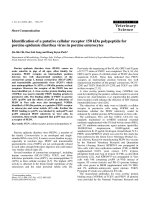
Fig. 1. Structure of female worms (a) whole body, (b) proboscis
with hooks, (c) anterior, (d) posterior, (e) egg (l, lemniscis; Vag,
vagina).
112 Jong-Tai Kim et al.
intermediate host of E gadi. Chu [3] and Cho [2] reported
upon the distribution of glycogen, mucopolysaccharides,
lipid and nucleic acid in the epicuticle of Echinorhynchus
gadi.
In this report, We report upon the identification of adult
worms that were found in ‘changran-pickles’ and
‘myungran-pickles’ by the Institute of Health &
Environment, Seoul Metropolitan Government, which
were respectively made from Gadus macrocophulus and
Theragra chalcogramma caught in the Korea, by
morphological observation and the measurement of
internal organs.
Materials and Methods
Echinorhynchus gadi were gathered from ‘changran-
pickles’ and ‘myungran-pickles’ by the Institute of Health
& Environment, Seoul Metropolitan Government, and
which were made from Gadus macrocophulus and
Theragra chalcogramma. The infected fishes were caught
in the Korea. Dead worms from ‘the pickles’ were fixed in
Fig. 2.
Structure of male worm (a) whole body, (b) proboscis
with hooks, (c) anterior (cg, cement gland; l, lemniscis; t, testis).
Fig. 3. Structure of female worms (a) anterior, (b) proboscis with
hooks, (c) posterior, (d) eggs.
Fig. 4. Structure of male worm (a) anterior, (b) testis, (c) cemen
t
glands.
Identification of acanthocephala discovered in changran-pickles and myungran-pickles 113
70% ethanol. Osmotic pressure and pH were not controlled
during fixation. Fixed worms were stained with Mayer’s
acid carmine and dehydrated through a graded series of
70%, 80%, 95% and 100% ethanol and transferred to
xylene. Finally, they were mounted in Canadian balsam.
In order to identify the worms, the length, and width of
the body and internal organs were measured. Species
identification was made according to Van Cleave [9] and
Yamaguti [11] classification of the acanthocephala.
Results
Morphology of male worms
All worms had a milk-white color, a smooth surface and
cylindrical body trunks. On the anterior, they had
cylindrical proboscis with hooks. The hooks on the
proboscis were symmetrically arranged in 18-19
longitudinal rows, with 4 hooks per row, which became
smaller toward the base of the proboscis. The hook
terminals were very sharp and the their roots were simple
and round. In each worms, there were two lateral claviform
protrusions, lemnisci, from the body wall at the base of
neck. In the middle of the trunk, two elliptical testes were
arranged linearly. Posterior to the testes, six cement glands,
pyriform in shape were arranged linearly, with ducts
leading into the sperm duct. Table 1 shows the
measurements of each male organ of E gadi.
Morphology of female worms
Female E gadi was usually larger than the males. The
proboscis and its arrangement, the shapes of the hooks on
the proboscis were similar to those of the males, and the
hooks on the proboscis were symmetrically arranged in
14-18 longitudinal rows, with 6-10 hooks per row that
became smaller toward the base of proboscis. There were
also two lateral lemnisci at the base of neck, which were
claviform or pyriform in shape. The body cavity of the
female worms was filled with eggs with a polar
prolongation of the middle shell, and of fusiform shape.
The uterine bell and the uterus in the posterior portion of
body. Table 2 shows the measurements of each female
organ E gadi.
Discussion
In the classification of acanthocephala by Van Cleave [9]
and Yamaguti [11], the body of Echinorhynchus was small
or middle-sized in the acanthocephala, and proboscis was
cylindrical in shape and the hooks on the proboscis were
arranged in 9-26 longitudinal rows, with 4-16 hooks per
Table 1.
Measurement of male worms
male worms
No. 1 No. 2
Body
L*(mm) 22 15
W** 0.8-0.6 0.6-0.4
Proboscis
L(
µ
) 284.8 524.9
W 227.6 151.4
Proboscis
sheath
L(
µ
) 1570.7 751.9
W 72.7 280.4
Lemnisci
L(
µ
) 2566.7 1085.6
W
Testis
L(
µ
) 2202.9-1860.5 1033-981.1
W 737.0-575.7 463.1-351.6
Cement gland
L(
µ
) 940.2 610.0
W 441.2 369.1
*L: length
**W: width
Table 2.
Measurement of female worms
female worms
No. 1 No. 2 No. 3 No. 4 No. 5 No. 6 No. 7
Body
L*(mm) 14 20 34 47 51 30 45
W** 0.3-0.7 0.8-0.3 0.9-0.5 2.1-1.3 1.9-0.9 2.2-1.4 0.8-0.7
Proboscis
L(
µ
) 466.1 - - - 331.7 - 252.0
W 259.9 - - - 254.0 - 268.9
Proboscis
sheath
L(
µ
) 506.0 1063.7 1550.7 1120.6 1018.9 1131.5 1313.7
W 298.8 231.1 275.9 257.9 266.9 295.8 261.0
Lemnisci
L(
µ
) 639.4 930.3 1177.3 1454.2 1267.9 1325.7 473.1
W
Egg
L(
µ
) 51.7 112.4 68.6 81.7 87.3 98.1 -
W 14.0 28.5 16.1 17.5 16.5 28.8 -
Uteriner bell
and Uterus
L(
µ
) 1358.5 1124.5 1229.1 - - - 34.9
W 69.0 49.8 84.7 - - - 654.4
*L: length
**W: width
114 Jong-Tai Kim et al.
row and became smaller towards the base of the proboscis.
Male worms had two testis arranged linearly in the middle
or posterior portion of the body with six cement glands
arranged linearly in the posterior portion of body. Van
Cleave [9] noted that worms of Echinorhynchus among the
genera possess a proboscis with fully developed hooks in
the cystacanth stage.
In these specimens, the hyperdermic nuclei and lacunar
system, which were described in the reports of Van Cleave
[9] and Yamaguti [11] could not be observed and
cystacanth neither could. However, the morphological
characteristic features of each organ of the specimens
corresponded to those listed by Van Cleave [9], Yamaguti
[11] and David [4]. The size, arrangement, number and
morphological character of the body, eggs, and hooks also
correspond to those listed by Arai [1] and Petrochenko [7].
According to the above morphological features, those
acanthocephala, which were collected from ‘changran-
pickles’ and ‘myungran-pickles’ were identified as
Echinorhynchus gadi.
References
1.
Arai, H. P.
Acanthocephala. In Guide to the parasites of
fishes of Canada. Prat III, L. Margolis and Z. Kabata (eds.).
Canadian Special Publication of Fisheries and Aquitic
Sciences 1989,
107
, 1-90.
2.
Cho, B. C.
Eletron microscopical and histochemical studies
on the epicuticle of E
chinorhynchus gadi
(Acanthocephala).
Korean J. Parasitol
1981,
19(1)
, 45-54.
3.
Chu, J. K., Kang, S. Y., Chu, J. P. and Sung, D. W.
Histological studies on
Echinorhynchus gadi
(Acanthocephala).
Korean J. Parasitol
1977,
15(1)
, 36-42.
4.
David, J. M.
Aeginina longicornis
(Amphipoda:
Caprellidea), New Intermediate Host for
Echinorhynchus
gadi
(Acanthocephala: Echinorhynchidae).
J. Parasitol
1994,
80(6)
, 1043-1045.
5.
Moller, H. and Anders, K.
Diseases and Parasites of marine
fishes. Verlag Moller, Kiel, Germany, 1986, pp. 365.
6.
Omar, M. A.
Key to the families and subfamilies of
Acanthocephala, with the erection of a new class
(Polyacanthocephala) and a new oder
(Polyacanthorhynchida).
J. Parasitol
1987,
73(6)
, 1216-
1219.
7.
Petrochenko, V. T.
Acanthocephala of domestic and wild
animals vol. I. Izadatel’stvo Academii Nauk S.S.S.R.,
Moscow, 1956, pp. 456. (English translation by Israel
program for scientific translations, Ltd., 1971)
8.
Soulsby, E. J. L.
Helminths, Arthropods and Protozoa of
Domesticated Animals. 7th Edit, Bailliere Tindall, London,
1982, 630-645
9.
Van Cleave, H. J.
The recognition of a new order in the
Acanthocephala.
J Parasitol
1936,
22
, 202-206.
10.
Whitefield, P.
Phylogenetic affinities of acanthocephal an
asessment of ultrastructural evidence.
Parasitology
1971,
63
,
49-58.
11.
Yamaguti, S.
Systema helminthum. Vol. V. Acanthocephala.
Interscience, New York and London, 1963.