Báo cáo khoa học: "Polysaccharides isolated from Phellinus gilvus enhances dermal wound healing in streptozotocin-induced diabetic rats" docx
Bạn đang xem bản rút gọn của tài liệu. Xem và tải ngay bản đầy đủ của tài liệu tại đây (373.35 KB, 4 trang )
JOURNAL OF
Veterinary
Science
J. Vet. Sci. (2005), 6(2), 161–164
Polysaccharides isolated from Phellinus gilvus enhances dermal wound
healing in streptozotocin-induced diabetic rats
Jae-Sung Bae, Kwang-Ho Jang, Hee-Kyung Jin*
College of Veterinary Medicine, Kyungpook National University, Daegu 702-701, Korea
Dermal wound healing is a complex process that
involved inflammation leading to re-epithelialization,
granulation tissue, and tissue remodeling. Previous studies
from our laboratory have shown that polysaccharides
isolated from fungus, Phellinus gilvus (PG) have various
anti-inflammatory activities. In present study, we have
assessed the effect of polysaccharides from PG on the
dermal wound healing of polysaccharides from PG in
streptozotocin-induced diabetic rat model. Six of 6-mm
circular wounds were created with biopsy punch on the
4th day after induction of diabetes. After 24 hours, each
test substance was applied to the wound twice a day for
next 5 days. Circular wounds treated with PG showed
significantly reduced wound contraction and complete re-
epithelialization, as compared to wounds of non-treated (p
< 0.05). These results show that polysaccharides isolated
from PG enhanced wound repair in diabetic impaired
healing, and could be developed as a wound healing agent
in such clinical settings.
Key words: diabetes, Phellinus gilvus, polysaccharides, rat,
wound healing
Tissue repair and wound healing are complex processes
involving inflammation, re-epithelialization, neovascularization,
fibroplasia, and wound contraction [8]. In the normal host,
wound healing is usually uncomplicated and proceeds at a
rapid rate. In contrast, most healing failures are associated
with some form of host impairment, including diabetes,
infection, immunosuppression, obesity, or malnutrition [14].
Among them, diabetes probably represents the prototype of
impaired healing model since contribution of impairment
such as peripheral vascular disease and lowered immunity
against infections. Therefore, we employed streptozotocin-
induced diabetic rat model for our study.
Several natural products have been investigated in the
promotion of wound healing of normal or abnormal host.
The extract of Centella asiatica (Madecassol; Dong Kook
Pharm, Korea) used in this study is a well-known medical
ointment in enhancing of dermal wound healing [15,16].
Recently, polysaccharides isolated from Phellinus spp. have
received special attention due to their potent pharmacological
activities such as anti-tumor [4,9] and anti-inflammatory
[11]. We have previously demonstrated that polysaccharides
isolated from the Phellinus gilvus (PG) have various
biological activities related to inflammation, including
inhibition of pulmonary inflammation [10], prevention of
intraperitoneal adhesion under infectious circumstances
[1,3,5], and promotion of dermal wound healing in normal
host [2]. In this work, we investigated whether polysaccharides
isolated from PG could enhance dermal wound healing in
streptozotocin-induced diabetic rats.
Ten male Sprague-Dawley rats (243-285 g) were purchased
from Charles River Laboratory (BioGenomics, Korea) and
used to carry out experiments after an acclimatization period
of 7 days in controlled room. The animals were fed with
commercial rat diet (Orient, Korea). Food and water were
provided ad libitum. All animals were used in compliance
with the Guidelines for Animal Care and Use [13]. Diabetes
was induced by a single intraperitoneal injection of
streptozotocin (70 mg/kg body weight, Sigma, USA) in
0.1 M citrate buffer, pH 4.0. Fasting blood glucose levels
were checked with glucose oxidase reagent strips before and
3 days after streptozotocin injection, and rats with consistent
blood glucose levels higher than 200 mg/dl were used for
this study.
The fruiting body of PG was kindly provided by
Gyeongbuk Agricultural Technology Administration (Daegu,
Korea) and grown rapidly for 3 months in artificial oak
sawduct cultures. It was homogenized, extracted by optimal
water extraction conditions, distilled water (1 : 25) at 100
C
for 10 hours, and concentrated at 80
C in a rotary
evaporator. The recovery procedure of the polysaccharides
solution from the fruiting body of PG followed an
established method in our previous studies [1-5]. The
concentration of 0.025, 0.25, and 2.5% polysaccharides
solutions was determined by total sugar according the
*Corresponding author
Tel:+82-53-950-5966; Fax: +82-53-950-5955
E-mail:
Short Communication
162 Jae-Sung Bae et al.
anthrone method [6] using glucose as the standard material.
These solutions formulated as a suspension in a small of
aqueous gel (Biosonic; Amitie, Korea) and Madecassol.
Wounds were created on the 4th day after induction of
diabetes. Animals were anesthetized using Rompun (10 mg/
kg) and Ketamine (200 mg/kg). Hair on the dorsal side of
the rats was shaved and the skin was cleaned with 70%
ethanol. A 6-mm skin biopsy punch was used to create six
of circular wounds under aseptic conditions. The wound
were made 1.25 cm to the right and left sides of the midline
and separated from cranial or caudal by 2.5 cm. At 24 hours
postoperatively, each test substance were applied to the
circular wound, respectively. Application of test substances
was continued twice a day for the next 5 days. Test
substances would be applied at a different wound location in
each animal. The substances were as follow: 0.025, 0.25,
and 2.5% polysaccharides isolated from PG (PG0.025,
PG0.25, and PG2.5 group), Madecassol (MC group),
aqueous gel (AG group) and no treatment (control group).
No dressing was placed on any of the wounds. Animals
were sacrificed 6 days postoperatively (5 days after initial
treatment by test substance). Wound radius was determined
by measuring craniocaudal and lateromedial distance. An
average of those two measurements was obtained and used
to predict wound diameter.
Wound tissue was collected at the time of euthanasia and
fixed in 10% buffered formalin. A 5 µm thick sections were
cut and stained with hematoxylin and eosin. All subsequent
analyses were performed by observers blinded to treatment.
Histological sections were used to observe the reepithelialization,
congestion, edema, infiltration of polymorphononuclear
leukocytes and monocytes, necrosis, fibroblastic proliferation,
collagen formation and angiogenesis. The degree of
reepithelialization was estimated as % of incision width
reepithelialized in each wound tissue. Analysis of differences
between treated groups and untreated groups was performed
using analysis of variance followed by multiple
comparisons and Fisher’s LSD test using the SAS statistical
package (release 8.1; SAS, USA). Differences at p < 0.05
were considered statistically significant.
Contraction of wound and re-epithelialization of tissue on
any of the treatment had no quantitative effect according to
location of wounds. Gross observation of the punch wounds
at the time of euthanasia showed that all PG treated wounds
were healthier, in that they were not oozing and appeared
well contracted unlike many control wounds in diabetic rats.
The most dramatic result of wound diameter were seen in
PG2.5 group compared to control group (p < 0.05).
Predicted wound diameter of control and each experimental
group by measuring craniocaudal and lateromedial distance
are presented in Table 1.
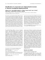
The microscopic examination of wound the dermis
showed proliferation of fibroblasts, hemorrhage, angiogenesis
and infiltration of polymorphonuclear leukocytes and
monocytes, degree of which parameters did not show
significant difference between each groups (Fig. 1). PG
treated wounds of diabetic rats showed an increase in the
rate of re-epithelialization. Complete re-epithelialization
was noted in PG2.5 group. In MC group, about 60% of
incision width was re-epithelialized. In control group, only
40% of the wound was re-epithelialized. Crust of necrotic
debris with inflammatory cells covered denuded epidermis
and the dermis below showed marked edema and
congestion (Fig. 1). The rate of re-epithelialization (%) of
the control group was 43 ± 8.3. The rate of PG2.5 (84 ±
11.9) and PG0.25 (80 ± 7.9) group was higher than that in
control and AG (60 ± 7.9) group (p < 0.05) (Fig. 2). PG0.025
(63 ± 7.6) and MC group (64 ± 11.9) was slightly higher
than control group. There was no statistically significant
difference in the rate of re-epithelialization of the AG and
control group.
The results of the present study showed that all
polysaccharides isolated from PG enhance significantly
wound healing in streptozotocin-induced diabetic rats.
Previous studies from our laboratory reported that the
polysaccharides have effect of dermal wound healing in the
normal host [2]. Thus, we conclude that the polysaccharides
may enhance wound healing both not only normal but also
diabetic host. PG used in our study is fungi belonging to the
Hymenochaetaceae basidiomycetes [7] and it is commonly
referred to as Sangwhang in Korea. The effects of PG related
to various pharmacological activities have demonstrated
continually. In addition, it has advantages over the other
Phellinus mushrooms in that it has a very short growth
period (3 months) and need it cheaper to produce. Therefore,
we can predict that use of PG will be increased gradually as
a functional food and medical supplements in the future.
We have previously demonstrated that polysaccharides
isolated from PG inhibit pulmonary inflammation [10] and
intraperitoneal adhesion related to intraperitoneal inflammation
[1,3,5]. These effects of PG related to anti-inflammatory
activities might be beneficial in the treatment of dermal
Tab le 1. Wound diameter predicted by measuring craniocaudal
and lateromedial distance at the time of euthanasia in diabetic
rats (n = 10) (mean ± SD)
Groups
Predicted wound diameter
(mm)
Control 4.24±0.57
MC 3.65±1.31
PG0.025 3.52±0.47
PG0.25 3.11±0.84
PG2.5 2.61±0.39
AG 3.74±1.23
p<0.05 compared with all groups was significant. p<0.05 versus control
group. Control group (no treatment), MC group (Madecassol), PG0.025,
PG0.25, and PG2.5 group (0.025, 0.25, and 2.5% polysaccharides gel
isolated from PG) and AG group (aqueous gel).
Enhancement of dermal wound healing by polysaccharides in diabetic rats 163
wound healing. In the studies, we showed that PG is a potent
macrophage stimulator that enhances macrophage cytotoxicity
and phagocytic capacity. Macrophages is known to play a
key role in wound repair. Leibovich and Ross [12] reported
that healing was delayed when wound macrophages were
depleted. Therefore we may conclude that dermal wound
healing may be promoted by modulating the macrophage
activity of polysaccharides isolated from PG.
In summary, polysaccharides isolated from PG enhanced
wound repair in diabetic impaired healing, and could be
developed as a wound healing agent in such clinical settings.
Additional studies regarding its mechanism of action will
help further reveal of its uses and limitations.
Acknowledgments
This work was supported by the Korea Research Foundation
Grant (KRF-2003-003-E00244). We are grateful to S.I.
Kim, J.K. Lee and H.B. Yoon for their skillful animal care.
Thanks are also due to Dr. J. Carter, Department of
Psychiatry and Behavioral Sciences, University College,
London, UK for helping with preparation of manuscript, and
Dr. S.C. Park, College of Veterinary Medicine, Kyungpook
National University for helping with the provision of
Phellinus gilvus.
References
1. Bae JS, Ahn SJ, Jang KH, Yim H, Jin HK. Prevention of
intraperitoneal adhesions and abscesses by polysaccharides
isolated from Phellinus spp. in a rat peritonitis model. Ann
Surg 2004, 241, 534-540.
2. Bae JS, Jang KH, Jin HK. Promotion of dermal wound
healing by polysaccharides isolated from Phellinus gilvus in
rats. J Vet Med Sci 2004, 67, 111-114.
Fig. 1. Histopathologic appearances of wounds at the time of euthanasia in streptozotocin-induced diabetic and normal rats. The wounds
showed dermal proliferation of fibroblasts and mononuclear inflammatory cells with angiogenesis. Re-epithelialization was only 40%
in control group (A) and about 60% in MC (Madecassol, B) and PG0.025 (C). Crust covered denuded epidermis and the dermis belo
w
showed marked edema. PG2.5 group (D) showed complete re-epithellialization with increased number of epithelial cell layer, thic
k
granular layer, hyperkeratosis and parakeratosis. H&E stain, ×100.
Fig. 2. Rate of re-epithelialization of control and experimental
wounds (n = 10). *p < 0.05 versus all groups and † p <0.05
versus AG and control group.
164 Jae-Sung Bae et al.
3. Bae JS, Jang KH, Jin HK. Comparison of intraperitoneal
anti-adhesive polysaccharides derived from Phellinus
mushrooms in a rat peritonitis model. World J Gastroenterol
2004, 11, 810-816.
4. Bae JS, Jang KH, Yim H, Jin HK. Polysaccharides isolated
from Phellinus gilvus inhibit melanoma growth in mice.
Cancer Lett 2004, 218, 43-52.
5. Bae JS, Jin HK, Jang KH. The effect of polysaccharides
and carboxylcellulose combination to prevent intraperitoneal
adhesion and abscess formation in a rat peritonitis model. J
Vet Med Sci 2004, 66, 1205-1211.
6. Bucci SJ, Scholz FG, Goldstein G, Meinzer FC, Sternberg
LDaS. Dynamic changes in hydraulic conductivity in
petioles of two savanna tree species: factors and mechanisms
contributing to the refilling of embolized vessels. Plant Cell
Environ 2003, 26, 1633-1645.
7. Dai YC, Xu MQ. Studies on the medicinal polypore,
Phellinus baumii, and its kin, P. linteus. Mycotaxon 1998,
67, 191-200.
8. Evans P. The healing process at cellular level: a review.
Physiotherapy 1980, 66, 256-259.
9. Han SB, Lee CW, Jeon YJ, Hong ND, Yoo ID, Yang KH,
Kim HM. 1999. The inhibitory effect of polysaccharides
isolated from Phellinus linteus on tumor growth and
metastasis. Immunopharmacology 1999, 41, 157-164.
10. Jang BS, Kim JC, Bae JS, Rhee MH, Jang KH, Song JC,
Kwon OD, Park SC. Extracts of Phellinus gilvus and
Phellinus baumii inhibit pulmonary inflammation induced by
lipopolysaccharide in rats. Biotechnol Lett 2004, 26, 31-33.
11. Kim GY, Kim SH, Hwang SY, Kim HY, Park YM, Park
SK, Lee MK, Lee SH, Lee TH, Lee JD. Oral administration
of proteoglycan isolated from Phellinus linteus in the
prevention and treatment of collagen-induced arthritis in
mice. Biol Pharm Bull 2003, 26, 823-831.
12. Leibovich SJ, Ross R. The role of the macrophage in wound
repair. A study with hydrocortisone and antimacrophage
serum. Am J Pathol 1975, 78, 71-100.
13. National Research Council. Guide for the Care and Use of
Laboratory Animals. pp. 1-70, National Academy Press,
Washington DC, 1996.
14. Sidhu GS, Mani H, Gaddipati JP, Singh AK, Seth P,
Banaudha KK, Patnaik GK, Maheshwari RK. Curcumin
enhances wound healing in streptozotocin induced diabetic
rats and genetically diabetic mice. Wound Repair Regen
1999, 7, 362-374.
15. Suguna L, Singh S, Sivakumar P, Sampath P,
Chandrakasan G.
Influence of Terminalia chebula on
dermal wound healing in rats. Phytother Res 2002, 16, 227-
231.
16. Suguna L, Sivakumar P, Chandrakasan G. Effects of
Centella asiatica extract on dermal wound healing in rats.
Indian J Exp Biol 1996, 34, 1208-1211.