Báo cáo khoa học: " Ultrasonographic evaluation of renal dimension and resistive index in clinically healthy Korean domestic short-hair cats" ppsx
Bạn đang xem bản rút gọn của tài liệu. Xem và tải ngay bản đầy đủ của tài liệu tại đây (1.44 MB, 5 trang )
JOURNAL OF
Veterinary
Science
J. Vet. Sci. (2008), 9(4), 415
419
*Corresponding author
Tel: +82-33-250-8681; Fax: +82-33-244-2367
E-mail:
Ultrasonographic evaluation of renal dimension and resistive index in
clinically healthy Korean domestic short-hair cats
In-Chul Park
1
, Hye-Sun Lee
1
, Jong-Taek Kim
2
, So-Jeong Nam
3
, Ran Choi
3
, Ki-Seok Oh
4
, Chang-Ho Son
4
,
Changbaig Hyun
3,
*
Sections of
1
Diagnostic Imaging,
2
Wildlife Medicine, and
3
Small Animal Internal Medicine, School of Veterinary Medicine and
Institute of Veterinary Science, Kangwon National University, Chuncheon 201-100, Korea
4
Section of Obstetrics, College of Veterinary Medicine, Chonnam National University, Gwangju 500-757, Korea
Renal length, height, width, resistive index (RI), size of
cortex, and medulla were determined by renal ultrasonography
in 50 healthy Korean domestic short-hair cats. In the
sagittal plane, the renal length was 3.83
±
0.51 cm (mean
±
SD) in the left kidney and 3.96
±
0.48 cm in the right
kidney, whereas the renal height was 2.42
±
0.27 cm in the
left kidney and 2.36
±
0.28 cm in the right kidney. In the
transverse plane, the renal height was 2.42
±
0.28 cm in
the left kidney and 2.38
±
0.27 cm in the right kidney,
whereas the renal width was: 2.65
±
0.35 cm in the left
kidney and 2.63
±
0.31 cm in the right kidney. In the
dorsal plane, the renal length was 3.84
±
0.53 cm in the
left kidney and 3.97
±
0.54 cm in the right kidney, whereas
the renal width was 2.65
±
0.34 cm in the left kidney and
2.66
±
0.33 cm in the right kidney. There were no significant
differences (p
>
0.05) among the same structure sizes measured
in different planes. In the sagittal plane, the size of the
renal cortex was 0.47
±
0.08 cm in the left kidney and 0.47
±
0.08 cm in the right kidney, whereas of the size of the
renal medulla was 0.55
±
0.30 cm in the left kidney and
0.50
±
0.07 cm in the right kidney. RI evaluated by pulsed
wave Doppler sonography was 0.52
±
0.05 in the left kidney
and 0.55
±
0.05 in the right kidney. The actual renal dimensions
determined by gross examination were not statistically
different from those determined by ultrasonography. Further-
more the renal dimensions and RI were statistically corre-
lated to the body weight of cats.
Keywords: kidney, Korean domestic cat, renal size, resistive
index, ultrasonography
Introduction
Ultrasonography is widely applied to detect the presence
of abnormal structures and morphological changes in solid
organs and is useful to narrow down the differential
diagnosis [6,13]. Ultrasonographic evaluation is especially
useful for assessing kidneys because important anatomic
information concerning the size, shape, and internal
architecture can be obtained even in the presence of impaired
renal function or abdominal fluid [1]. Compared to
conventional survey radiology, ultrasonography can better
visualize kidneys in emaciated animals and those with
retroperitoneal fluid. Subcapsular fluid, localized perirenal
fluid, small renal or perirenal masses, and pelvic or ureteral
dilation can be also easily detected in ultrasonography.
Ultrasound-guided interventional procedures and Doppler
ultrasound imaging enable us to better assess renal
functional status [7].
Reduced renal artery diastolic flow indicates a generalized
increase in renal vascular resistance. Although this finding
is nonspecific, it is a good indicator for acute renal disease,
acute tubular necrosis, and renal obstruction [11]. To
determine this renal vascular resistance, in practice, resistive
index (RI) as determined by Doppler ultrasonography is
widely applied. Signals from the arteries near the renal hilum
(segmental or interlobar) and corticomedullary junction
(arcuate) are used for the RI determination. Because of the
inaccessibility to these small vessels, the frequency shift is
used to provide a relative assessment of blood flow velocity
during systole and diastole [12]. Because the normal
reference range of RI is less than 0.70, any increased RI value
suggests that the increased renal vascular resistance is due
to certain renal disease.
Although a few studies have been already reported about
normal renal dimensions and RI, the comparison between
the actual and ultrasonographic dimension is rarely
reported [4,5]. Also most of the cat populations enrolled in
416 In-Chul Park et al.
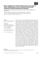
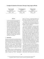
Fig. 1. Determination of renal length and height on the sagittal
p
lane. When two bright parallel bars formed by cross sectioned
p
elvic diverticulum were clearly visible, the renal length an
d
height were measured. L: length, H: height, C: cortex, M: medulla.
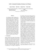
Fig. 2. Determination of renal length and height on the transvers
e
p
lane. When the "C"-sign of the renal crest was clearly visible,
the renal length and height were measured. H: height, W: width.
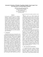
Fig. 3. Determination of renal length and height on the dorsal
p
lane. L: length, W: width.
previous studies included a wide range of cat breeds.
Therefore in this study, we restricted our research to a
single breed of cat (domestic short-hair cat) and evaluated
the renal dimensions and RI in a clinically healthy cat
population. Furthermore to provide better reference index,
we compared the actual renal dimensions determined by
necropsy to those determined by ultrasonography.
Materials and Methods
Animals
Fifty healthy adult cats (23 females and 27 males),
weighing 2.1∼5.5 kg were used. The cats were considered
healthy on the basis of physical examination and normal
CBC, serum urea nitrogen concentration, and routine
urinalysis. The health status of the cats were re-evaluated 2
weeks after the initial evaluation. Only healthy cats were
enrolled in this study. Our study was approved by the
Animal Ethics Committee of Kangwon National University
and was performed under strict adherence to the guidelines
which included animal care, euthanasia, and disposal of
dead animals.
Preparation and anesthesia
For each cat, preparations for an ultrasound scan included
a 12-h fast, the availability of water at all times, and a tepid
water enema 1 to 2 h before the procedure. The ventral
abdominal hair coat was clipped from the costal arch to the
iliac wings. The cat was anesthetized by atropine (0.03
mg/kg, SC), ketamine (10 mg/kg, IM) and xylazine (1
mg/kg, IM).
Ultrasonography
Cats were placed in dorsal recumbency for survey
ultrasonography. A water-soluble coupling gel was applied
liberally to the ventral abdomen to permit sound conduction.
Ultrasound scans were performed, using a static B-mode
articulated scan arm and a 4∼9 MHz transducer (Sonoace
8000SE; Medison, Korea). The longitudinal axis of the left
kidney was located by use of a survey scan. Sagittal scans
were begun at the medial margin of the kidney, and a serial
sequence of sagittal scans was made at 0.5-cm intervals
until the most lateral margin of the kidney was no longer
visible (Fig. 1). The scan arm was then rotated 90
o
. Cross
sectional (transverse) images were obtained, beginning at
the cranial pole of the left kidney, and a serial sequence of
transverse scans was made at 0.5-cm steps until the caudal
pole was no longer visible (Fig. 2). Dorsal scans were made as
described in sagittal scans after the scan probe move
laterally (Fig. 3). The procedure was repeated for the right
kidney.
Resistive index (RI)
To obtain the RI, a renal interlobar or arcuate artery was
Ultrasonographic evaluation of renal dimension and resistive index in clinically healthy Korean domestic short-hair cats 417
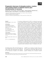
Fig. 4. Resistive index (RI) was measured by using pulsed wave
Doppler echocardiography.
Tabl e 1 . Comparison of renal dimensions of 50 Korean domestic
short-hair cats measured in different ultrasonographic angles
Renal
dimensions
Sagittal
plane
Transverse
plane
Dorsal
plane
Length
Height
Width
L
R
L
R
L
R
3.83 ± 0.51
3.96 ± 0.48
2.42 ± 0.27
2.36 ± 0.28
N/A
N/A
N/A
N/A
2.42 ± 0.28
2.38 ± 0.27
2.65 ± 0.35
2.63 ± 0.31
3.84 ± 0.53
3.97 ± 0.54
N/A
N/A
2.65 ± 0.34
2.66 ± 0.33
Unit: cm; Mean ± SD, L: left, R: right, N/A: not applicable.
Table 2. Comparison of renal dimensions of 27 Korean domestic
short-hair cats as measured by gross and ultrasonographic exam-
inations
Renal
dimensions
Actual
size
Sagittal
plane
Transverse
plane
Dorsal
plane
Length
Height
Width
L
R
L
R
L
R
3.92 ± 0.48
4.06 ± 0.49
2.38 ± 0.32
2.32 ± 0.27
2.79 ± 0.36
2.73 ± 0.30
3.89 ± 0.51
3.98 ± 0.43
2.40 ± 0.28
2.35 ± 0.29
N/A
N/A
N/A
N/A
2.40 ± 0.30
2.37 ± 0.27
2.73 ± 0.37
2.68 ± 0.32
3.89 ± 0.51
4.02 ± 0.44
N/A
N/A
2.73 ± 0.36
2.69 ± 0.36
Unit: cm; Mean ± SD, L: left, R: right, N/A: not applicable.
identified with color Doppler. The Doppler tracing was
then obtained and recorded by placing a gate of 2.5 mm
width (adjusted when necessary) over the artery, setting the
wall filter to 125 Hz, and selecting the smallest scale that
displayed the flow without aliasing (Fig. 4). In most cats,
one five-second waveform strip from one artery for each
kidney was recorded. The peak systolic and end diastolic
velocities were measured by the methods used in a
previous report [4].
Gross measurement by necropsy
To measure the actual dimensions of the kidneys, twenty-
seven of 50 cats were necropsied after ultrasonographic
examination. Both kidneys were removed and measured
using Vernier calipers (Mitutoyo, Japan).
Data analysis
The correlation coefficiency for the group means for body
weight and renal dimension were calculated and compared
using statistical software packages (SAS Ver 8.2; SAS
Institute, USA). Renal dimensions obtained through each
different ultrasonographic plane and gross measurements
(necropsy) were compared using paired t-test.
Results
Comparison of renal dimensions in different sonographic
planes and RIs
Renal dimensions measured by ultrasonography in 50
domestic shorthair cats including renal length, height, and
width were summarized in Table 1. No statistically
significant difference between the renal dimensions
measured in different songraphic angles was observed (p
> 0.05). The mean thicknesses of renal cortex and medulla
were 0.47 ± 0.08 cm (mean ± SD) in the left kidney and
0.47 ± 0.08 cm in the right kidney, and 0.55 ± 0.30 cm in the
left kidney and 0.50 ± 0.07 cm in the right kidney,
respectively. The means of RI of both kidneys were 0.52 ±
0.05 in the left kidney and 0.55 ± 0.05 in the right kidney.
Comparison of renal dimensions between gross and
sonographic measurements
Renal dimensions measured by gross examination in 27
domestic shorthair cats including renal length, height, and
width were summarized in Table 2. No statistically
significant difference between the renal dimensions as
measured by two different measurements was observed (p
> 0.05).
Statistical analysis for bodyweight
Since the cats enrolled in this study had a wide range of
body weight (2.1∼5.5 kg), the measured renal dimensions
were statistically analyzed to identify the correlation index
(Table 3). Although a high degree of correlation to body
weight has been observed in the renal dimensions (renal
length, height, and width) and the renal cortical thickness,
a low degree of correlation has been observed in the renal
medullary thickness and RI (Table 3).
418 In-Chul Park et al.
Tabl e 3 . Coefficient of correlation of body weight to renal
dimensions
Renal
dimensions
Sagittal
plane
Transverse
plane
Dorsal
plane
Length
Height
Width
Cortical
thickness
Medullary
thickness
RI
L
R
L
R
L
R
L
R
L
R
L
R
0.711
0.615
0.657
0.621
N/A
N/A
0.604
0.648
0.182
0.291
0.031
-0.033
N/A
N/A
0.670
0.660
0.627
0.651
N/A
N/A
N/A
N/A
N/A
N/A
0.675
0.593
N/A
N/A
0.621
0.610
N/A
N/A
N/A
N/A
N/A
N/A
If coefficient of correlation is below 0.3, we regarded the correlatio
n
is weak. N/A; not applicable, L: left, R: right, RI: resitive index.
Discussion
Normal echocardiographic structure of the kidney is
influenced by the echocardiographic angle (plane),
breathing patterns of animals, degree of interference by other
organs (e.g. liver and spleen), and the skills of the
practitioner, although the high-quality ultrasound machine
and appropriate transducer were used in the examinations.
In addition, the slight pressure on abdomen by transducer can
displace the location and orientation of the kidney. However
it can not be avoided, since feline kidneys are extremely
mobile. In the dorsal plane, the correct measurement of renal
width is problematic, because of the abundance of
connective tissues in the renal hillus. This can be overcome
in the cross measurement by using the transverse plane.
Generally the caudal and cranial pole of kidney is sometimes
unclear. These can be better visualized by using the dorsal
plane. As in previous literature which had mentioned the
problems encountered in the measurement of renal
dimensions, we measured renal dimensions at the three
different angles and measured three times in each case to
minimize factors affecting correct measurement in this
study.
The renal dimensions that we measured in this study were
similar to previous reports from those measured by others
[14], because the weight ranges of the cats were not
significantly different. Furthermore the discrepancy of the
renal dimensions measured by gross (necropsy) and
ultrasonographic examination was statistically insignificant.
However, the renal cortical thickness was not similar to
previous reports of the measurement by others [14], although
the medullary thickness was not different. However, in the
study by others [14], cortical dimensions were slightly
greater because their measurement included the bright
diverticular echoes, which could contribute to the discrepancy
from our results. However, because the cortical and
medullary dimensions were different even in the same cats
depending on the anatomical location measured and the
dimensions of the renal medulla were not able to be clearly
defined due to the unclear borders of renal sinuses in the
transverse plane, the clinical application of renal cortical and
medullary dimensions is limited.
Because renal function is dependent on renal blood flow,
glomerular and tubular function, and urine flow, the
measurement for renal blood flow (e.g. RI) may help for
the diagnosis, treatment, and prognosis of renal disease.
Renal arterial RI is the ratio of systolic to diastolic velocity
and is used to estimate vascular resistance. Since increased
vascular resistance decreases diastolic velocity, the
increased renal arterial RI implies reduced renal blood
flow. A significant relationship between RI and acute renal
failure has been reported in veterinary literature [4].
However, in humans, the reliability of renal arterial RI
measurements is controversial [2,3], although it has
proven to be useful in humans for evaluating renal
transplant complications [10]. Because RI is influenced by
age, the patency of urinary tract, and the animal’s
circulatory status and because the normal reference range
of RI in cats are too wide, clinical application of RI still
limited, although one study reported a high specificity of
RI for canine renal diseases [4]. As noticed in previous
studies [4,5], the range of RI in healthy cats was wide (left:
0.42∼0.71, right: 0.41∼0.73). The means of RI in both
kidneys were similar to previous reports [9], although the
different anesthetic protocol was used in this study. One
study found RI was not markedly influenced by deep
sedation, as reported previously [8]. However, this study
used a different anesthetic protocol, which might
potentially affect RI in our study population, although the
means of RI in both kidneys were similar to others [9].
Probably, the actual RI in our study population might be
higher than others [9], since it might be underestimated due
to influence from the hypotensive effect from xylazine
(used in this study). Otherwise, xylazine might minimally
influence the RI in our study population, so that the mean
RI was similar to others [9]. Because we did not clarify this
issue prior to study, the mean RI found in this study might
be different from the RI in cats without chemical restraints.
Therefore, future studies should be directed to clarify the
effects on RI from different type of chemical restraints.
Although the cats enrolled in this study have a wide range
of body weights, the measured renal dimensions except
renal medullary thickness were statistically closely
correlated. Probably this result was because there were no
severely obese and emaciated cats included in this study.
In summary, renal dimensions and RI measured by
Ultrasonographic evaluation of renal dimension and resistive index in clinically healthy Korean domestic short-hair cats 419
ultrasonography in 50 Korean cats were similar to those
measured by others and gross examinations. The renal
dimensions and RI were statistically correlated to the body
weight of cats.
Acknowledgments
This study was supported from Institute of Veterinary
Science, Kangwon National University.
References
1. Armbrust LJ, Biller DS, Hoskinson JJ, Meier HT, Lora-
Michiels M. The basics of renal ultrasonography. Vet Med
2001, 96, 114-133.
2. Genkins SM, Sanfilippo FP, Carroll BA. Duplex Doppler
sonography of renal transplants: lack of sensitivity and spe-
cificity in establishing pathologic diagnosis. Am J Roentgenol
1989, 152, 535-539.
3. Jurriaans E, Dubbins PA. Renal transplantation: the nor-
mal morphological and Doppler ultrasound examination. J
Clin Ultrasound 1992, 20, 495-506.
4. Morrow KL, Salman MD, Lappin MR, Wrigley R.
Comparison of the resistive index to clinical parameters in
dogs with renal disease. Vet Radiol Ultrasound 1996, 37,
193-199.
5. Nyland TG, Fisher PE, Doverspike M, Hornof WJ,
Olander HJ. Diagnosis of urinary tract obstruction in dogs
using duplex Doppler ultrasonography. Vet Radiol Ultrasound
1993, 34, 348-352.
6. Osborne CA, Finco DR. Canine and Feline Nephrology and
Urology. 8th ed. pp. 370-464, Williams & Wilkins, Baltimore,
1995.
7. Platt J F. Duplex Doppler evaluation of native kidney dys-
function: obstructive and nonobstructive disease. AJR Am J
Roentgenol 1992, 158, 1035-1042.
8. Pollard R, Nyland TG, Bernsteen L, Gregory CR,
Hornof WJ. Ultrasonographic evaluation of renal auto-
grafts in normal cats. Vet Radiol Ultrasound 1999, 40, 380-
385.
9. Quarto di Palo F, Rivolta R, Elli A, Castagnone D. The
well-functioning renal graft evaluated by color Doppler
flowmetry. Nephron 1995, 70, 314-318.
10. Rifkin MD, Needleman L, Pasto ME, Kurtz AB, Foy PM,
McGlynn E, Canino C, Baltarowich OH, Pennell RG,
Goldberg BB. Evaluation of renal transplant rejection by
duplex Doppler examination: value of the resistive index.
AJR Am J Roentgenol 1987, 148, 759-762.
11. Rivers BJ, Walter PA, Letourneau JG, Finlay DE,
Ritenour ER, King VL, O'Brien TD, Polzin DJ. Duplex
Doppler estimation of resistive index in arcuate arteries of
sedated, normal female dogs: implications for use in the di-
agnosis of renal failure. J Am Anim Hosp Assoc 1997, 33,
69-76.
12. Rivers BJ, Walter PA, O'Brien TD, Polzin DJ. Duplex
Doppler estimation of Pourcelot resistive index in arcuate ar-
teries of sedated normal cats. J Vet Intern Med 1996, 10,
28-33.
13. Walter PA, Feeney DA, Johnston GR, Fletcher TF. Feline
renal ultrasonography: quantitative analyses of imaged
anatomy. Am J Vet Res 1987, 48, 596-599.
14. Walter PA, Johnston GR, Feeney DA, O'Brien TD. Renal
ultrasonography in healthy cats. Am J Vet Res 1987, 48,
600-607.