Báo cáo toán học: "Expressions of EphA2 and EphrinA-1 in early squamous cell cervical carcinomas and their relation to prognosis" pps
Bạn đang xem bản rút gọn của tài liệu. Xem và tải ngay bản đầy đủ của tài liệu tại đây (1.18 MB, 6 trang )
Int. J. Med. Sci. 2008, 5
121
International Journal of Medical Sciences
ISSN 1449-1907 www.medsci.org 2008 5(3):121-126
© Ivyspring International Publisher. All rights reserved
Research Paper
Expressions of EphA2 and EphrinA-1 in early squamous cell cervical
carcinomas and their relation to prognosis
Ruth Holm
1
, Gregg Van de Putte
2
, Zhenhe Suo
1,3
,
A Kathrine Lie
1
, Gunnar B Kristensen
4,5
1. Division of Pathology, The Norwegian Radium Hospital, Rikshospitalet University Hospital, Oslo, Norway.
2. Department of Gynaecology, Ziekenhuizen Oost Limburg, Genk, Belgium
3. Faculty Division The Norwegian Radium Hospital, Medical Faculty, University of Oslo, Norway.
4. Division of Obstetrics and Gynaecology, The Norwegian Radium Hospital, Rikshospitalet University Hospital, Oslo,
Norway.
5. Institute of Medical Informatics, The Norwegian Radium Hospital, Rikshospitalet University Hospital, Oslo, Norway.
Correspondence to: Ruth Holm, PhD, Division of Pathology, The Norwegian Radium Hospital Montebello, 0310 Oslo, Norway. Telephone:
(47) 22934207; Fax: (47) 22930164; E-mail:
Received: 2008.03.31; Accepted: 2008.06.04; Published: 2008.06.05
By using immunohistochemistry we investigated the expression of EphA2 and EphrinA-1 in 217 early squamous
cell cervical carcinomas and examine their prognostic relevance. For EphA2 expression, 21 tumors (10%) showed
negative, 108 (50%) weak positive, 69 (32%) moderate positive and 19 (9%) strong positive, whereas for
EphrinA-1 expression, 33 tumors (15%) showed negative, 91 (42%) weak positive, 67 (31%) moderate positive and
26 (12%) strong positive. In univariate analysis high expression (strong staining) of EphrinA-1 was associated
with poor disease-free (P = 0.033) and disease-specific (P = 0.039) survival. However, in the multivariate analyses
neither EphrinA-1 nor EphA2 was significantly associated to survival. The increased levels of EphA2 and
EphrinA-1 in a relative high number of early stage squamous cell carcinomas suggested that these two proteins
may play an important role in the development of a subset of early cervical cancers. However, EphA2 and
EphrinA-1 were not independently associated with clinical outcome.
Key words: Immunohistochemistry, early cervical cancer, EphA2 and EphrinA-1
Introduction
Worldwide, cervical carcinoma is the second
most frequent malignancy among women and the
death rate is 8 per 100,000 [1]. Nearly half of patients
present with stage I disease and about one third of
patients treated with surgery received adjuvant
treatment [2]. This adjuvant treatment is accompanied
by considerable morbidity [3]. Therefore, it is
important to make individualized treatment
procedures in order to reduce negative side effects for
patients with a good prognosis. Tumor diameter,
pelvic lymph node metastasis, vascular invasion, deep
stromal invasion and close resection margins are most
often used to target adjuvant treatment to those
patients with highest risk of relapse [4-7]. New
molecular markers that improve the prediction of
clinical outcome could be of considerable value to
design individualized treatment procedures.
Receptor tyrosine kinases have important roles in
the development and progression of human tumors
[8]. Eph receptors represent the largest known family
of receptors tyrosine kinases and are broadly divided
into subclasses, EphA and EphB. The EphA receptors
(A1-A9) bind to EphrinA ligands, a group of glycosyl
phosphatidylinositol-linked membrane proteins and
EphB receptors (B1-B6) interact with EphrinB ligands
[9]. The normal function of EphA2 is not fully
understood, but it is thought to function in the
regulation of cell growth, angiogenesis, survival and
migration [9-11]. EphA2 is overexpressed in a variety
of human cancers, including breast [12], prostate [13,
14], oesophagus [15], renal [16], lung [17], pancreas
[18] and ovarian [19, 20]. Furthermore, increase
expression of both EphA2 and EphrinA-1 have been
observed in vulvar carcinomas [21], cervical
carcinomas [22], bladder carcinomas [23], oesophageal
carcinomas [24], gastric carcinomas [25] and ovarian
carcinomas [26]. In various human cancers poor
outcome for the patients has been associated with
increase expression of EphA2 and EphrinA-1 [16, 17,
19, 24, 26]. In our previous study of cervical
carcinomas FIGO stage I-IV we found that increase
expression of EphA2 and EphrinA-1 was significantly
associated with shorter overall survival in multivariate
analysis [22]. The prognostic significance of EphA2
and EphrinA-1 expression in early stage cervical
carcinoma has to our knowledge not been examined.
The aims of our study were to investigate the
Int. J. Med. Sci. 2008, 5
122
expression of EphA2 and EphrinA-1 in a series of 217
early squamous cell cervical carcinomas and to
identify predictive markers of the clinical outcome for
these patients.
Materials and methods
Patient materials
A retrospective study of 217 patients with clinical
squamous cervical carcinomas FIGO stage IB, treated
by radical hysterectomy and bilateral pelvic
lymphadenectomy at The Norwegian Radium
Hospital in the period January 1987 to December 1993,
was performed. The median age at diagnosis was 39
years, ranging from 34 to 51 years. All patients were
examined under general anesthesia and tumor
diameter was assessed by inspection and palpation.
Postoperatively, adjuvant treatment was given in case
of large tumor diameter, lymph node metastasis, or
invasion into the parametria. A median of 26 lymph
nodes were removed. Radiotherapy was given to 32
patients, radiation and chemotherapy to 12, and
another 31 received chemotherapy only. All patients
were followed until death or July 2001. Forty-four
(20.3%) patients suffered a relapse and 37 (17.1%) died
of cervical cancer. Median follow-up for patients
without relapse was 128 months (range from 112 to 146
months); for patients still alive, median follow-up was
129 months (range from 114 to 147).
Histological specimens were reevaluated blindly
by an experienced pathologist (A. K. L.) for
histopathological diagnosis according to the World
Health Organization [27]. Table 1 provides a detailed
description of the tumor characteristics. As controls,
we used samples of 10 normal cervices from patients
who underwent amputation of the cervix for prolapse
(age range, 31-49 years, median age 43 years). The
Regional Committee for Medical Research Ethics
South of Norway (S-06381a), The Social- and Health
Directorate (06/4509 and 06/4417) and The Data
Inspectorate (06/01467-3) approved the study.
Table 1 Ephrin A-1 and EphA2 immunostaining in relation to clinicopathological variables.
Variables Total EphA2* Ephrin A-1*
n Low High (%) p
‡
Low High (%) p
‡
Tumor size (cm) 0.812 0.442
< 2 97 88 9 (9) 85 12 (12)
2.0 – 3.9 86 78 8 (9) 78 8 (9)
≥ 4
34
32
2 (6)
28
6 (18)
Depth of invasion (mm)
0.236
0.005
≤ 10 122 113 9 (7) 111 11 (9)
11-15
48
45
3 (6)
45
3 (6)
> 15 47 40 7 (15) 35 12 (25)
Vascular invasion
0.716
0.669
Absent 117 106 11 (9) 104 13 (11)
Present
100
92
8 (8)
87
13 (13)
Parametrial invasion
0.011
<0.001
Absent 202 187 15 (7) 183 19 (9)
Present 15 11 4 (27) 8 7 (47)
Lymph node metastasis 0.680 0.298
Absent 175 159 16 (9) 156 19 (11)
Present 42 39 3 (7) 35 7 (17)
Numbers in parentheses are percentages.
*
Low: Negative/weak/moderate staining; High: Strong staining.
‡
Pearson chi-square.
Immunostaining method
Sections were immunostained using the Dako
EnVision
TM
+ System, Peroxidase (DAB) (K4011, Dako
Corporation, CA, U. S.A.) and Dakoautostainer. After
microwaving in 10 mM citrate buffer pH 6.0 to unmask
the epitopes the sections were treated with 0.03%
hydrogen peroxide (H
2
O
2
) for 5 minutes to block
endogenous peroxidase. Polyclonal rabbit antibodies
EphA2 (sc-924, 1:400, 0.5μg IgG/ml) and EphrinA-1
(sc-911, 1:300, 0.7 μg IgG/ml) both from Santa Cruz
Biotechnology Inc., CA, U.S.A. were applied on the
sections for 30 minutes at room temperature followed
by incubation with peroxidase labeled polymer
conjugated to goat anti-rabbit for 30 minutes. The
peroxidase reaction was developed using
3`3-diaminobenzidine tetrahydrochloride (DAB) as
chromogen. Human cervical carcinomas that had been
shown to express EphA2 and EphrinA-1 have been
included in all series as positive controls. Negative
controls included substitution of the polyclonal
antibody with normal rabbit IgG of the same
concentration as the polyclonal antibody. All controls
gave satisfactory results. The specificity of anti-EphA2
and anti-EphrinA-1 has been tested previously [26].
Cytoplasmic staining was considered positive. Four
Int. J. Med. Sci. 2008, 5
123
semiquantitative classes were used to describe the
intensity of staining; no staining, weak staining,
moderate staining and strong staining. Since tumor
cells stained uniformly across the samples we did not
considered the fraction of tumor cells with positive
staining. Sections were scored by two independent
observers (R. H. and Z. S.) without knowledge of
clinical data. Conflicting results were reviewed until
final agreement was achieved. Based on our previous
published study of cervical carcinomas FIGO stage
I-IV two different cutoffs were used [22]. EphA2 and
EphrinA-1 expression were defined as high when
strong staining was seen in the tumor cells or,
alternatively, when strong/moderate staining was
identified.
Statistical analyses
Pearson chi-square test with the Yates continuity
correction for 2 x 2 tables or Fisher`s Exact test were
used to test the association between the expressed
proteins and between protein expression and
clinicopathological parameters. Using the method
described by Kaplan and Meier, disease-free and
disease-specific survival curves were obtained from
date of diagnosis to relapse or death, respectively, or to
July 17, 2001. The log rank test with a test for trend in
case of ordered variables was used for univariate and a
Cox proportional hazards regression model was used
for multivariate analysis of survival. In multivariate
analysis, a backward stepwise regression with a P < 0.1
in the univariate analysis as the inclusion criterion was
used. The hazard proportionality was verified by
computing the log minus log against time. All
calculations were performed using the SPSS 15.0
statistical software package (SPSS, Chicago, IL).
Statistical significance was considered as P < 0.05.
Results
In normal squamous epithelium weak
cytoplasmic staining for EphA2 and EphrinA-1 was
present in 4/10 (40%) and 6/10 (60%) of the cases,
respectively. EphA2 and EphrinA-1 staining were seen
in basal, parabasal and middle layers. For EphA2
cytoplasmic expression was observed in 196/217 (90%)
of the cervical cancers, whereas, for EphrinA-1
cytoplasmic expression was seen in 184/217 (85 %) of
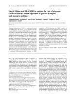
the cases (Table 2, Figure 1).
Table 2 Immunostaining results for EphA2 and Ephrin A-1.
Staining intensity Number of cases
EphA2 (%) Ephrin A-1 (%)
Negative 21 (10) 33 (15)
Weak 108 (50) 91 (42)
Moderate 69 (32) 67 (31)
Strong 19 (9) 26 (12)
Figure 1. Immunohistochemical analysis for EphA2 (A-D) and
EphrinA-1 (E-H) showing cases with no staining (A, E), weak
staining (B, F), moderate staining (C, G) and strong staining (D,
H).
In previous reports [28, 29] protein expression of
p27, p21, p16, cyclin A, cyclin E and cyclin D3 were
studied with immunohistochemistry in the same
patient populations. We therefore make a comparison
between these proteins and EphA2 and EphrinA-1.
There was a clear correlation between expression of
EphA2 and EphrinA-1 (P < 0.0001). No other
significant relationship concerning protein levels was
found (Table 3).
High EphA2 expression (strong staining) was
significantly correlated to the present of parametrial
invasion (P = 0.011). There was a clear correlation
between high EphrinA-1 expression (strong staining)
and deep invasion (P = 0.005) and presence of
parametrial invasion (P < 0.001) (Table 1).
Furthermore, there was a significant association
between high EphrinA-1 and large tumor size (P =
0.043) when grouping the staining pattern
moderate/strong staining as high.
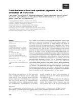
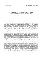
In the univariate analysis high expression of
EphrinA-1 (strong staining) was associated with poor
disease-free (P = 0.033) (Figure 2) and disease-specific
(P = 0.039) survival (Figure 3, Table 4). No significant
Int. J. Med. Sci. 2008, 5
124
correlation was obtained when using strong/moderate
staining as cutoff for EphrinA-1 or strong and
strong/moderate staining as cutoff for EphA2
expression. In the multivariate analyses EphrinA-1
was not an independent prognostic factor for
disease-free (P = 0.139) and disease-specific (P = 0.222)
survival.
Table 3. Correlations.
Variables EphrinA-1 EphA2 p27
†
p21
†
p16
†
Cyclin A
†
Cyclin E
†
Cyclin D3
†
EphrinA-1 1 <0.0001
*
0.883 0.581 0.449 0.160 0.943 0.695
EphA2 1 0.497 0.825 0.152 0.883 0.944 0.771
*
Correlation is significant at 0.01 level.
†
Anti-p27 (Transduction Laboratories, Lexington, USA), anti-p21 (Oncogene Science, MA, USA), anti-p16 (Neomarkers, CA, USA), anti-cyclin
A and anti-cyclin E (both from Novocastra Laboratories, Newcastle, UK) and anti-cyclin D3 (Dako, Glostrup, Denmark). Protein levels were
defined as high when ≥5% of tumor cells were positive for p21, cyclin E and cyclin D3 and ≥50% of tumor cells were positive for p27, p16 and
cyclin A [28, 29].
Table 4 5-years disease-free and disease-specific survival.
Variables
Total
Relapses
N=44
DFS
*
(%)
P
Deaths
N=37
DSS
†
(%)
P
EphA2 0.547 0.748
Low
(negative/weak/moderate)
198
39
81
33
85
High (strong) 19 5 84 4 89
EphrinA-1 0.033 0.039
Low
(negative/weak/moderate)
191 35 83 29 87
High (strong) 26 9 73 8 77
*
Disease-free survival.
†
Disease-specific survival.
Figure 2. Disease-free survival in relation to EphrinA-1
expression.
Figure 3. Disease-specific survival in relation to EphrinA-1
expression.
Discussion
In the present work, weak immunostaining for
EphA2 and EphrinA-1 was present in 40% and 60% of
normal squamous epithelium, respectively. The
positive overall staining (weak/moderate/strong) of
EphA2 and EphrinA-1 was observed in 90% and 85%
Int. J. Med. Sci. 2008, 5
125
of squamous cell cervical carcinomas FIGO stage IB,
respectively. This is in line with our previous study
identifying EphA2 and EphrinA-1 in 88% and 92% of
squamous cell cervical carcinomas FIGO stage I-IV,
respectively [22]. Furthermore, high expression
(moderate/strong staining) for EphA2 and EphrinA-1
was found in 41% and 43% of the cases, respectively.
Previously, a wide range of EphA2 overexpression
(34-87%) [15, 17, 19, 22, 24-26] and EphrinA-1
overexpression (41-61%) [22, 24, 26] has been reported
in many other human cancer types FIGO stage I-IV,
including squamous cell cervical carcinomas were 43%
and 46% of the cases overexpressed EphA2 and
EphrinA-1, respectively [22]. The increased levels of
EphA2 and EphrinA-1 in a relative high number of
early stage squamous cell carcinomas suggested that
these two proteins may be important factors in the
development of a subset of early cervical cancers. This
is in agreement with the study of Zheng and
coworkers [14] where EphA2 protein was suggested to
play an important role in the early stage of prostate
carcinogenesis.
Our findings that EphrinA-1 overexpression
tended to be associated with large tumor size and deep
invasion are consistent with a recent report by Holm et
al. [21] in vulvar carcinoma. However, in oesophagus
cancer [24] and gastric cancer [25] EphrinA-1
expression did not correlate with tumor size.
Increased expression of EphA2 was not of
prognostic significance, whereas overexpression of
EphrinA-1 (strong staining) was associated with poor
survival in squamous cell cervical carcinomas FIGO
stage IB in univariate analysis. However, EphrinA-1
overexpression was not significantly associated with
reduced survival when multivariate analysis was
applied. Contrary, in our previous study of squamous
cell cervical carcinomas FIGO stage I-IV we found that
increased expression of EphA2 and EphrinA-1 was
significantly associated with shorter overall survival in
multivariate analysis [22]. This discrepancy could not
be explained by use of different conditions, because in
these two studies primary antibodies, detection
system, pretreatment and other technical aspects as
well as evaluation and cutoffs of
immunohistochemical results were exactly the same.
Therefore, our results may indicate that EphA2 and
EphrinA-1 could be used as an independent prognostic
factor in squamous cell cervical carcinomas FIGO stage
I-IV [22], but not when only evaluating early
squamous cell cervical carcinomas. In other human
cancers FIGO stage I-IV, high EphA2 [15] and
EphrinA-1 [21, 24, 30] expressions have been
correlated with short survival in univariate analysis,
but not in multivariate analysis. Furthermore, a high
EphA2 level has been correlated with poor survival in
univariate analysis as well as in multivariate analysis
in patients with ovarian carcinomas FIGO stage I-IV
[19, 26] and oesophagus carcinomas FIGO stage I-IV
[24]. In contrast, neither EphrinA-1 nor EphA2 was
associated with survival in patients with carcinomas
FIGO stage I-IV of ovarian [26] and vulvar [21],
respectively. Taken together, these studies showed
that the prognostic significance of EphA2 and
EphrinA-1 in different human cancers is conflicting
and no clear picture can be drawn.
Previously, EphA2 and EphrinA-1 have been
found to be regulated by p53, p63 and p73 in non-small
cell lung carcinoma cell line and breast
adenocarcinoma cell line [31]. In contrast, there was no
correlation between EphA2 or EphrinA-1 and p53 in
vulvar cancer [21]. However, EphA2 overexpression
has been found to be associated with high cyclin A
level, whereas, increase expression of EphrinA-1
correlated with high cyclin A and p21 levels in vulvar
cancer [21]. We did not identify any correlation
between EphA2 or EphrinA-1 and the cell cycle
proteins p21, p27, p16, cyclin A, cyclin E and cyclin D3
in early squamous cell cervical carcinomas. These
studies may indicate that the mechanisms of
expression EphA2 and EphrinA-1 are different in
various types of cancers.
In conclusion, the increased levels of EphA2 and
EphrinA-1 in a relative high number of early stage
squamous cell carcinomas suggested that these two
proteins may play an important role in the
development of a subset of early cervical cancers.
However, EphA2 and EphrinA-1 were not
independently associated with clinical outcome.
Acknowledgements
We thank Mai Thi Phuong Nguyen, Liv Inger
Håseth and Ellen Hellesylt for excellent technical
assistance. This study was supported in part by grant
from the Norwegian Cancer Society.
References
1. Globocan 2000. Cancer Incidence, Mortality and Prevalence
Worldwide; IARC CancerBase No 5(Version 1-0). Lyon, France:
IARCPress. 2001.
2. Benedet JL, Odicino F, Maisonneuve P, et al. Carcinoma of the
cervix uteri. J Epidemiol Biostat. 2001; 6: 7-43.
3. Landoni F, Maneo A, Colombo A, et al. Randomised study of
radical surgery versus radiotherapy for stage Ib-IIa cervical
cancer. Lancet. 1997; 350: 535-40.
4. Delgado G, Bundy BN, Fowler WCJr., et al. A prospective
surgical pathological study of stage I squamous carcinoma of the
cervix: a Gynecologic Oncology Group Study. Gynecol Oncol.
1989; 35: 314-20.
5. Inoue T, Okumura M. Prognostic significance of parametrial
extension in patients with cervical carcinoma Stages IB, IIA, and
IIB. A study of 628 cases treated by radical hysterectomy and
lymphadenectomy with or without postoperative irradiation.
Int. J. Med. Sci. 2008, 5
126
Cancer. 1984; 54: 1714-9.
6. Kamura T, Tsukamoto N, Tsuruchi N, et al. Multivariate
analysis of the histopathologic prognostic factors of cervical
cancer in patients undergoing radical hysterectomy. Cancer.
1992; 69: 181-6.
7. Sevin BU, Nadji M, Lampe B, et al. Prognostic factors of early
stage cervical cancer treated by radical hysterectomy. Cancer.
1995; 76: 1978-86.
8. Nakamoto M, Bergemann AD. Diverse roles for the Eph family
of receptor tyrosine kinases in carcinogenesis. Microsc Res Tech.
2002; 59: 58-67.
9. Zhang J, Hughes S. Role of the ephrin and Eph receptor tyrosine
kinase families in angiogenesis and development of the
cardiovascular system. J Pathol. 2006; 208: 453-61.
10. Andres AC, Reid HH, Zurcher G, et al. Expression of two novel
eph-related receptor protein tyrosine kinases in mammary gland
development and carcinogenesis. Oncogene. 1994; 9: 1461-7.
11. Holder N, Klein R. Eph receptors and ephrins: effectors of
morphogenesis. Development. 1999; 126: 2033-44.
12. Zelinski DP, Zantek ND, Stewart JC, et al. EphA2 overexpression
causes tumorigenesis of mammary epithelial cells. Cancer Res.
2001; 61: 2301-6.
13. Walker-Daniels J, Coffman K, Azimi M, et al. Overexpression of
the EphA2 tyrosine kinase in prostate cancer. Prostate. 1999; 41:
275-80.
14. Zeng G, Hu Z, Kinch MS, et al. High-level expression of EphA2
receptor tyrosine kinase in prostatic intraepithelial neoplasia.
Am J Pathol. 2003; 163: 2271-6.
15. Miyazaki T, Kato H, Fukuchi M, et al. EphA2 overexpression
correlates with poor prognosis in esophageal squamous cell
carcinoma. Int J Cancer. 2003; 103: 657-63.
16. Herrem CJ, Tatsumi T, Olson KS, et al. Expression of EphA2 is
prognostic of disease-free interval and overall survival in
surgically treated patients with renal cell carcinoma. Clin Cancer
Res. 2005; 11: 226-31.
17. Kinch MS, Moore MB, Harpole DHJr. Predictive value of the
EphA2 receptor tyrosine kinase in lung cancer recurrence and
survival. Clin Cancer Res. 2003; 9: 613-8.
18. Mudali SV, Fu B, Lakkur SS, et al. Patterns of EphA2 protein
expression in primary and metastatic pancreatic carcinoma and
correlation with genetic status. Clin Exp Metastasis. 2006; 23:
357-65.
19. Thaker PH, Deavers M, Celestino J, et al. EphA2 expression is
associated with aggressive features in ovarian carcinoma. Clin
Cancer Res. 2004; 10: 5145-50.
20. Lin YG, Han LY, Kamat AA, et al. EphA2 overexpression is
associated with angiogenesis in ovarian cancer. Cancer. 2007;
109: 332-40.
21. Holm R, Knopp S, Suo Z, et al. Expression of EphA2 and
EphrinA-1 in vulvar carcinomas and its relation to prognosis. J
Clin Pathol. 2007; 60: 1086-91.
22. Wu D, Suo Z, Kristensen GB, et al. Prognostic value of EphA2
and EphrinA-1 in squamous cell cervical carcinoma. Gynecol
Oncol. 2004; 94: 312-9.
23. Abraham S, Knapp DW, Cheng L, et al. Expression of EphA2
and Ephrin A-1 in carcinoma of the urinary bladder. Clin Cancer
Res. 2006; 12: 353-60.
24. Xu F, Zhong W, Li J, et al. Predictive value of EphA2 and
EphrinA-1 expression in oesophageal squamous cell carcinoma.
Anticancer Res. 2005; 25: 2943-50.
25. Nakamura R, Kataoka H, Sato N, et al. EPHA2/EFNA1
expression in human gastric cancer. Cancer Sci. 2005; 96: 42-7.
26. Han L, Dong Z, Qiao Y, et al. The clinical significance of EphA2
and Ephrin A-1 in epithelial ovarian carcinomas. Gynecol Oncol.
2005; 99: 278-86.
27. Poulsen HE, Taylor CW, and Sobin LH. Histological typing of
female genital tract tumours. Geneva: World Health
Organization. 1975.
28. Van de Putte G, Holm R, Lie AK, et al. Expression of p27, p21,
and p16 protein in early squamous cervical cancer and its
relation to prognosis. Gynecol Oncol. 2003; 89: 140-7.
29. Van de Putte G, Kristensen GB, Lie AK, et al. Cyclins and
proliferation markers in early squamous cervical carcinoma.
Gynecol Oncol. 2004; 92: 40-6.
30. Herath NI, Spanevello MD, Sabesan S, et al. Over-expression of
Eph and ephrin genes in advanced ovarian cancer: ephrin gene
expression correlates with shortened survival. BMC Cancer.
2006; 6: 144.
31. Dohn M, Jiang J, Chen X. Receptor tyrosine kinase EphA2 is
regulated by p53-family proteins and induces apoptosis.
Oncogene. 2001; 20: 6503-15.