Báo cáo y học: "Monoclonal Antibodies against Nucleophosmin Mutants: Potentials for the Detection of Acute Myeloid Leukemia" ppt
Bạn đang xem bản rút gọn của tài liệu. Xem và tải ngay bản đầy đủ của tài liệu tại đây (470.16 KB, 6 trang )
Int. J. Med. Sci. 2011, 8
309
I
I
n
n
t
t
e
e
r
r
n
n
a
a
t
t
i
i
o
o
n
n
a
a
l
l
J
J
o
o
u
u
r
r
n
n
a
a
l
l
o
o
f
f
M
M
e
e
d
d
i
i
c
c
a
a
l
l
S
S
c
c
i
i
e
e
n
n
c
c
e
e
s
s
2011; 8(4):309-314
Research Paper
Monoclonal Antibodies against Nucleophosmin Mutants: Potentials for the
Detection of Acute Myeloid Leukemia
Shi Tan
1
, Ling Zhang
1
, Xiao-Ming Zhong
1
, Zai-Lin Yang
2
, Liu-Yang Zhao
1
, Yu-Jie Gao
1
, Hui-Yuan Shao
1
,
Feng-Xian Qin
1
, Xian-Chun Chen
1
, Hui-Juan Zhang
1
, Hui Chen
3
, Li Wang
4
1. Key Laboratory of Laboratory Medical Diagnostics, Ministry of Education, Department of Laboratory Medicine, Chong-
qing Medical University, Chongqing 400016, China
2. Center for Hematology, Southwest Hospital, Third Military Medical University, Chongqing 400016, China
3. Department of Laboratory Medicine, the First Affiliated Hospital, Chongqing Medical University, Chongqing 400016,
China
4. Department of Hematology, the First Affiliated Hospital, Chongqing Medical University, Chongqing 400016, China
Corresponding author: Ling Zhang, Department of Laboratory Medicine, Chongqing Medical University, 1#, Yixueyuan
Road, Chongqing, 400016, China. Tel: +86 023-68485223, Fax: +86 023-68485005; Email:
© Ivyspring International Publisher. This is an open-access article distributed under the terms of the Creative Commons License (
licenses/by-nc-nd/3.0/). Reproduction is permitted for personal, noncommercial use, provided that the article is in whole, unmodified, and properly cited.
Received: 2011.04.26; Accepted: 2011.05.11; Published: 2011.05.17
Abstract
Nucleophosmin (NPM1) gene mutations resulting in cytoplasmic delocalization of Nucleo-
phosmin (NPMc+) are the most common genetic alteration in acute myeloid leukemia (AML).
Here, we attempted to prepare monoclonal antibodies (mAbs) against NPM1 mutation A
(NPM-mA) and investigated the mAbs’ clinical utility in immunohistochemical detection of
NPMc+AML. The pET-32a-NPM-mA vector with the whole open reading frame of the
NPM-mA gene was constructed. E.coli BL21 transformed with the vector were induced to
express the NPM-mA recombinant protein. BALB/c mice were immunized with the recom-
binant NPM-mA. Positive clones were selected by indirect ELISA and the mAbs were ob-
tained. Immunohistochemistry was performed to detect the NPMc+ in bone marrow smears
from 10 AML patients with NPM-mA. The results showed that the pET-32a-NPM-mA vector
was successfully constructed and the NPM-mA recombinant protein was used to immunize
the mice. Two positive clones (2G3 and 3F9) were selected. The mAbs against NPM-mA were
raised, but did cross-react with wild type NPM1. The mAbs can be used to detect the cyto-
plasmic dislocation of NPM1 in all AMLs carrying NPM-mA. Our results show that an-
ti-NPM-mA mAbs were produced. Though they would cross-react with wild type NPM1, the
mAbs may still have potential in the detection of NPMc+AMLs.
Key words: acute leukemia, nucleophosmin mutants, recombinant protein, monoclonal antibody
1. Introduction
Nucleophosmin (NPM1) is an ubiquitously ex-
pressed nucleo-cytoplasmic shuttling protein with
prominent nucleolar localization [1, 2]. Previous
studies have demonstrated that mutations of the
NPM1 gene leading to aberrant cytoplasmic NPM1
expression (NPMc+) occur in about one-third of acute
myeloid leukemias (AML) and 45% to 64% of AML
with normal karyotype cases [3, 4]. The most common
molecular variant of the NPM1 gene is mutation A,
accounting for about 75-85% of cases. It is due to a
duplication of TCTG tetranucleotide at the
C-terminus of the NPM1 gene, which generates a nu-
clear export signal (NES) motif responsible for cyto-
plasmic accumulation of NPM1 [5-7]. Many observa-
Ivyspring
International Publisher
Int. J. Med. Sci. 2011, 8
310
tions indicate that the NPM1 mutation A (NPM-mA)
is not only an AML-specific genetic event, but also
remains stable during the course of the disease [6, 8,
9]. Meanwhile, the AML with cytoplasmic NPM1
(NPMc+AML) exhibits distinctive biological and
clinical features and has been included as a new pro-
visional entity in the 2008 World Health Organization
(WHO) classification of myeloid neoplasms [5, 10-13].
Thus, the analysis of NPM1 mutations may emerge as
an initial screening step in the diagnostic/prognostic
work-up of AML and could also serve to monitor
minimal residual disease (MRD) [14].
Over the past five years, several qualitative and
quantitative molecular assays for identifying NPM1
mutations have been developed. Currently available
screening of NPM1 mutations using conventional
polymerase chain reaction (PCR) followed by capil-
lary electrophoresis is rather time-consuming, tech-
nical-demanding and laborious [15]. Alternatively,
the simple, inexpensive and specific immunohisto-
chemical tests (IHC) which indirectly detect aberrant
cytoplasmic accumulation of NPM1 proteins can
serve as a surrogate to molecular studies [16-18]. To
popularize IHC detection of cytoplasmic NPM1 in
clinical diagnosis/prognosis of NPMc+AML, we need
to prepare the anti-NPM-mA monoclonal antibodies
(mAbs) as the primary antibody in IHC assay.
In 1999, Cordell et al prepared the first panel of
mAbs associated with NPM1 protein, two of which
recognized the N-terminal portion of NPM1 present
in NPM-ALK fusion protein and the third was specific
for wild-type NPM1 (NPM-wt). Their main purpose
was to detect the NPM-ALK fusion protein created by
the t(2;5) chromosomal translocation in anaplastic
large-cell lymphoma (ALCL) [19]. Nowadays, exten-
sive detection of cytoplasmic dislocation of NPM1 by
IHC has been performed using aspecific antibodies
that bind both the NPM-wt and NPM-mA proteins. In
IHC assay labeling with this kind of mAbs, the cyto-
plasmic subcellular localization of NPM1 may not be
closely associated with NPM1 gene mutations proba-
bly because of NPM1 diffusion during the tissue fixa-
tion and the influence of fixatives [20]. Thus, produc-
tion of anti-NPM-mA mAbs for routine diagnostic of
NPMc+AML is of critical importance.
To date, most detections of cytoplasmic NPM1
by IHC have been carried out in bone marrow biop-
sies. However, not all hematological centers, espe-
cially in developing countries, adopt bone marrow
biopsy as a frontline diagnostic procedure for AML.
Hence, the ability to detect cytoplasmic NPM1 on
bone marrow smears would be advantageous. In view
of this, we attempted to produce the mAbs that were
specific for NPM-mA protein and preliminarily ex-
plore the application of IHC labeling with these mAbs
on bone marrow smears of AML patients with NPM1
mutations.
2. Materials and Methods
2.1 PCR for amplification of NPM-mA gene
According to the published sequence of the
NPM-mA in GenBank (no.AY740634), a pair of spe-
cific primers were designed to amplify the ORF of
NPM-mA gene from pEGFP-C1-NPM-mA vectors,
which were kindly provided by Dr. B Falini (Institute
of Hematology, University of Perugia, Perugia, Italy).
The forward and backward primers were:
5’-CGGGATCCATCGAAGGTCGTGAAGATTCGAT
GGACAT-3’, and 5’-CGCGCGACCGAGCGGAA
GCTTCTATTTTCTTAAAGAGAC-3’. Underlined
nucleotides represent the BamH I and Hind III site,
respectively. PCR conditions included
pre-denaturation at 98°C for 5 min; 32 cycles of de-
naturation at 98°C for 20 sec, annealing at 56°C for 20
sec, and extension at 72°C for 80 sec; followed by a
final extension at 72°C for 5 min.
2.2 Construction of expressing vector
pET-32a-NPM-mA
After being checked by using 1% agarose gel
electrophoresis and retrieved utilizing the MinElute
Gel Extration Kit (Tiangen, Beijing, China), the ampli-
fication products (NPM-mA gene) were cloned into
the BamH I and Hind III site of the pET-32a plasmids
creating fusion vectors pET-32a-NPM-mA in the
presence of T4 DNA Ligase (TaKara, Tokyo, Japan).
The fusion vectors were subsequently transformed
into E. coli DH5α cloning vectors and E. coli BL21
(DE3) expression bacteria and then grown overnight
at 37°C in Luria-Bertani (LB) medium with ampicillin
(100 μg/ml). The positive expression clones were
screened out by colony PCR. After extracted by a
commercial kit (Huashun, Shanghai, China),
pET-32a-NPM-mA was further identified by re-
striction enzyme digestions and DNA sequencing
(Invitrogen, Shanghai, China). The positive expres-
sion BL21 (DE3) was stored in LB containing 15%
glycerine at -80°C.
2.3 Expression and Purification of NPM-mA
protein
Overnight culture of pET-32a-NPM-mA trans-
formed BL21 (1 ml) was inoculated to 1000 ml
LB/amp and cultured at 37°C for 3-4 h at 200 rpm
until OD600 reached 0.3-0.4, then 0.1 mM IPTG
(TaKara, Tokyo, Japan) was added to induce protein
expression. The culture was incubated for 4 h at 37°C
at 200 rpm before harvesting the cells by centrifuga-
Int. J. Med. Sci. 2011, 8
311
tion (15,000×g, 20 min, 4°C) and the cell pellets were
washed and lysed by sonication on ice. After centri-
fuged at 15,000×g for 20 min, the supernatant was
analyzed by SDS-PAGE as the soluble fraction and the
remaining cell pellet as the insoluble fraction to de-
termine whether native or denaturing conditions
were necessary for protein purification. The superna-
tant was loaded to His-Bind-Resins affinity column
(Novagen, Darmstadt, Germany) to purify the fusion
protein. The purified protein was dialysed against
phosphate-buffered saline (PBS) overnight at 4°C and
stored at -80°C before analyzed by SDS-PAGE and
quantitated by using the BCA Protein Assay Kit (Be-
yotime, Shanghai, China).
2.4 Immunizations
Five-week old female BALB/c mice initially re-
ceived subcutaneous injection of purified NPM-mA
fusion protein (100 μg) emulsified in an equal volume
of Freund’s complete adjuvant (Sigma, St. Luis, MO,
USA). A second injection of the same dose of
NPM-mA protein in incomplete Freund’s adjuvant
was administered 2 weeks later. 10-14 days after the
second booster, the mice were then given NPM-mA
fusion protein without adjuvant intraperitoneally. An
additional intraperitoneal injection of 100 μg of anti-
gen was given 2 days before harvesting the spleen
cells. Experiments with injected mice were performed
under the guidelines for care and use of experimental
animals.
2.5 Cellular fusions
When the anti-NPM-mA antibodies titre of mice
serum reached 1:1024 checked by indirect en-
zyme-linked immunosorbent assay (ELISA), myeloma
cells line SP2/0 (10
6
) were fused with splenocytes
(10
7
) by the addition of 45% polyethylene glycol
(PEG-4000). Hybridomas were selected in HAT me-
dium (Gibco, Carlsbad, CA, USA) and cultured in
96-well plates with BALB/c (8 weeks old) peritoneal
macrophages cells as feeder cells at 37°C in 5% CO
2
in
air. When single colonies of cells were visualized, cell
culture supernatants were obtained and screened for
the presence of anti-NPM-mA antibodies using indi-
rect ELISA. Selected positive hybridomas were ex-
panded and subcloned by limiting dilution.
2.6 Purification and characterization of mAbs
After typed by mouse monoclonal antibody iso-
typing kit (Sigma, St. Luis, MO, USA), the prepared
mAbs were purified from cell-culture supernatant by
affinity chromatography. Indirect ELISA was then
carried out on NPM-wt and NPM-mA coated plates to
check the antigenic characterization of mAbs.
2.7 Patients
Bone marrow/peripheral blood smears were
obtained from de novo AML patients, who were from
Southwest Hospital of Third Military Medical Uni-
versity and The First Affiliated Hospital of Chongqing
Medical University (Chongqing, China) between 2008
and 2009. Informed consent was obtained from all
patients, and the study was approved by the ethics
committees of the participating institutions. Ten posi-
tive samples with NPM-mA were selected by direct
sequencing.
2.8 Immunohistochemistry
Slides were incubated with the anti-NPM anti-
body we prepared (1:100 in Tris-buffered saline)
overnight at 4°C. Immunohistochemistry was per-
formed using the Streptavidin-Peroxidase (SP)-9000
kit (Zhongshan, Beijing, China) according to the
manufacturer’s instructions. Peroxidase activity was
revealed with 3-3-diaminobenzidine-copper sulphate
(Sigma, St. Luis, MO, USA) to obtain brownblack
granules. The subcellular distribution of NPM-mA
was assessed after counterstained with hematoxylin.
PBS was used as a negative control for the anti-NPM
antibody.
3. Results
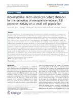
3.1 PCR for amplification of NPM-mA gene
Using a pair of primers specific for NPM-mA
gene, a DNA fragment of approximately 900 bp size
was amplified from the pEGFP-C1-NPM-mA plas-
mids by PCR technique (Figure 1), which corre-
sponded to the full length of open reading frame
(ORF) of the NPM-mA gene (935 bp).
Figure 1. PCR amplifying the full sequence of ORF of the
NPM-mA gene. The PCR products amplified with a pair of
primers against the NPM-mA gene were analyzed by 1%
agarose gel electrophoresis.1: DL2000 markers; 2-3:
products of PCR.
Int. J. Med. Sci. 2011, 8
312
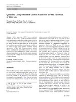
3.2 Construction of recombinant vector
pET-32a-NPM-mA
To generate a recombinant human encoding the
NPM-mA protein, the pET-32a-NPM-mA vector was
cloned. As shown in Figure 2, the pET-32a-NPM-mA
vector was successfully constructed as verified by
bacterial colony PCR (Figure 2A), restriction enzyme
digestions (Figure 2B) and DNA sequencing (data not
shown).
Figure 2. Cloning of the recombinant vector pET-32a-NPM-mA. A, Bacterial colony PCR for the detection of the BL21
(DE3) clones with the target prokaryotic expression vector pET-32a-NPM-mA. 1-7: 7 colonies of bacteria selected on LB
medium with ampicillin; 8: DL2000 markers. B, Double endonuclease digestion of the prokaryotic expression vector
pET-32a-NPM-mA. 1-2: pET-32a-NPM-mA; 3: DL15000 markers; 4: DL2000 markers; 5-6: double digestion with the BamH
I and Hind III.
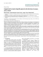
3.3 Expression and purification of recombinant
NPM-mA antigen
The NPM-mA fusion protein was resoluble and
detected in the culture supernatants. SDS-PAGE
analysis of the fusion protein is displayed in Figure 3.
Expression and purification of the NPM-mA antigen
were performed as described in Materials and Meth-
ods. The concentration of the purified recombinant
protein was 1.95 μg/μl determined by BCA protein
assay.
Figure 3. SDS-PAGE assay of the purified NPM-mA fusion
protein. 1: protein size markers; 2-3: recombinant NPM-mA
fusion protein.
3.4 Production of the anti-NPM mAbs
For selecting the clones with mAbs against
NPM-mA, the supernatants of fused cells were as-
sayed by indirect ELISA. Two clones were found to be
positive in the ELISA screen of culture supernatants
(designated as 2G3 and 3F9). The 2G3 clones which
exhibited good growth characteristics and antibody
production were subjected to subcloning. Antibodies
secreted by the 2G3 clones were found to be IgG iso-
type. The specificity of the mAbs against NPM-mA
was assessed by indirect ELISA, and the mAbs were
able to react with both NPM-wt and NPM-mA.
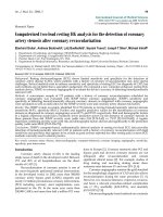
3.5 Immunohistochemical staining for the cases
with NPM1 mutations
Ten AML samples had been confirmed to bear
NPM-mA by direct sequencing (data not shown). To
validate the mAbs against NPM-mA as a diagnostic
tool for AML patients, bone marrow or peripheral
blood samples were analyzed by IHC, using the 2G3
mAb. The cytoplasmic dislocation of NPM1 was ob-
served in all 10 samples with NPM1 mutations,
without staining in the cytoplasm of leukemic blasts
in the negative control. Figure 4 shows representative
results from a NPMc+AML patient.
Int. J. Med. Sci. 2011, 8
313
Figure 4. Immunohistochemistry analyses of NPMc+AML samples using the 2G3 mAb. A, The cytoplasmic dislocation of
NPM1 protein was observed in a representative bone marrow smear from NPMc+AML patients. Brownblack coarse
granules in the cytoplasm of leukemic cells are shown. B, Negative control; the bone marrow from the same case as in (A)
was stained with PBS substituting for the 2G3 mAb.
4. Discussion
Mutations involving the NPM1 gene are the
most frequent genetic aberrations of AML, and define
a clinically distinct subset of AML [21, 22]. NPM1
gene mutations always result in cytoplasmic disloca-
tion of NPM1, which is the immunohistochemical
hallmark of NPMc+AML. Immunohistochemistry
may be a simple, rapid screening test for putative
NPM1 gene alterations in a wide range of human
hematological malignancies [17, 21]. The crucial step
of immunohistochemical detection is to generate
mAbs directed against NPM1 mutants.
Currently, immunohistochemistry is usually
performed with mAbs that recognize wild-type and
mutated NPM1 proteins. In the present study, we
attempted to prepare mAbs against NPM-mA. The
specific mAbs reacting with NPM-mA have the ad-
vantage of directly detecting the NPM-mA protein in
leukemic cells. Firstly, some technical factors, such as
NPM1 diffusion during tissue fixation and the use of
different fixatives, may result in the incomplete con-
cordance between NPMc+ and NPM mutations status
in some cases [20, 23]. Furthermore, because NPM1 is
a nuclear-cytoplasmic shuttling protein and highly
expressive in proliferative cells, the small fraction of
NPM-wt protein may pathologically present in the
cytoplasm of tumor cells [24]. As a result, IHC label-
ing with the mAbs against NPM-mA and NPM-wt
may detect the NPM-wt existing in the cytoplasm and
cause false positives. In this study, we analyzed the
antigen epitope of NPM-mA protein and confirmed it
may exist in the C-terminal domain of the NPM-mA
by using the Protean module of DNAstar analysis
software. However, our results revealed that the ob-
tained mAbs did cross-react with NPM-wt. A possible
explanation is that the distinction between NPM-mA
and NPM-wt is small (only a tetranucleotide insertion
located at the C-terminus of NPM-mA) [25]. So the
2G3 mAb we obtained may not interat with a specific
epitope generated by the NPM1 mutation. Recently,
Gruszka et al [26] have raised a mAb (T26) only
against NPM1 mutants by using a 19-aminoacid pol-
ypeptide immunogen (CLAVEEVLSRK) containing
the unique C-terminus of the NPM-mA protein. It
indicated that the specific polypeptide generated by
the C-terminus of the NPM1 (type A) mutation may
be an optimal immunogen.
Over the past five years, IHC detection of
NPMc+ on bone marrow biopsies has been widely
carried out. However, as bone marrow biopsies are
not always performed for the diagnosis of AML, es-
pecially in developing countries, to detect NPMc+ on
bone marrow smears would be more advantageous.
IHC assay was performed using the 2G3 mAb on bone
marrow/peripheral blood smears of 10 AML patients
with NPM-mA, and significant correlation was found
between NPMc+ and NPM1 mutations status, which
is not consistent with the finding of Mattsson et al
[27]. They reported that the immunocytochemical
staining should not be used as a surrogate for NPM1
mutations in AML, due to the high false positive and
negative rates for NPMc+ in cell smears. The possible
reasons for the two different results include the dif-
Int. J. Med. Sci. 2011, 8
314
ferent anti-NPM antibodies (2G3 mAb or NA24 mAb)
and the different method used (SP method or immu-
noalkaline phosphatase method).
In summary, we put forward the production of
mAbs that specifically recognize NPM1. Although the
mAbs prove to react with NPM-mA and NPM-wt, this
result provides valuable information in that the mAbs
against NPM-mA cannot be raised using the recom-
binant NPM-mA protein as immunogen. Further-
more, the complete correlation between NPMc+ in cell
smears and NPM1 mutations status has been found in
clinical samples by IHC using the 2G3, which would
be utilized for other potential techniques,such as
immunofluorescence, flow cytometry, etc.
Acknowledgements
We would like to thank Dr. Falini B in University
of Perugia for the gift of pEGFP-C1-NPM-mA vectors.
This project was supported by a grant from National
Natural Science Foundation of China (No. 30872418)
and Natural Science Foundation Project of CQ CSTC
(No. 2010BB5363).
Conflict of Interest
The authors have declared that no conflict of in-
terest exists.
References
1. Borer RA, Lehner CF, Eppenberger HM, et al. Major nucleolar
proteins shuttle between nucleus and cytoplasm. Cell. 1989; 56:
379-90.
2. Nishimura Y, Ohkubo T, Furuichi Y, et al. Tryptophans 286 and
288 in the C-terminal region of protein B23.1 are important for
its nucleolar localization. Biosci Biotechnol Biochem. 2002; 66:
2239-42.
3. Schnittger S, Schoch C, Kern W, et al. Nucleophosmin gene
mutations are predictors of favorable prognosis in acute mye-
logenous leukemia with a normal karyotype. Blood. 2005; 106:
3733-9.
4. Boissel N, Renneville A, Biggio V, et al. Prevalence, clinical
profile, and prognosis of NPM mutations in AML with normal
karyotype. Blood. 2005; 106: 3618-20.
5. Falini B, Nicoletti I, Martelli MF, et al. Acute myeloid leukemia
carrying cytoplasmic/mutated nucleophosmin (NPMc+ AML):
biologic and clinical features. Blood. 2007; 109: 874-85.
6. Liso A, Bogliolo A, Freschi V, et al. In human genome, genera-
tion of a nuclear export signal through duplication appears
unique to nucleophosmin (NPM1) mutations and is restricted
to AML. Leukemia. 2008; 22: 1285-9.
7. Falini B, Bolli N, Shan J, et al. Both carboxy-terminus NES motif
and mutated tryptophan(s) are crucial for aberrant nuclear ex-
port of nucleophosmin leukemic mutants in NPMc+ AML.
Blood. 2006; 107: 4514-23.
8. Chou WC, Tang JL, Lin LI, et al. Nucleophosmin mutations in
de novo acute myeloid leukemia: the age-dependent incidences
and the stability during disease evolution. Cancer Res. 2006; 66:
3310-6.
9. Palmisano M, Grafone T, Ottaviani E, et al. NPM1 mutations
are more stable than FLT3 mutations during the course of dis-
ease in patients with acute myeloid leukemia. Haematologica.
2007; 92:1268-9.
10. Falini B, Martelli MP, Bolli N, et al. Acute myeloid leukemia
with mutated nucleophosmin (NPM1): is it a distinct entity?
Blood. 2011; 117: 1109-20.
11. Becker H, Marcucci G, Maharry K, et al. Favorable prognostic
impact of NPM1 mutations in older patients with cytogenet-
ically normal de novo acute myeloid leukemia and associated
gene- and microRNA-expression signatures: a Cancer and
Leukemia Group B study. J Clin Oncol. 2010; 28: 596-604.
12. Vardiman JW, Thiele J, Arber DA, et al. The 2008 revision of the
World Health Organization (WHO) classification of myeloid
neoplasms and acute leukemia: rationale and important
changes. Blood. 2009; 114:937-51.
13. Pasqualucci L, Liso A, Martelli MP, et al. Mutated nucleo-
phosmin detects clonal multilineage involvement in acute my-
eloid leukemia: Impact on WHO classification. Blood. 2006;
108:4146-55.
14. Papadaki C, Dufour A, Seibl M, et al. Monitoring minimal
residual disease in acute myeloid leukaemia with NPM1 muta-
tions by quantitative PCR: clonal evolution is a limiting factor.
Br J Haematol. 2009; 144: 517-23.
15. Noguera NI, Ammatuna E, Zangrilli D, et al. Simultaneous
detection of NPM1 and FLT3-ITD mutations by capillary elec-
trophoresis in acute myeloid leukemia. Leukemia. 2005;
19:1479-82.
16. Falini B, Mason DY. Proteins encoded by genes involved in
chromosomal alterations in lymphoma and leukemia: clinical
value of their detection by immunocytochemistry. Blood. 2002;
99: 409-26.
17. Falini B, Mecucci C, Tiacci E, et al. Cytoplasmic nucleophosmin
in acute myelogenous leukemia with a normal karyotype. N
Engl J Med. 2005; 352: 254-66.
18. Falini B, Martelli MP, Bolli N, et al. Immunohistochemistry
predicts nucleophosmin (NPM) mutations in acute myeloid
leukemia. Blood. 2006; 108:1999-2005.
19. Cordell JL, Pulford KA, Bigerna B, et al. Detection of normal
and chimeric nucleophosmin in human cells. Blood. 1999; 93:
632-42.
20. Konoplev S, Huang X, Drabkin HA, et al. Cytoplasmic locali-
zation of nucleophosmin in bone marrow blasts of acute mye-
loid leukemia patients is not completely concordant with
NPM1 mutation and is not predictive of prognosis. Cancer.
2009; 115: 4737-44.
21. Thiede C, Koch S, Creutzig E, et al. Prevalence and prognostic
impact of NPM1 mutations in 1485 adult patients with acute
myeloid leukemia (AML). Blood. 2006; 107: 4011-20.
22. Falini B, Sportoletti P, Martelli MP. Acute myeloid leukemia
with mutated NPM1: diagnosis, prognosis and therapeutic
perspectives. Curr Opin Oncol. 2009; 21: 573-81.
23. Falini B, Martelli MP, Pileri SA, et al. Molecular and alternative
methods for diagnosis of acute myeloid leukemia with mutated
NPM1: flexibility may help. Haematologica. 2010; 95: 529-34.
24. Ochs R, Lischwe M, O'Leary P, et al. Localization of nucleolar
phosphoproteins B23 and C23 during mitosis. Exp Cell Res.
1983; 146:139-49.
25. Falini B, Bolli N, Liso A, et al. Altered nucleophosmin transport
in acute myeloid leukaemia with mutated NPM1: molecular
basis and clinical implications. Leukemia. 2009; 23:1731-43.
26. Gruszka AM, Lavorgna S, Consalvo MI, et al. A monoclonal
antibody against mutated nucleophosmin1 for the molecular
diagnosis of acute myeloid leukemias. Blood. 2010; 116:
2096-102.
27. Mattsson G, Turner SH, Cordell J, et al. Can cytoplasmic nu-
cleophosmin be detected by immunocytochemical staining of
cell smears in acute myeloid leukemia? Haematologica. 2010;
95: 670-3.