Báo cáo y học: "Dissection of a locus on mouse chromosome 5 reveals arthritis promoting and inhibitory genes" ppsx
Bạn đang xem bản rút gọn của tài liệu. Xem và tải ngay bản đầy đủ của tài liệu tại đây (849.61 KB, 12 trang )
Open Access
Available online />Page 1 of 12
(page number not for citation purposes)
Vol 11 No 1
Research article
Dissection of a locus on mouse chromosome 5 reveals arthritis
promoting and inhibitory genes
Therese Lindvall
1
, Jenny Karlsson
1,3
, Rikard Holmdahl
1
and Åsa Andersson
2
1
Department of Experimental Medical Science, Unit for Medical Inflammation Research, BMC I11, Lund University, S-221 84 Lund, Sweden
2
Department of Pharmacology and Pharmacotherapy, Faculty of Pharmaceutical Sciences, Copenhagen University, Universitetsparken 2, DK-2100
Copenhagen Ø, Denmark
3
Current address: Pathology and Laboratory Medicine, University of California Los Angeles, 675 S. Charles E. Young Drive, Los Angeles, CA 90095,
USA
Corresponding author: Therese Lindvall,
Received: 18 Jul 2008 Revisions requested: 9 Aug 2008 Revisions received: 2 Dec 2008 Accepted: 20 Jan 2009 Published: 20 Jan 2009
Arthritis Research & Therapy 2009, 11:R10 (doi:10.1186/ar2597)
This article is online at: />© 2009 Lindvall et al.; licensee BioMed Central Ltd.
This is an open access article distributed under the terms of the Creative Commons Attribution License ( />),
which permits unrestricted use, distribution, and reproduction in any medium, provided the original work is properly cited.
Abstract
Introduction In a cross between two mouse strains, the
susceptible B10.RIII (H-2
r
) and resistant RIIIS/J (H-2
r
) strains, a
locus on mouse chromosome 5 (Eae39) was previously shown
to control experimental autoimmune encephalomyelitis (EAE).
Recently, quantitative trait loci (QTL), linked to disease in
different experimental arthritis models, were mapped to this
region. The aim of the present study was to investigate whether
genes within Eae39, in addition to EAE, control development of
collagen-induced arthritis (CIA).
Methods CIA, induced by immunisation with bovine type II
collagen, was studied in Eae39 congenic and sub-interval
congenic mice. Antibody titres were investigated with ELISA.
Gene-typing was performed by micro-satellite mapping and
statistics was calculated by standard methods.
Results Experiments of CIA in Eae39 congenic- and sub-
interval congenic mice, carrying RIIIS/J genes on the B10.RIII
genetic background, revealed three loci within Eae39 that
control disease and anti-collagen antibody titres. Two of the loci
promoted disease and the third locus was protected against
CIA development. By further breeding of mice with small
congenic fragments, we identified a 3.2 mega base pair (Mbp)
interval that regulates disease.
Conclusions Disease-promoting and disease-protecting genes
within the Eae39 locus on mouse chromosome 5 control
susceptibility to CIA. A disease-protecting locus in the telomeric
part of Eae39 results in lower anti-collagen antibody responses.
The study shows the importance of breeding sub-congenic
mouse strains to reveal genetic effects on complex diseases.
Introduction
Rheumatoid arthritis (RA) and multiple sclerosis (MS) are com-
plex inflammatory autoimmune disorders in which genetic and
environmental factors contribute to disease development [1].
RA is characterised by peripheral joint inflammation, cartilage
and bone destruction and, subsequently, joint deformation. In
MS, the myelin and axons are affected by inflammation within
the CNS often leading to severe neurological dysfunction. The
disease-causing mechanisms remain unknown, although it is
known that the aetiology is dependent on multiple genetic and
environmental factors. To date, only a few genes have been
associated with susceptibility to RA [2-4] and MS [5,6].
The most commonly used animal model for RA is collagen-
induced arthritis (CIA) [7]. The B10.RIII (H-2
r
) mouse strain
develops poly-arthritis after immunisation with bovine type II
collagen, whereas the RIIIS/J mouse strain, having the same
major histocompatibility complex (MHC) haplotype (H-2
r
), is
resistant to poly-arthritis development. Induction of CIA is
dependent on genes within the MHC, but as previously shown
in crosses between B10.RIII and RIIIS/J mice, non-MHC
AUC: area under curve; BSA: bovine serum albumin; CIA: collagen-induced arthritis; CNS: central nervous system; EAE: experimental autoimmune
encephalomyelitis; ELISA: enzyme-linked immunosorbent assay; FP: front primer; IFA: incomplete Freund's adjuvant; Ig: immunoglobulin; Mbp: mega
base pairs; MHC: major histocompatibility complex; MS: multiple sclerosis; PBS: phosphate-buffered saline; PCR: polymerase chain reaction; QTL:
quantitative trait locus; RA: rheumatoid arthritis; RP: reverse primer.
Arthritis Research & Therapy Vol 11 No 1 Lindvall et al.
Page 2 of 12
(page number not for citation purposes)
genes also play an important role in susceptibility to disease
[8-10].
Experimental autoimmune encephalomyelitis (EAE) is an
inflammatory demyelinating disease of the central nervous sys-
tem (CNS), widely used as an animal model for MS. The
B10.RIII strain is susceptible to EAE induced by the myelin
basic protein (MBP) peptide 89–101 [11]. From studies of
crosses between B10.RIII and RIIIS/J (resistant to EAE devel-
opment), a number of non-MHC quantitative trait loci (QTLs),
linked to EAE susceptibility, have been reported [12-14]. In
one study, the Eae39 locus on mouse chromosome 5 was
linked to acute EAE [13]. The inheritance pattern showed that
RIIIS/J genes were dominantly protective. The Eae39 locus is
the only QTL linked to EAE on mouse chromosome 5, but six
QTLs linked to disease in arthritis models have been identified
on this chromosome: Cia13, Cia14 and Cia27 for CIA
[15,16], Pgia16 for proteoglycan-induced arthritis [17], and
Bbaa3 and Bbaa2 for Borrelia burgdorferi-associated arthritis
[18].
The Eae39 locus was identified as a genetic region of about
30 mega base pairs (Mbp). In order to further investigate the
genetic control of disease in the B10.RIII/RIIIS/J model, we
have studied CIA in Eae39 and Eae39 sub-interval congenic
mice. We observed three different inheritance patterns asso-
ciated with arthritis development, which argues that there are
at least three genes in Eae39 that are important for the devel-
opment of inflammatory disease. Two of the loci, located within
a distance of a few Mbp, contain genes that, depending on the
allele, either protect from or promote disease. This suggests a
balancing effect by closely located genes on disease suscep-
tibility that is revealed when QTLs are split into smaller frag-
ments.
Materials and methods
Animals
C57Bl/10.RIII (B10.RIII) were originally provided by J. Klein
(Tübingen, Germany), and kept in the breeding colony at the
Department of Medical Inflammation Research, Lund Univer-
sity. RIIIS/J animals were purchased from The Jackson Labo-
ratory (Bar Harbor, ME). The Eae39 congenic mice (C1,
Figure 1) were produced by marker selected backcrossing of
the RIIIS/J (donor) mice to the B10.RIII (recipient) strain. All
experiments were approved by the local ethical authorities in
Malmö-Lund, Sweden (permit numbers: M70-04, M75-04,
M107-07 and M109-07).
Induction and evaluation of collagen-induced arthritis
Bovine type II collagen was prepared from calf nasal cartilage
by pepsin digestion and was purified as previously described
[19]. CIA was induced by intra-dermal immunisation at the
base of the mouse's tail with 100 μg bovine CII emulsified in
incomplete Freund's adjuvant (IFA) (Difco, Detroit, MI, USA).
The mice were boosted 35 days later with 50 μg bovine CII
emulsified in IFA. The mice, ranging in age between 10 and 24
weeks, were all immunised the same day. Clinical disease was
monitored once or twice a week according to a scoring system
based on the number of inflamed joints. Each inflamed toe or
knuckle was given a score of one and an inflamed wrist or
ankle was given five points. Each mouse could in total get 15
points per limb and a maximum score of 60. The area under the
curve (AUC) was calculated as the sum of scores for each
individual mouse during a defined test period. The mean max-
imum score, representing disease severity, was calculated as
the mean of the maximum score of all sick mice in the respec-
tive groups.
Antibody measurement
Blood was collected on day 14, 21 or day 54 after immunisa-
tion. Sera were prepared and stored at -20°C until assayed.
ELISA was used to determine levels of antibodies against col-
lagen type II. Plates (Nunc maxisorp, Roskilde, Denmark) were
coated with bovine CII (10 μg/ml) in PBS (pH 9) and blocked
with 1% BSA. Immunoglobulin (Ig) M, IgG1, IgG2c, IgG3 and
total Ig levels were measured using biotinylated secondary
antibodies: goat anti-mouse IgM (No. 1020-08); IgG1
(No.1070-08); IgG2c (No. 1079-04); IgG3 (No. 1100-08);
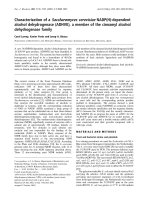
Figure 1
The Eae39 locus on mouse chromosome 5The Eae39 locus on mouse chromosome 5. The C1 congenic fragment
is derived from RIIIS/J and bred on to the B10.RIII background by
marker selected back-crossing. Black = two B10.RIII alleles; white =
two RIIIS/J alleles. Mbp = mega base pairs (positions according to
Ensembl release 49).
Available online />Page 3 of 12
(page number not for citation purposes)
and total Ig (No. 1010-08) (Southern Biotechnologies Associ-
ates, Birmingham, AL, USA). Binding of biotinylated antibod-
ies was revealed by Extravidin Peroxidase (No. E-2886)
(Sigma-Aldrich, St Louis, MO, USA). Plates were developed
with ABTS: 2,2'-Azino-di-[3-ethylbenzthiazoline sulfonate (6)]
diammonium salt (Roche, Mannheim, Germany). Pooled sera
were used as a standard and the antibody levels were meas-
ured as arbitrary concentrations.
Genotyping and linkage analysis
Genomic DNA was isolated from toe or tail biopsies. The biop-
sies were dissolved in 500 μl of 50 mM sodium hydroxide for
one to two hours at 95°C, and subsequently neutralised with
100 μl 1 mM Tris-HCl (pH 8). To perform a standard 10 μl
PCR, 1 μl of the solution was used. The PCR products were
analysed on a MegaBACE DNA analysis system 1000 (Amer-
sham Pharmacia Biotech, Little Chalfont, UK), according to
the manufacturer's protocol. Fifteen informative fluorescence-
labelled micro-satellite markers (Interactiva Biotechnologie,
Ulm, Germany and MWG Biotech, Ebersberg, Germany) were
used to genotype the Eae39 congenic fragment. Linkage anal-
ysis and permutation tests were conducted as previously
described [13]. Sub-congenic mice were genotyped with
additional micro-satellite markers, where some markers are
made in-house: D5acacbhm4 (114.42 Mbp, forward primer
(FP) 5'-CCCTGTAGAAGACTGGGAATTG-3, reverse primer
(RP) 5'-TCCAGGACAGTCAGGGCTAC-3'), D5taokhm12
(117.53 Mbp, FP: 5'-TCAGGGCTCCATGCACTT-3', RP: 5'-
CACAAGTGGCTCTCAGTGCT-3), D5sdshm18 (120.74
Mbp, FP: 5'-GGGGAACACAAGGAGTTTGA-3', RP: 5'-
ATTCAAGGGCATGTGTGTGA-3').
Results
Eae39 controls collagen-induced arthritis
The Eae39 locus on mouse chromosome 5 (Figure 1) was pre-
viously described in a genetic linkage analysis based on a
backcross between the RIIIS/J and B10.RIII strains [13]. The
confidence interval for Eae39 extended from the micro-satel-
lite marker D5Mit259 (90 Mbp) to D5Mit136 (119 Mbp). This
locus was shown to control incidence of acute EAE in male
mice, with a dominant effect of RIIIS/J alleles on protection
from disease development.
To further investigate the Eae39 locus, a 65 Mbp RIIIS/J frag-
ment was bred into the B10.RIII genome to establish a BR.
Eae39
RIIIS/J
congenic strain (Figure 1). This region contains a
number of QTLs linked to the development of disease in exper-
imental models for arthritis, and we wanted to investigate
whether Eae39 controls the susceptibility to CIA in the
B10.RIII and RIIIS/J strain combination. Thus, Eae39 congenic
mice were immunised with bovine collagen type II in IFA. As
shown in Figure 2 and Tables 1 and 2, genes within Eae39
control CIA. Figure 2 shows the development of disease in
mice with the congenic Eae39 fragment shown in Figure 1.
Mice with the C1 congenic fragment had higher incidence of
disease (64%) and higher accumulated arthritis score (AUC
(d50-73) = 125 ± 50) compared with the non-congenic litter-
mates (incidence of disease 24%, AUC (d50-73) = 57 ± 29).
(Incidence, p = 0.0271, Chi squared test; AUC (d50-73), p =
0.0379, Mann–Whitney U test).
Next, we intercrossed C1 heterozygous mice in order to get
offspring with overlapping congenic fragments (Figure 3) to
pinpoint smaller intervals within Eae39 linked to the disease
phenotype. The mice were investigated for development of
CIA and each individual was genotyped with markers span-
ning the congenic fragment. We observed that RIIIS/J alleles
at marker D5Mit113 (78 Mbp) promoted disease incidence
(Table 1). In contrast, one RIIIS/J allele at the marker
D5Mit136, in the telomeric part of the fragment, protected
against CIA development (Table 1). There was no difference in
mean maximum score of the affected mice, except for females
with RIIIS/J alleles at marker D5Mit136, which had signifi-
cantly lower CIA scores compared with littermate controls
(Table 1). This is in line with the male mice carrying hetero-
zygous alleles at D5Mit136, where none of the mice devel-
oped arthritis.
In Table 2, correlation between the disease severity phenotype
AUC, day 50 to 73 after immunisation, and genotype is shown.
The AUC is the sum of scores for each individual mouse dur-
ing a defined test period and describes the development of
disease in terms of onset, duration and severity. In line with the
disease incidence data (Table 1), RIIIS/J alleles at D5Mit113
promoted disease, whereas one RIIIS/J allele at about 120
Mbp (D5Mit136, D5Mit367) almost completely protected
from CIA (Table 2). From these results we conclude that
Eae39 harbors genes that, in addition to controlling EAE, are
important for susceptibility to CIA, and that the region contains
Figure 2
Collagen-induced arthritis (CIA) development in mice with the C1 con-genic fragmentCollagen-induced arthritis (CIA) development in mice with the C1 con-
genic fragment. a/a (area under the curve (AUC) (d50-73) = 125 ± 50,
incidence = 64%), b/b (AUC (d50-73) = 57 ± 29, incidence = 24%),
(AUC (d50-73) p = 0.0379, Mann-Whitney U test, incidence p =
0.0271, Chi squared test).
Arthritis Research & Therapy Vol 11 No 1 Lindvall et al.
Page 4 of 12
(page number not for citation purposes)
Table 1
Incidence and mean maximum score of collagen-induced arthritis (CIA) in Eae39 congenic mice
a
Phenotype Marker Mbp
b
Group a/a
c
a/b b/b p-value
d
Incidence D5Mit113 77.68 Total 10/14 (71%) 7/12 (58%) 8/42 (19%) 0.0005
Males 3/5 (60%) 4/9 (44%) 2/21 (10%) 0.0222
Females 7/9 (78%) 3/3 (100%) 6/21 (29%) 0.0082
D5Mit136 119.18 Total 11/15 (73%) 3/19 (16%) 11/34 (32%) 0.0019
Males 3/5 (60%) 0/10 (0%) 6/20 (30%) 0.0345
Females 8/10 (80%) 3/9 (33%) 5/14 (36%) 0.0573
Mean max score
e
D5Mit113 77.68 Total 23 ± 5 21 ± 5 21 ± 7 0.7766
Males 31 ± 15 20 ± 10 14 ± 2 0.3280
Females 20 ± 5 22 ± 10 24 ± 9 0.9604
D5Mit136 119.18 Total 21 ± 5 6 ± 4 27 ± 15 0.1034
Males 31 ± 15 18 ± 3 0.1948
Females 18 ± 5 6 ± 4 37 ± 6 0.0317
a
Shows the mean incidence and the mean maximum score of mice with the respective genotypes on markers D5Mit113 and D5Mit136.
Calculations were made on all mice in Figures 1 and 3 (a to l), and littermate controls. The sub-interval congenic mice were generated by
intercrossing the C1 congenic mice (Figure 1).
b
Mbp = mega base pairs. The Mbp position is according to Ensembl release 49.
c
a/a = homozygous RIIIS/J alleles; b/b = homozygous B10.RIII alleles; a/b = heterozygous.
d
Statistics for incidence was calculated with Chi squared test. Statistics for severity was calculated with Kruskal-Wallis test and Mann-Whitney U
test.
e
Mean of the maximum score for all affected mice in Figures 1 and 3.
Table 2
CIA severity in Eae39 congenic mice
AUC (d50-73)
a
Marker Mbp
b
a/a
c
(n) a/b (n) b/b (n) p-value
d
D5Mit113 77.68 126 ± 41 (14) 87 ± 35 (12) 30 ± 15 (42) 0.0006
D5Mit157 101.06 118 ± 39 (15) 41 ± 17 (27) 46 ± 24 (26) 0.0071
D5Mit240 109.52 118 ± 39 (15) 33 ± 16 (27) 55 ± 25 (26) 0.0075
D5Mit136 119.18 118 ± 39 (15) 4 ± 4 (19) 66 ± 22 (34) 0.0023
D5Mit367 120.31 111 ± 37 (16) 5 ± 4 (17) 66 ± 22 (34) 0.0077
D5Mit137 123.73 107 ± 44 (13) 21 ± 13 (22) 68 ± 22 (33) 0.1014
D5Mit95 125.31 99 ± 41 (14) 22 ± 14 (21) 68 ± 22 (33) 0.1799
D5Mit161 127.40 106 ± 44 (13) 24 ± 14 (35) 64 ± 21 (20) 0.2488
D5Mit59 128.20 99 ± 41 (14) 25 ± 15 (19) 66 ± 22 (34) 0.2322
D5Mit31 139.00 125 ± 50 (11) 47 ± 34 (8) 48 ± 16 (49) 0.0423
a
Area under curve (AUC) is the mean ± standard error of the total sum of scores for mice with the respective genotypes (day 50 until day 73). All
mice in Figures 1 and 3 (a to l), and littermate controls (b/b) are included in the calculations. The sub-interval congenic mice were generated by
intercrossing the C1 congenic mice (Figures 1 and 2).
b
Mbp = mega base pairs. The Mbp position is according to Ensembl release 49.
c
a/a = homozygous RIIIS/J alleles; b/b = homozygous B10.RIII alleles; a/b = heterozygous.
d
Statistics calculated with Kruskal-Wallis test.
Available online />Page 5 of 12
(page number not for citation purposes)
genes operating in different directions in the disease develop-
ment.
In humans, women are more affected by RA than men. The sex
influence on susceptibility to CIA is, however, normally the
opposite in mice. In the first investigation of CIA development
in mice with overlapping Eae39 sub-interval congenic frag-
ments, we observed that female congenic mice had the same,
or slightly higher, incidence of disease compared with male
mice (Table 1). Severity of CIA (mean maximum score) was the
same, except for females homozygous for B10.RIII alleles (b/
b) at marker D5Mit136, in which the severity was higher (p <
0.05) compared with male mice (Table 1). From the original
mapping experiment, Eae39 was linked to development of
acute EAE in male mice [13]. For this reason, and in order to
keep the number of mice used to a minimum, we decided to
continue the present study with male mice only.
The collagen type II antibody response is controlled by
genes in the Eae39 locus
In an F2 cross between the arthritis susceptible DBA/1J and
the resistant FVB/N strains, it was recently shown that the
Cia27 locus controls anti-collagen type II IgG2a antibody lev-
els [16]. To investigate the corresponding region within the
Eae39 locus for disease phenotypes, we produced mice with
smaller, overlapping congenic fragments (C2 to C5) (Figure 4)
and studied the anti-collagen antibody response after immuni-
sation. The IgG1, IgG2c, IgG3 and IgM anti-collagen serum
levels at day 14 after immunisation were significantly lower in
mice with the C2 congenic fragment (Figure 4) compared with
littermate controls (Table 3). By comparing antibody levels in
mice with the C3 and C4 congenic fragments, we found that
the anti-collagen type II serum titres of the IgG2c isotype were
significantly lower in mice with the C4 fragment compared
with littermates and to mice with the C3 fragment. Mice with
the C5 fragment (spanning from D5Mit317 (112 Mbp) to
D5Mit367 (120 Mbp)) had significantly lower IgG1, IgG2c,
IgG3 and total Ig serum levels compared with littermate con-
trols (Table 3). This confirms the effect on the antibody
response observed with the C2 fragment and shows that
genes in this region control antibody responses to type II col-
lagen.
Collagen-induced arthritis development and antibody
responses to type II collagen in the C5, C6, C9, C10, and
C11 congenic mice
Investigation of CIA development in C5 congenic mice
showed that mice with one RIIISJ allele in this interval are pro-
tected from disease development compared with littermate
controls (C5 congenics, incidence = 19%, mean maximum
score = 24 ± 9; littermate controls, incidence = 50%, and
mean maximum score = 31 ± 3; Table 4 and Figure 5b).
To further dissect the C5 region within the Eae39 locus, we
bred congenic mice with overlapping fragments spanning the
C5 region (C6, C9-C11) (Figure 5a). The new congenic mice
were investigated for CIA and antibody responses to type II
collagen. When splitting up the C5 fragment, we observed
Figure 3
Eae39 sub-interval congenic miceEae39 sub-interval congenic mice. The sub-interval congenic mice were generated by intercrossing heterozygous C1 congenic mice. Black = two
B10.RIII alleles; white = two RIIIS/J alleles; grey = heterozygous. Mbp = mega base pairs (positions according to Ensembl release 49).
Arthritis Research & Therapy Vol 11 No 1 Lindvall et al.
Page 6 of 12
(page number not for citation purposes)
two different disease patterns. Mice with the C9 congenic
fragment, which in contrast to C5 does not include the
D5Mit317 marker, had a similar non-severe disease pattern to
mice with the C5 fragment (Table 4, Figure 5d). Mice with the
C6 fragment, covering the centromeric part but lacking the
most telomeric part of the C5 fragment, developed more
severe arthritis compared with mice with the C9 fragment
(Table 4, Figures 5c,d). In mice with the C10 and C11 con-
genic fragments, a different pattern of disease development
was observed because these mice were no longer protected
from CIA, but instead developed more severe disease com-
pared with littermate controls (Figures 5e and 5f, Table 4).
The anti-collagen type II antibody titre was not significantly
lower in mice with the C6 and C9 fragments compared with
the controls (Table 5). In the C10 and C11 congenic mice, the
collagen type II antibody levels followed the disease course
and the antibody concentrations were significantly higher
compared with the littermate controls (Table 5).
In conclusion, when splitting up the C5 fragment into smaller
intervals, we suggest a disease-controlling gene (or genes)
close to the D5Mit317 marker in the upper part of the frag-
ment. This part of C5 is shared with the disease promoting
congenic fragments C10 and C11. Although not statistically
significant for mice with the C6 fragment, the results from the
C6 and C9 congenic mice suggest that a gene conferring pro-
tection against CIA development when one RIIIS/J allele is
present, is located close to the D5Mit136 marker. In contrast
to the C6 fragment, the C9 does not include the promoting
gene/genes around the D5Mit317 marker, which could
explain why the C9 congenic mice are more protected from
disease development. Another possibility would be that there
is another protecting gene close to the D5Mit367 marker,
which is not present in the C6 fragment.
Discussion
This study demonstrates that genes within the Eae39 on
mouse chromosome 5 control development of CIA, and that
this locus contains sub-loci that balance out each other in sus-
ceptibility to disease. By subdividing the original locus into
smaller congenic intervals, we observe stronger effects on the
disease phenotype in either direction. The original locus was
defined in EAE, but here we show that Eae39 additionally con-
trols CIA. Several QTLs for disease development in arthritis
models have been mapped to this region: CIA (Cia13, Cia14
and Cia27) [15,16], proteoglycan-induced arthritis (Pgia16)
[17] and Borrelia burgdorferi-associated arthritis (Bbaa3 and
Bbaa2) [18]. The homologous regions in rats and humans
have been linked to EAE development [20], pristane-induced
arthritis [21], CIA [22] and RA [23,24], MS [25-27] and type
1 diabetes, respectively [28]. This suggests a shared genetic
pathway in autoimmune diseases that is controlled by genes in
this region.
The Eae39 locus was previously identified in a backcross
between the B10.RIII and RIIIS/J mouse strains and was
shown to control acute EAE in male mice. The inheritance pat-
tern showed that one RIIIS/J allele conferred protection from
EAE [13]. We, and others, have previously demonstrated that
loci linked to the development of polygenic diseases can con-
sist of several sub-QTLs, operating in an additive fashion or in
different directions in the control of the disease trait [9,10,29-
32]. In the present study, we suggest that the original Eae39
locus harbors at least three genes that are involved in disease
development (Figure 6). This could explain why the Eae39
locus was not found in previous EAE and CIA experiments
with B10.RIII/RIIIS/J crosses, where the number of mice did
not allow for the density of genetic recombinations needed to
reveal a disease protecting or enhancing locus [8,14]. Mice
with a small heterozygous Eae39 congenic fragment in the tel-
omeric part of Eae39 were protected from disease. In con-
Figure 4
Schematic outline of congenic fragments in the Eae39 locusSchematic outline of congenic fragments in the Eae39 locus. C2
(D5Mit412 – D5Mit59); C3 (D5Mit412 – D5Mit317); C4 (D5Mit317 –
D5Mit95); C5 (D5Mit317 – D5Mit367). The C3 and C4 fragments
were generated by backcrossing the C2 fragment to the parental
B10.RIII strain and subsequently intercrossing the offspring. The C5
fragment was generated by backcrossing the C4 fragment to the
parental B10.RIII strain and subsequently intercrossing the offspring.
Black = two B10.RIII alleles; white = two RIIIS/J alleles; grey = hetero-
zygous.
Available online />Page 7 of 12
(page number not for citation purposes)
trast, homozygous RIIIS/J alleles in the complete Eae39 region
or one RIIIS/J allele at the D5Mit113 (77.7 Mbp) marker, in the
centromeric part of the fragment, promoted disease. This sup-
ports a complex inheritance pattern where RIIIS/J alleles in the
centromeric part of Eae39 promote disease, whereas one
RIIIS/J allele in the telomeric part of Eae39 protects against
disease. The data could be explained by a strong dominant
disease-promoting RIIIS/J gene close to D5Mit113, which
overcomes the effect of the protecting RIIIS/J alleles in the tel-
omeric part of the fragment. We have previously reported a
similar inheritance effect between two QTLs, Cia26 and
Cia30, within the Eae2 locus on mouse chromosome 15 [10].
Splitting up the disease protecting C5 fragment (Figure 5) into
smaller congenic intervals, revealed opposing effects on CIA
development. RIIIS/J alleles in the upper part of C5 (110.1 to
114.6 Mbp) strongly enhanced the disease, whereas mice
carrying congenic fragments including the D5Mit136 marker
(119.8 Mbp) were protected from disease development. The
observation that the C5 fragment, sharing the disease-promot-
ing parts with the C10 and C11 fragments, is protective, could
be explained by a gene close to D5Mit136 that has a stronger
effect on disease compared with the disease promoting gene
located close to D5Mit317. The length of the protective region
is 3.2 Mbp (117.7 to 121.0 Mbp). Except for the nitric oxide
synthase 1 (Nos1), this interval contains no genes known to be
directly involved in inflammation, but includes genes important
in cell signalling, regulation (Taok3, Wsb2, Rfc5, Ksr2, Tesc)
and development (Tbx3, Tbx5, Lhx5).
In addition to studies of CIA development in the Eae39 con-
genic mice, we investigated the antibody response to type II
collagen after immunisation. We observed that the C5 con-
genic mice had lower antibody responses to collagen type II
and were protected from disease development. In contrast,
mice carrying the disease promoting C10 and C11 congenic
fragments had enhanced anti-collagen antibody titres. This
may suggest that the same gene(s) influence anti-collagen
Table 3
Anti-collagen type II antibody responses in C2, C3, C4, and C5 congenic mice
Congenic fragment
a
Day after immunisation Antibody isotype a/a
b
a/b
c
b/b
d
p-value
e
C2 14 IgG1 65
f
± 7
g
115 ± 13 0.0006
IgG2c 80 ± 8 114 ± 12 0.0172
IgG3 97 ± 12 176 ± 25 0.0017
IgM 113 ± 9 171 ± 15 0.0003
C3 54 IgG1 187 ± 30 381 ± 118 0.2667
IgG2c 1468 ± 405 987 ± 373 0.6634
IgG3 238 ± 46 275 ± 62 0.2852
IgM 102 ± 15 155 ± 57 0.4862
C4 54 IgG1 162 ± 57 272 ± 99 0.3465
IgG2c 350 ± 73 797 ± 162 0.0280
IgG3 176 ± 52 214 ± 86 0.4118
IgM 81 ± 9 89 ± 17 >0.999
C5 14 IgG1 139 ± 20 258 ± 37 335 ± 63 0.0172
IgG2c 240 ± 71 368 ± 73 1201 ± 409 0.0076
IgG3 89 ± 18 142 ± 19 189 ± 24 0.0132
IgM 306 ± 68 633 ± 98 875 ± 142 0.0041
a
Congenic fragments according to Figure 4.
b
Male mice with homozygous RIIIS/J alleles in C3 (n = 22), C4 (n = 12) and C5 (n = 10).
c
Male mice with heterozygous alleles in C2 (n = 45) and C5 (n = 10).
d
Male mice with homozygous B10.RIII alleles in C2 (n = 28), C3 (n = 9), C4 (n = 8) and C5 (n = 7).
e
Statistics was calculated with Mann-Whitney U test and Kruskal-Wallis test.
f
Arbitrary antibody concentration. Anti-collagen type II antibodies in non-immunised mice are not detectable.
g
Mean ± standard error of the mean.
Arthritis Research & Therapy Vol 11 No 1 Lindvall et al.
Page 8 of 12
(page number not for citation purposes)
Figure 5
Collagen-induced arthritis (CIA) in Eae39 congenic miceCollagen-induced arthritis (CIA) in Eae39 congenic mice. (a) A schematic outline of overlapping congenic fragments confined to the C5 interval. Black
= two B10.RIII alleles; grey = heterozygous. (b - f) CIA development in C5, C6, C9, C10 and C11 congenic mice, and littermate controls. The littermate
control group comprises all mice homozygous for B10.RIII alleles (b/b) from the breeding of the congenic mice. The different littermate control groups'
data were pooled because they had similar disease progression. The C5 and C9 congenic fragment have been generated from C4 (Figure 4) by back-
crossing to the B10.RIII parental strain and subsequently intercrossing the offspring. The C6, C10 and C11 were generated by backcrossing C5 con-
genic mice to the B10.RIII parental strain followed by intercrossing of the offspring. Stars indicate significant differences in mean arthritis score: * p <
0.05, ** p < 0.01.
Available online />Page 9 of 12
(page number not for citation purposes)
Table 4
CIA phenotypes in C5, C6, C9, C10, and C11 congenic mice
Phenotype
a
B10.RIII
b
C5 B10.RIII
c
C6 C9 C10 C11
Incidence 22/44 (50%) 3/17 (19%)* 32/67 (48%) 6/16 (38%) 1/10 (10%)* 10/15 (67%) 14/19 (74%)*
Onset
d
37 ± 2
e
49 ± 3 34 ± 2 34 ± 4 36 32 ± 1.5 29 ± 1
Severity
f
31 ± 3 24 ± 9 35 ± 3 24 ± 6 24 46 ± 3 48 ± 3*
AUC
g
101 ± 20 13 ± 8* 80 ± 13 40 ± 19 10 ± 10* 137 ± 28 166 ± 27**
a
Values for incidence, onset, mean maximum score and area under the curve (AUC) are the mean phenotype values for the respective congenic
mice. The congenic intervals are outlined in Figure 5a. Statistics was calculated with Chi squared test for incidence and Mann-Whitney U test for
onset, severity and AUC. * p < 0.05, ** p < 0.01.
b
B10.RIII = littermate controls for mice with the C5 congenic (a/b) fragment.
c
B10.RIII = littermate controls for mice with the C6, C9, C10 and C11 congenic (a/b) fragment.
d
The day for onset of disease.
e
Mean ± standard error of the mean
f
Severity is the mean of the maximum score of all affected mice in the respective group.
g
AUC is the mean of the total sum of scores for mice in the respective group (day 21 to 69).
Table 5
Anti-collagen type II antibody responses in C6, C9, C10 and C11 congenic mice
Antibody isotype B10.RIII
a
C6 C9 C10 C11
IgG1 2046
b
± 276
c
1567 ± 265 1354 ± 193 2520 ± 388* 2868 ± 576*
IgG2c 2337 ± 262 1754 ± 212 2031 ± 420 2859 ± 477 4096 ± 692**
IgG3 1873 ± 141 1364 ± 273 2144 ± 473 2483 ± 370 3021 ± 409**
IgTot 2705 ± 300 2038 ± 291 2439 ± 661 4130 ± 551** 3696 ± 543**
a
B10.RIII = littermate controls (n = 64) for mice with the different congenic fragments. The congenic intervals are outlined in Figure 5a. C6 (n =
14), C9 (n = 9), C10 (n = 15), C11 (n = 17).
b
Arbitrary antibody concentration in serum. Blood was collected day 21 after immunisation. Anti-collagen type II antibodies in non-immunized mice
are not detectable.
c
Mean ± standard error of the mean
Statistics was calculated with Mann-Whitney U test, * p < 0.05, **p < 0.01
Arthritis Research & Therapy Vol 11 No 1 Lindvall et al.
Page 10 of 12
(page number not for citation purposes)
antibody titres together with the disease phenotype. Interest-
ingly, Yu and colleagues [33] recently reported that the Cia27
locus on mouse chromosome 5 controls anti-collagen IgG2a
antibody titres and the CIA disease phenotype. Although
Cia27 was defined in an arthritis model with a different dis-
ease-inducing protocol and with mouse strains different from
the strains used in the present study, it could be speculated
that the same gene is operating in the two different models.
Cia27 has been confined to 4.1 Mbp, and the peak marker is
located at 120 Mbp, which corresponds to the genetic region
found to control CIA disease phenotypes and anti-collagen
type II antibody responses.
Male mice are normally more susceptible to CIA compared
with female mice. In the present study, Eae39 sub-interval con-
genic female mice had as high an incidence of disease as male
mice. Gender differences in susceptibility to CIA are believed
to be dependent on hormones, genetic factors and behaviour
[34]. We recently reported the identification of QTLs linked to
CIA susceptibility in multiparous female mice [35], but this
study did not reveal any linkage to mouse chromosome 5.
Interestingly, the Eae39 locus includes two genes that are
involved in the effects of oestrogen signalling; the G protein-
coupled oestrogen receptor 1 (Gper, 139.9 Mbp) and oestro-
gen sulfotransferase (Sult1e1, 88.0 Mbp). The studies of
smaller congenic fragments within Eae39 were performed
with male mice and the gender susceptibility was not studied
further. Investigations addressing any role for polymorphisms
between B10.RIII and RIIIS/J in those genes would possibly
contribute to the understanding of gender discrepancies in
susceptibility to CIA.
This study demonstrates that breeding of mice with sub-con-
genic intervals, containing a limited number of genes, is inform-
ative in the dissection of QTLs defined in two-generation-
crosses. Furthermore, it demonstrates that genes within the
same disease pathways are located a close distance apart in
the genome and possibly inherited together. Disease-protec-
tive polymorphisms have balancing effects, while a polymor-
phism in a different genetic context could increase the risk for
disease.
Conclusion
We have located a region in the telomeric part of Eae39 on
mouse chromosome 5 that contains genes that control inci-
dence and severity of CIA and serum levels of anti-collagen
type II antibodies. In addition, we suggest that this region is
influenced by a locus close to the marker D5Mit113 (77.7
Mbp), where B10.RIII alleles together with one RIIIS/J allele at
marker D5Mit136 (119.2 Mbp) result in protection from dis-
ease. The disease-protecting region in the telomeric part of
Eae39 is 3.2 Mbp and includes about 20 genes. Further stud-
ies will focus on the role of the genes within this sub-locus in
the control of inflammatory disease- and sub-phenotypes.
Competing interests
The authors declare that they have no competing interests.
Authors' contributions
TL was responsible for the breeding of congenic mice, carried
out the CIA and sub-phenotyping experiments, participated in
the design of the study and drafted the manuscript. JK partici-
pated in the initial breeding of the congenic mice. RH partici-
pated in the design of the study and helped to draft the
manuscript. ÅA participated in the design and coordination of
the study, and helped to draft the manuscript.
Acknowledgements
We thank I. Bohlin for help with animal care. This work was supported
by grants from the King Gustaf V 80-years Foundation, The Swedish
Rheumatism Foundation, The Crafoord Foundation, The Danish Rheu-
matism Foundation, The Novo Nordisk Foundation, Greta and Johan
Figure 6
Collagen-induced arthritis (CIA) promoting- and protecting sub-loci within Eae39Collagen-induced arthritis (CIA) promoting- and protecting sub-loci
within Eae39. The arrows indicate whether RIIIS/J alleles in this region
enhanced or suppressed disease.
Available online />Page 11 of 12
(page number not for citation purposes)
Kocks Foundation, Österlunds Foundation (ÅA), and from the European
Union Grants Autocure (LSHB-2006-018661) and Neuropromise
(LSHM-LT-018637) (RH).
References
1. Marrack P, Kappler J, Kotzin BL: Autoimmune disease: why and
where it occurs. Nat Med 2001, 7:899-905.
2. Begovich AB, Carlton VE, Honigberg LA, Schrodi SJ, Chokkalin-
gam AP, Alexander HC, Ardlie KG, Huang Q, Smith AM, Spoerke
JM, Conn MT, Chang M, Chang SY, Saiki RK, Catanese JJ, Leong
DU, Garcia VE, McAllister LB, Jeffery DA, Lee AT, Batliwalla F,
Remmers E, Criswell LA, Seldin MF, Kastner DL, Amos CI, Sninsky
JJ, Gregersen PK: A missense single-nucleotide polymorphism
in a gene encoding a protein tyrosine phosphatase (PTPN22)
is associated with rheumatoid arthritis. Am J Hum Genet 2004,
75:330-337.
3. Suzuki A, Yamada R, Chang X, Tokuhiro S, Sawada T, Suzuki M,
Nagasaki M, Nakayama-Hamada M, Kawaida R, Ono M, Ohtsuki
M, Furukawa H, Yoshino S, Yukioka M, Tohma S, Matsubara T,
Wakitani S, Teshima R, Nishioka Y, Sekine A, Iida A, Takahashi A,
Tsunoda T, Nakamura Y, Yamamoto K: Functional haplotypes of
PADI4, encoding citrullinating enzyme peptidylarginine deimi-
nase 4, are associated with rheumatoid arthritis. Nat Genet
2003, 34:395-402.
4. Tokuhiro S, Yamada R, Chang X, Suzuki A, Kochi Y, Sawada T,
Suzuki M, Nagasaki M, Ohtsuki M, Ono M, Furukawa H,
Nagashima M, Yoshino S, Mabuchi A, Sekine A, Saito S, Takahashi
A, Tsunoda T, Nakamura Y, Yamamoto K: An intronic SNP in a
RUNX1 binding site of SLC22A4, encoding an organic cation
transporter, is associated with rheumatoid arthritis. Nat Genet
2003, 35:341-348.
5. Gregory SG, Schmidt S, Seth P, Oksenberg JR, Hart J, Prokop A,
Caillier SJ, Ban M, Goris A, Barcellos LF, Lincoln R, McCauley JL,
Sawcer SJ, Compston DA, Dubois B, Hauser SL, Garcia-Blanco
MA, Pericak-Vance MA, Haines JL, Multiple Sclerosis Genetics
Group: Interleukin 7 receptor alpha chain (IL7R) shows allelic
and functional association with multiple sclerosis. Nat Genet
2007, 39:1083-1091.
6. Lundmark F, Duvefelt K, Iacobaeus E, Kockum I, Wallström E,
Khademi M, Oturai A, Ryder LP, Saarela J, Harbo HF, Celius EG,
Salter H, Olsson T, Hillert J: Variation in interleukin 7 receptor
alpha chain (IL7R) influences risk of multiple sclerosis. Nat
Genet 2007, 39:1108-1113.
7. Trentham DE, Townes AS, Kang AH: Autoimmunity to type II col-
lagen an experimental model of arthritis. J Exp Med 1977,
146:857-868.
8. Jirholt J, Cook A, Emahazion T, Sundvall M, Jansson L, Nordquist
N, Pettersson U, Holmdahl R: Genetic linkage analysis of colla-
gen-induced arthritis in the mouse. Eur J Immunol 1998,
28:
3321-3328.
9. Johannesson M, Karlsson J, Wernhoff P, Nandakumar KS,
Lindqvist AK, Olsson L, Cook AD, Andersson A, Holmdahl R: Iden-
tification of epistasis through a partial advanced intercross
reveals three arthritis loci within the Cia5 QTL in mice. Genes
Immun 2005, 6:175-185.
10. Karlsson J, Johannesson M, Lindvall T, Wernhoff P, Holmdahl R,
Andersson A: Genetic interactions in Eae2 control collagen-
induced arthritis and the CD4+/CD8+ T cell ratio. J Immunol
2005, 174:533-541.
11. Jansson L, Olsson T, Hojeberg B, Holmdahl R: Chronic experi-
mental autoimmune encephalomyelitis induced by the 89–101
myelin basic protein peptide in B10RIII (H-2r) mice. Eur J
Immunol 1991, 21:693-699.
12. Jirholt J, Lindqvist AB, Holmdahl R: The genetics of rheumatoid
arthritis and the need for animal models to find and under-
stand the underlying genes. Arthritis Res 2001, 3:87-97.
13. Karlsson J, Zhao X, Lonskaya I, Neptin M, Holmdahl R, Andersson
A: Novel quantitative trait loci controlling development of
experimental autoimmune encephalomyelitis and proportion
of lymphocyte subpopulations. J Immunol 2003,
170:1019-1026.
14. Sundvall M, Jirholt J, Yang HT, Jansson L, Engstrom A, Pettersson
U, Holmdahl R: Identification of murine loci associated with
susceptibility to chronic experimental autoimmune encepha-
lomyelitis. Nat Genet 1995, 10:313-317.
15. Adarichev VA, Valdez JC, Bardos T, Finnegan A, Mikecz K, Glant
TT: Combined autoimmune models of arthritis reveal shared
and independent qualitative (binary) and quantitative trait loci.
J Immunol 2003, 170:2283-2292.
16. Bauer K, Yu X, Wernhoff P, Koczan D, Thiesen HJ, Ibrahim SM:
Identification of new quantitative trait loci in mice with colla-
gen-induced arthritis. Arthritis Rheum 2004, 50:3721-3728.
17. Otto JM, Chandrasekeran R, Vermes C, Mikecz K, Finnegan A,
Rickert SE, Enders JT, Glant TT: A genome scan using a novel
genetic cross identifies new susceptibility loci and traits in a
mouse model of rheumatoid arthritis. J Immunol 2000,
165:5278-5286.
18. Weis JJ, McCracken BA, Ma Y, Fairbairn D, Roper RJ, Morrison TB,
Weis JH, Zachary JF, Doerge RW, Teuscher C: Identification of
quantitative trait loci governing arthritis severity and humoral
responses in the murine model of Lyme disease. J Immunol
1999, 162:948-956.
19. Carlsen S, Hansson AS, Olsson H, Heinegard D, Holmdahl R:
Cartilage oligomeric matrix protein (COMP)-induced arthritis
in rats. Clin Exp Immunol 1998, 114:477-484.
20. Bergsteinsdottir K, Yang HT, Pettersson U, Holmdahl R: Evidence
for common autoimmune disease genes controlling onset,
severity, and chronicity based on experimental models for
multiple sclerosis and rheumatoid arthritis. J Immunol 2000,
164:1564-1568.
21. Vingsbo-Lundberg C, Nordquist N, Olofsson P, Sundvall M, Saxne
T, Pettersson U, Holmdahl R: Genetic control of arthritis onset,
severity and chronicity in a model for rheumatoid arthritis in
rats. Nat Genet 1998, 20:401-404.
22. Griffiths MM, Wang J, Joe B, Dracheva S, Kawahito Y, Shepard JS,
Reese VR, McCall-Vining S, Hashiramoto A, Cannon GW, Rem-
mers EF, Wilder RL: Identification of four new quantitative trait
loci regulating arthritis severity and one new quantitative trait
locus regulating autoantibody production in rats with colla-
gen-induced arthritis. Arthritis Rheum 2000, 43:1278-1289.
23. Jawaheer D, Seldin MF, Amos CI, Chen WV, Shigeta R, Monteiro
J, Kern M, Criswell LA, Albani S, Nelson JL, Clegg DO, Pope R,
Schroeder HW Jr, Bridges SL Jr, Pisetsky DS, Ward R, Kastner
DL, Wilder RL, Pincus T, Callahan LF, Flemming D, Wener MH,
Gregersen PK: A genomewide screen in multiplex rheumatoid
arthritis families suggests genetic overlap with other autoim-
mune diseases. Am J Hum Genet 2001, 68:927-936.
24. MacKay K, Eyre S, Myerscough A, Milicic A, Barton A, Laval S, Bar-
rett J, Lee D, White S, John S, Brown MA, Bell J, Silman A, Ollier
W, Wordsworth P, Worthington J: Whole-genome linkage anal-
ysis of rheumatoid arthritis susceptibility loci in 252 affected
sibling pairs in the United Kingdom. Arthritis Rheum 2002,
46:632-639.
25. Vitale E, Cook S, Sun R, Specchia C, Subramanian K, Rocchi M,
Nathanson D, Schwalb M, Devoto M, Rohowsky-Kochan C: Link-
age analysis conditional on HLA status in a large North Amer-
ican pedigree supports the presence of a multiple sclerosis
susceptibility locus on chromosome 12p12. Hum Mol Genet
2002, 11:295-300.
26. Xu C, Dai Y, Fredrikson S, Hillert J: Association and linkage anal-
ysis of candidate chromosomal regions in multiple sclerosis:
indication of disease genes in 12q23 and 7ptr-15. Eur J Hum
Genet 1999, 7:110-116.
27. Xu C, Dai Y, Lorentzen JC, Dahlman I, Olsson T, Hillert J: Linkage
analysis in multiple sclerosis of chromosomal regions syn-
tenic to experimental autoimmune disease loci. Eur J Hum
Genet 2001, 9:458-463.
28. McAleer MA, Reifsnyder P, Palmer SM, Prochazka M, Love JM,
Copeman JB, Powell EE, Rodrigues NR, Prins J-B, Serreze DV,
DeLarato NH, Wicker LS, Peterson LB, Schork NJ, Todd JA, Leiter
EH: Crosses of NOD mice with the related NON strain. A poly-
genic model for IDDM. Diabetes 1995, 44:1186-1195.
29. Ahlqvist E, Bockermann R, Holmdahl R: Fragmentation of two
quantitative trait loci controlling collagen-induced arthritis
reveals a new set of interacting subloci. J Immunol 2007,
178:3084-3090.
30. Hill NJ, Lyons PA, Armitage N, Todd JA, Wicker LS, Peterson LB:
NOD Idd5 locus controls insulitis and diabetes and overlaps
the orthologous CTLA4/IDDM12 and NRAMP1 loci in humans.
Diabetes 2000, 49:1744-1747.
31. Morel L, Blenman KR, Croker BP, Wakeland EK: The major
murine systemic lupus erythematosus susceptibility locus,
Arthritis Research & Therapy Vol 11 No 1 Lindvall et al.
Page 12 of 12
(page number not for citation purposes)
Sle1, is a cluster of functionally related genes. Proc Natl Acad
Sci USA 2001, 98:1787-1792.
32. Otto JM, Cs-Szabo G, Gallagher J, Velins S, Mikecz K, Buzas EI,
Enders JT, Li Y, Olsen BR, Glant TT: Identification of multiple loci
linked to inflammation and autoantibody production by a
genome scan of a murine model of rheumatoid arthritis.
Arthritis Rheum 1999, 42:2524-2531.
33. Yu X, Bauer K, Wernhoff P, Koczan D, Moller S, Thiesen HJ, Ibra-
him SM: Fine mapping of collagen-induced arthritis quantita-
tive trait Loci in an advanced intercross line. J Immunol 2006,
177:7042-7049.
34. Brand DD, Kang AH, Rosloniec EF: The mouse model of colla-
gen-induced arthritis. Methods Mol Med 2004, 102:295-312.
35. Liljander M, Sällström MA, Andersson S, Andersson Å, Holmdahl
R, Mattsson R: Identification of collagen-induced arthritis loci
in aged multiparous female mice. Arthritis Res Ther 2006,
8:R45.