Báo cáo y học: "Bone marrow lesions from osteoarthritis knees are characterized by sclerotic bone that is less well mineralized" ppsx
Bạn đang xem bản rút gọn của tài liệu. Xem và tải ngay bản đầy đủ của tài liệu tại đây (746.04 KB, 9 trang )
Open Access
Available online />Page 1 of 9
(page number not for citation purposes)
Vol 11 No 1
Research article
Bone marrow lesions from osteoarthritis knees are characterized
by sclerotic bone that is less well mineralized
David J Hunter
1,2
, Lou Gerstenfeld
2
, Gavin Bishop
2
, A David Davis
2
, Zach D Mason
2
,
Tom A Einhorn
2
, Rose A Maciewicz
3
, Pete Newham
3
, Martyn Foster
4
, Sonya Jackson
4
and
Elise F Morgan
2
1
Division of Research, New England Baptist Hospital, 125 Parker Hill Avenue, Boston, MA 02120, USA
2
Orthopedic Department, Boston University, 715 Albany Street, Boston, MA 02118, USA
3
Respiratory & Inflammation AstraZeneca, Macclesfield, Cheshire SK10 4TF, UK
4
AstraZeneca R&D, Charnwood, Loughborough, Leicestershire LE11 5RH, UK
Corresponding author: David J Hunter,
Received: 30 Sep 2008 Revisions requested: 6 Nov 2008 Revisions received: 23 Dec 2008 Accepted: 26 Jan 2009 Published: 26 Jan 2009
Arthritis Research & Therapy 2009, 11:R11 (doi:10.1186/ar2601)
This article is online at: />© 2009 Hunter et al.; licensee BioMed Central Ltd.
This is an open access article distributed under the terms of the Creative Commons Attribution License ( />),
which permits unrestricted use, distribution, and reproduction in any medium, provided the original work is properly cited.
Abstract
Introduction Although the presence of bone marrow lesions
(BMLs) on magnetic resonance images is strongly associated
with osteoarthritis progression and pain, the underlying
pathology is not well established. The aim of the present study
was to evaluate the architecture of subchondral bone in regions
with and without BMLs from the same individual using bone
histomorphometry.
Methods Postmenopausal female subjects (n = 6, age 48 to 90
years) with predominantly medial compartment osteoarthritis
and on a waiting list for total knee replacement were recruited.
To identify the location of the BMLs, subjects had a magnetic
resonance imaging scan performed on their study knee prior to
total knee replacement using a GE 1.5 T scanner with a
dedicated extremity coil. An axial map of the tibial plateau was
made, delineating the precise location of the BML. After surgical
removal of the tibial plateau, the BML was localized using the
axial map from the magnetic resonance image and the lesion
excised along with a comparably sized bone specimen adjacent
to the BML and from the contralateral compartment without a
BML. Cores were imaged via microcomputed tomography, and
the bone volume fraction and tissue mineral density were
calculated for each core. In addition, the thickness of the
subchondral plate was measured, and the following quantitative
metrics of trabecular structure were calculated for the
subchondral trabecular bone in each core: trabecular number,
thickness, and spacing, structure model index, connectivity
density, and degree of anisotropy. We computed the mean and
standard deviation for Teach parameter, and the unaffected
bone from the medial tibial plateau and the bone from the lateral
tibial plateau were compared with the affected BML region in
the medial tibial plateau.
Results Cores from the lesion area displayed increased bone
volume fraction but reduced tissue mineral density. The samples
from the subchondral trabecular lesion area exhibited increased
trabecular thickness and were also markedly more plate-like
than the bone in the other three locations, as evidenced by the
lower value of the structural model index. Other differences in
structure that were noted were increased trabecular spacing
and a trend towards decreased trabecular number in the cores
from the medial location as compared with the contralateral
location.
Conclusions Our preliminary data localize specific changes in
bone mineralization, remodeling and defects within BMLs
features that are adjacent to the subchondral plate. These BMLs
appear to be sclerotic compared with unaffected regions from
the same individual based on the increased bone volume
fraction and increased trabecular thickness. The mineral density
in these lesions, however, is reduced and may render this area
to be mechanically compromised, and thus susceptible to
attrition.
BML: bone marrow lesion; MRI: magnetic resonance imaging; OA: osteoarthritis; PBS: phosphate-buffered saline; SMI: structure model index; TKR:
total knee replacement.
Arthritis Research & Therapy Vol 11 No 1 Hunter et al.
Page 2 of 9
(page number not for citation purposes)
Introduction
Osteoarthritis (OA) is best modeled as a disease of organ fail-
ure, in which injury to one joint component leads to damage of
other components, and collectively to joint failure and the clin-
ical manifestations of OA. Using magnetic resonance imaging
(MRI) as a measurement tool, we have previously demon-
strated that bone marrow lesions (BMLs) are an important
source of OA symptoms and are also involved in the etiopatho-
genesis of the disease [1-4]. BMLs are characterized as ill-
defined hyperintensities seen on short T1 inversion-recovery
images and on fat-suppressed proton density and T2-
weighted fast spin echo magnetic resonance images [5].
Findings from the Boston Osteoarthritis of the Knee Study, a
natural history study of knee osteoarthritis, have demonstrated
that BMLs are strongly associated with the presence of pain in
knee osteoarthritis [1], are potent predictors of progression on
radiographs [2], and are also predictive of cartilage loss meas-
ured semiquantitatively on MRI [4]. There are conflicting data,
albeit from smaller studies with different methods, suggesting
no relation of BMLs to pain [6,7]; however, the balance of data
would support a strong relation of BMLs to pain. Fifty-seven
percent of knees in the Boston Osteoarthritis of the Knee
Study symptomatic knee OA cohort had a BML at baseline;
and of these lesions, 99% remained the same or increased in
size at follow-up. Knee compartments with a higher baseline
BML score and knee compartments with an increase in BML
size were both strongly associated with further worsening of
cartilage score. Enlarging or new BMLs occurred mostly in
malaligned limbs on the side of the malalignment.
In the clinical research setting, it is clear that MRI of BMLs is
useful in that these lesions can be used to identify persons at
highest risk for compartment-specific OA progression and
those with increased likelihood of having symptoms. Given the
strong relationship between BML and mechanical alignment,
local mechanical factors may predispose to the development
of these lesions.
BMLs in osteoarthritic knees display a number of noncharac-
teristic histologic abnormalities. In a study by Zanetti and col-
leagues, 16 consecutive patients referred for total knee
replacement (TKR) were examined with sagittal short-inver-
sion-time inversion-recovery and T1-weighted and T2-
weighted turbo spin-echo MRI 1 to 4 days before surgery [5].
Tibial plateau abnormalities on magnetic resonance images
were compared quantitatively with those on histologic maps.
The BMLs (identified as ill-defined and hyperintense on short
T1 inversion-recovery images and hypointense on T1-
weighted magnetic resonance images) mainly consisted of
normal tissue (53% of the area was fatty marrow, 16% was
intact trabeculae, and 2% was blood vessels) and a smaller
proportion of several abnormalities (bone marrow necrosis
(11% of area), necrotic or remodeled trabeculae (8%), bone
marrow fibrosis (4%), bone marrow edema (4%), and bone
marrow bleeding (2%)). Importantly, edema was not a major
constituent of MRI signal intensity abnormalities in these
knees.
BMLs have also been found at histologic examinations per-
formed after core decompression in the proximal femora with
abnormal MRI findings [8]. MRI–histologic correlation studies
of these lesions have demonstrated fat cell destruction and
fibrovascular regeneration in the lesion area [9]. In addition,
histologic samples obtained in two patients with MRI signal
abnormalities in the tibia (that is, similar to those termed here
as BML) revealed focal marrow fibrosis and new bone forma-
tion, with foci of devitalized bone [10], suggestive of increased
remodeling.
These small studies have provided initial insights into the
pathology of these lesions although a true understanding of
their structure is lacking. The trabecular structure of subchon-
dral bone has been shown previously to be altered in osteoar-
thritic knees as compared with healthy knees [11].
Specifically, increased bone volume fraction and trabecular
thickness, and decreased structure model index (indicating a
more plate-like as opposed to rod-like structure), has been
observed in the subchondral region of osteoarthritic knees
[11]. Whether BMLs themselves are associated with particu-
lar abnormalities in trabecular structure, however, is not known
at present.
We would hypothesize that BMLs in osteoarthritic knees rep-
resent local areas of increased remodeling in subchondral
bone, and that contained within the lesion are alterations in
trabecular structure. This hypothesis needs to be clarified if we
are to maximize our understanding of these lesions, as poten-
tially this knowledge could lead to the definition of therapeutic
strategies for the treatment of both the symptoms and struc-
tural deterioration associated with knee OA. The aim of the
present study was to evaluate the trabecular structure of
subchondral bone in regions with and without BMLs using
bone histomorphometry.
Materials and methods
Study population
We recruited six, postmenopausal, female subjects (age range
48 to 90 years, body mass index range 24.4 to 38.7) with pre-
dominantly medial tibiofemoral compartment OA (one partici-
pant had predominantly lateral tibiofemoral OA) who were on
a waiting list for TKR. The visual analog scale pain in the signal
knees of participants ranged from 50 to 100, and the Kellgren
and Lawrence grade in that knee ranged from grade 3 to grade
4.
The institutional review board of Boston University Medical
Center approved the study. Informed consent was obtained
from all study participants.
Available online />Page 3 of 9
(page number not for citation purposes)
Magnetic resonance imaging
Subjects had a MRI scan performed of their study knee prior
to TKR (within 2 weeks of their surgery date) on a 1.5 T scan-
ner with dedicated extremity coil (1.5 T Twin Speed Excite
scanner; GE Healthcare, Waukesha, WI, USA). The MRI
examination consisted of two localizer scans, a sagittal proton
density/T2-weighted fat-suppressed series, and a high-resolu-
tion coronal three-dimensional SPGR spoiled gradient
recalled acquisition with water excitation. The following imag-
ing sequence was used for BML localization on each patient:
sagittal dual-echo fast spin echo fat suppressed with TR/TE of
~4,000 milliseconds/15 milliseconds, 60 milliseconds, 2.5 to
3 mm slices, no skip/gap, 256 × 256 matrix, 12 cm field of
view (for distal femur and proximal tibia), and acquisition time
4.50 minutes.
Images from the MRI visit of each participant were obtained
and uploaded for analysis using Efilm Workstation Software
(Merge Healthcare, Milwaukee, WI USA). An axial map of the
tibial plateau delineating the precise location of the BML was
generated from these images in order to facilitate specimen
harvest.
Specimen harvest
At the time of the TKR the tibial plateau was removed as a
block with an osteotome. The medial and lateral tibial plateaus
were identified and the specimen was transferred immediately
to the laboratory for specimen harvest. The BML map on the
axial MRI was provided to the laboratory technician, who over-
laid this map on the specimen prior to coring. Depending on
the size of the BML, between four and 10 cylindrical cores
from the medial and lateral tibial condyle were obtained from
each specimen using a 6 mm internal diameter trephine (34
cores in total). The locations of the cores within each tibial pla-
teau were specified according to a predefined algorithm (Fig-
ure 1) to ensure consistent harvesting between individuals.
Cores were taken from the BML area, from another area within
the medial tibiofemoral compartment not affected by BML, and
from the lateral tibiofemoral compartment as well as from
matched locations from the lateral compartment. Core length
ranged from 0.3 to 8.2 mm and contained both the cortical-like
subchondral plate and some underlying subchondral bone.
Micro-computed tomography
Specimens were fixed for 5 days in 4% paraformaldehyde in
PBS at 4°C. They were then scanned in a high-resolution,
desktop microcomputed tomography system (75 kVp, 140
mA, 200 ms integration time; Scanco CT40; Scanco Medical
AG, Basserdorf, Switzerland) at a resolution of 12 microns/
voxel. Reconstructed three-dimensional images were seg-
mented using a global threshold determined by an iterative
technique [12].
The bone volume fraction and average tissue mineral density,
or the degree of mineralization, were calculated for the entire
core (subchondral plate and subchondral trabecular bone). To
compute the mineral density, the X-ray attenuation of each
voxel was converted to mineral density using a calibration
curve that was generated from a scan of a set of five hydroxya-
patite phantoms of known density (0, 100, 200, 400, and 800
mg hydroxyapatite/cm) provided by the system manufacturer.
The tissue mineral density was calculated only for voxels
exceeding the threshold (that is, only for voxels occupied by
mineralized tissue) – and to minimize partial volume effects, a
two-voxel-thick layer was excluded from all trabecular sur-
faces.
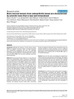
Figure 1
Representative core sampling map as applied to the tibial plateau of a study participantRepresentative core sampling map as applied to the tibial plateau of a study participant. (a) Bone marrow lesions (BML) identified in the medial tibial
plateau (arrow). (b) Regions from the BML area, from another area within the medial tibiofemoral compartment not affected by BMLs, and from the
lateral tibiofemoral compartment as well as from matched locations from the lateral compartment were defined. (c) Multiple cores were machined
from each region.
Arthritis Research & Therapy Vol 11 No 1 Hunter et al.
Page 4 of 9
(page number not for citation purposes)
For each core the region containing only trabecular bone was
identified manually, and the following structural parameters
were quantified: bone volume fraction, trabecular number,
trabecular spacing, trabecular number, structural model index
(SMI), connectivity density, and degree of anisotropy. The tis-
sue mineral density was also calculated for the trabecular
region.
We note that for the analyses of subchondral trabecular bone
we required the superior–inferior length of the region contain-
ing only trabecular bone to be at least 5 mm. This ensured that
the region analyzed would contain a sufficient number of
trabeculae for adequate sampling of the trabecular structure.
This length criterion resulted in exclusion of 12 cores (one to
four cores per donor). Finally, the thickness of the subchondral
plate was calculated as the average of measurements made
from the microcomputed tomography image data at four
equally spaced locations across the superior surface of the
plate.
Statistical analysis
Cores were classified according to location: lesion area
(lesion); contralateral (medial or lateral, depending on which
compartment contained the lesion) compartment, location
matched to lesion area (matched); medial compartment, out-
side the lesion area or matched area (medial); and lateral com-
partment, outside the matched area or lesion area (lateral).
Repeated-measures analyses of variance with Tukey post hoc
tests were used to determine differences in bone structure
and tissue mineral density among the four locations. When
multiple cores were available from a given location for a given
donor, each core was treated as an individual measurement for
the statistical analyses; measurements were not averaged
prior to the analyses of variance.
Results
We collected specimens from six postmenopausal female OA
patients following MRI scan and total knee joint replacement.
Histomorphometry measures were obtained on bone core
samples from medial (or lateral) tibia affected by BMLs and
bone medial (or lateral) regions unaffected by BMLs as well as
from control regions from the lateral (or medial) tibial plateau.
In order to compare histomorphometric parameters and delin-
eate features specific to BML while controlling for medial-spe-
cific and lateral-specific bone features, we categorized bone
samples as described above (Figure 1): lesion (bone sample
core obtained from medial or lateral tibia affected by BMLs),
matched, medial and lateral. The bone volume fraction, trabec-
ular thickness, trabecular spacing, tissue mineral density and
other architectural features of BML-affected bone tissue cores
were therefore compared with control regions contralateral to
the BML and with unaffected regions immediately adjacent to
the BML.
Cores from the lesion area displayed increased bone volume
fraction but reduced tissue mineral density (P < 0.04; Figure
2). With respect to the subchondral trabecular structure, the
samples from the lesion area exhibited increased trabecular
thickness as compared with samples from the matched area
and lateral location (P = 0.02; Figure 3a). The subchondral
bone in the lesion area was also markedly more plate-like than
the bone in the other three locations, as evidenced by the
lower value of the SMI (P = 0.009) (Figure 3b). Other differ-
ences in structure that were noted were increased trabecular
spacing (P = 0.02) and a trend (P = 0.07) towards decreased
trabecular number in the cores from the medial location as
compared with matched location (Figure 3c,d). No differences
among locations were found in connectivity density, degree of
anisotropy, or tissue mineral density of the subchondral
trabecular bone (P > 0.10). In addition, no differences among
locations were found in the subchondral plate thickness (P =
0.10) (Table 1).
Discussion
Research into the etiology and progression of knee OA has
focused on the destruction of articular cartilage. However, it is
clear that knee OA is an organ-level failure of the joint and
involves pathological changes in subchondral bone as well as
in articular cartilage [13]. We have found that BMLs, which are
strongly associated with OA symptoms and disease progres-
sion, have specific changes in bone mineralization and trabec-
ular structure. The BML area, when compared with bone
samples within the same knee but outside the lesion area,
appeared to be sclerotic, based on the increased bone volume
fraction and increased trabecular thickness. The trabecular
architecture within the lesions was also more plate-like; how-
ever, the tissue mineral density was reduced relative to medial
tibial bone outside the BML.
Our findings are consistent with those of prior work suggest-
ing that hypomineralization of trabecular bone in OA occurs
subjacent to the thickened cortical plate [14-16]. This reduced
mineralization is possibly linked to abnormal bone cell behavior
in OA joints, reported as imbalances in bone resorption, bone
formation or both [17]. Recent studies have confirmed that
increased bone resorption plays an integral role in the disease
process, with increased levels of bone resorption markers,
including type I collagen [18] and deoxypryidinoline [19],
reported in patients with radiographic evidence of knee OA.
Urinary excretion of pyridinium cross-links is significantly
increased in patients with large joint OA and hand OA, sug-
gesting an increased rate of bone turnover [20]. Data from the
population based Chingford study demonstrated that urinary
collagen cross-link excretion (urine C-telopeptide and N-tel-
opeptide) levels were significantly elevated in knee OA sub-
jects [21]. Elevated levels of urinary N-telopeptide indicate
elevated human bone resorption [22], and our own data sug-
gest their levels are increased in persons with BMLs [23]. It is
important to note that these findings are not consistent with
Available online />Page 5 of 9
(page number not for citation purposes)
previous research such as that showing the bone turnover
markers were decreased in patients with knee OA compared
with control individuals (-36%, -38%, and -52%, P < 0.0001
for serum osteocalcin, serum and urinary C-terminal telopep-
tide of type 1 collagen, respectively) [24].
Moreover, our data are in agreement with previous findings
from early OA tibial bone specimens. This previous research
indicates that the trabeculae in the medial compartment of OA
joints are significantly thicker and more plate-like than normal
trabeculae [11,25,26], but that the affected trabecular bone is
less stiff than normal bone at both the apparent level [25] and
the tissue level [27].
Our data also extend previous findings, however, in that they
uniquely identify OA BMLs as foci of bone architecture pathol-
ogy. While prior studies have compared the subchondral
trabecular structure between osteoarthritic knees and normal
knees [11,25,27], the present study provides a comparison of
the architecture in BML-affected area with that in other areas
within the same tibial plateau. Results of the latter comparison
indicate that the affected region is one of pronounced abnor-
malities in structure and mineralization. These structural abnor-
malities are evident in the quantitative analysis of trabecular
architecture (Figure 3) as well as through qualitative examina-
tion of the three-dimensional images of the specimens (Figure
2c). In particular, SMI values in the lesion areas ranged from -
2.20 to 0.89 (mean = -1.14), while those in the other three
areas ranged from -0.38 to 2.78 (mean = 1.24). A SMI of 3
indicates an ideal cylindrical rod structure, an SMI of 0 indi-
cates an ideal plate structure, and values less than zero indi-
cate a structure in which the plates are curved and begin to
close off the pores from one another [28]. The results of the
present study therefore indicate that the BML architecture is
an extreme representation of the changes that occur through-
out the affected compartment in OA.
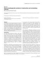
Figure 2
Bone volume fraction and average tissue mineral density for four locations from the entire coreBone volume fraction and average tissue mineral density for four locations from the entire core. (a) Bone volume fraction (BV/TV) and (b) average
tissue mineral density (TMD) for the entire core for each of the four locations. HA, hydroxyapatite. Each bar represents the mean, and error bars rep-
resent one standard deviation. *Significant differences between groups (P < 0.05). Cores from the lesion area exhibited the highest volume fraction
but lowest mineral density. (c) Longitudinal cut-away views of cores from each of the four locations. Each row contains cores from one donor.
Arthritis Research & Therapy Vol 11 No 1 Hunter et al.
Page 6 of 9
(page number not for citation purposes)
The changes in trabecular structure within the BML-affected
area are in opposition to the typical age-related changes in
trabecular structure in the proximal tibia that involve trabecular
thinning and progression from a plate-like structure to a rod-
like structure [29-31]. These OA-related changes, however,
do not necessarily imply any mitigation of age-related degrada-
tion in mechanical properties.
Previous studies have found that while bone volume fraction
and bone mineral density can increase substantially in the early
stages of OA [27,32,33], these changes are associated with
either no change or a slight decrease in apparent Young's
modulus and compressive strength [25,27]. Studies on the
microstructural and mechanical properties of tibial cancellous
bone by Ding and colleagues have revealed that the SMI and
the bone volume fraction can be primary determinants of can-
cellous bone mechanical properties [34]. Importantly, plate-
like cancellous bone is associated with increased relative
strength relative to rod-like bone; however, the converse is
true in osteoarthritic bone [11]. In addition, studies on cancel-
lous bone from the femoral head of OA and osteoporosis
patients revealed that the stiffness of osteoarthritic bone
increased more slowly with apparent density and that its mate-
rial density was significantly reduced (associated with 12%
reduction in mineral mass fraction). Intriguingly, the authors
reported there was also greater site-to-site variation of both
apparent and material density in the osteoarthritic bone, sug-
gesting an altered sensitivity to applied load [35].
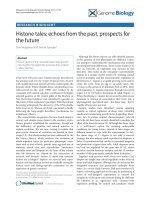
Figure 3
Quantitative measures of the trabecular structure for each of the four locationsQuantitative measures of the trabecular structure for each of the four locations. (a) Trabecular thickness (Tb.Th*). (b) Structure model index (SMI).
(c) Trabecular spacing (Tb.Sp*). (d) Trabecular number (Tb.N*). Cores from the lesion area exhibited the highest Tb.Th* but lowest SMI. Differences
in trabecular structure were also noted between the matched and medial locations. Each bar represents the mean, and error bars represent one
standard deviation. *Significant differences between groups (P < 0.05).
#
A trend (0.05 P < 0.10).
Table 1
Subchondral plate thickness by location
Location n Mean (standard deviation) (mm)
Lateral 11 0.49 (0.14)
Lesion 5 0.77 (0.24)
Matched 5 0.56 (0.10)
Medial 9 0.66 (0.34)
Available online />Page 7 of 9
(page number not for citation purposes)
These collective findings have led to the hypothesis that the
trabecular tissue is mechanically compromised in OA, proba-
bly as a result of poor trabecular organization [11,27]. The
reduced tissue mineral density and mineral:collagen ratio in
OA tissue [15,36] is consistent with this hypothesis. Although
the cellular basis for the reduced mineralization is not clear at
present, prior studies have noted in OA that increases in oste-
oid volume occur as a result of trabecular thickening that is
usually not accompanied by increased bone mineralization
[16,37].
The histological analyses for this study are ongoing. Prelimi-
nary data from parallel histopathological studies on OA BML
cores indicate a mixed pathology within the BML including
granulation, edema, diffuse necrosis, fibrinoid deposition and
hyperplasia of blood vessel walls (see Figure 4). Some of
these features have previously been reported [5,9]; however,
taken as a whole, our preliminary data point towards pathology
consistent with a localized infarction reaction. Although these
data need further validation and cross-reference versus addi-
tional tissue sets, our early findings may point to towards a
localized oxygen deficit in the BML – which may contribute to
the focal bone remodeling reactions observed in OA BMLs.
There are a number of important limitations of the present
study that warrant mention. This is a small sample of six post-
menopausal women and thus the findings cannot be general-
ized to men and those with OA in other parts of the joint.
Further, given the small sample size, this work should be
extended and replicated in other samples. An additional rate-
limiting step in this approach is that the depth of the cut in the
tibial plateau from TKR provides small specimens that did not
always permit quantification of the subchondral trabecular
structure. Despite these limitations, however, our data are
striking in that statistically significant differences in several his-
tomorphometric parameters were identified.
Conclusion
We have localized specific changes in bone mineralization,
remodeling and defects within BML features that are adjacent
to the subchondral plate. The mineral density of these BMLs is
reduced, and they appear to be sclerotic compared with unaf-
fected regions from the same individual based on the
increased bone volume fraction, increased trabecular thick-
ness, and decreased SMI. Further work is required to deter-
mine how these changes in composition and structure affect
the mechanical properties of the BML subchondral bone, and
thus whether these changes render the bone susceptible to
attrition. In addition, future studies are required to evaluate
Figure 4
Histopathological analyses of bone marrow lesion cores indicating a mixed pathologyHistopathological analyses of bone marrow lesion cores indicating a mixed pathology. (a) Diffuse granulation reaction in the marrow compartment.
All blood vessels show signs of secondary remodeling with thickened walls. Some vessels show evidence of focal fibrinoid adhesion to the endothe-
lium. (b) High-power view of focal granulation reaction. (c) Regional granulation reaction continuous with a focal fibrinoid reaction with thrombus
inclusions. There is evidence of a low-grade inflammation peripheral to the fibrinoid edge. The marked vessel remodeling and the presence of fibri-
noid inclusions in the granulation zone are consistent with a focal infarction. (d) Vascular leak with multiple thrombus inclusions. There is fibrinoid
occupation and casting of the marrow stroma.
Arthritis Research & Therapy Vol 11 No 1 Hunter et al.
Page 8 of 9
(page number not for citation purposes)
whether these observations are caused by an increase in syn-
thesis or a decrease in resorption of the bone, and how these
relate to histopathological features.
Competing interests
The authors declare that they have no competing interests.
Authors' contributions
DJH conceived of the study, participated in acquisition of the
data, data analysis and interpretation, and manuscript prepa-
ration, and approved the final manuscript. LG, TAE, and RAM
participated in data analysis and interpretation, manuscript
preparation and approved the final manuscript. GB, ADD,
ZDM, PN, EFM participated in acquisition of the data, data
analysis and interpretation, and manuscript preparation, and
approved the final manuscript. MF and SJ participated in
acquisition of the data and manuscript preparation.
Acknowledgements
The authors would like to thank the participants and staff of this study
without whose help this would not have been possible. In particular, they
would like to acknowledge the support of Paula McCree and Sasha
Goldberg. Supported by U01 AR50900-02 NIH/NIAMS Biomarkers in
Osteoarthritis MRI Studies and AstraZeneca.
References
1. Felson DT, Chaisson CE, Hill CL, Totterman SM, Gale ME, Skinner
KM, Kazis L, Gale DR: The association of bone marrow lesions
with pain in knee osteoarthritis [see comments]. Ann Intern
Med 2001, 134:541-549.
2. Felson DT, McLaughlin S, Goggins J, LaValley MP, Gale ME, Tot-
terman S, Li W, Hill CL, Gale DR: Bone marrow edema and its
relation to progression of knee osteoarthritis. Ann Intern Med
2003, 139:330-336.
3. Lo GH, Hunter DJ, Zhang Y, McLennan CE, LaValley MP, Kiel DP,
McLean RR, Genant HK, Guermazi A, Felson DT: Bone marrow
lesions in the knee are associated with increased local bone
density. Arthritis Rheum 2005, 52:2814-2821.
4. Hunter D, Zhang Y, Niu J, Goggins J, Amin S, LaValley M, Guermazi
A, Genant HK, Gale D, Felson DT: Increase in bone marrow
lesions is associated with cartilage loss: a longitudinal MRI
study in knee osteoarthritis. Arthritis Rheum 2006,
54:1529-1535.
5. Zanetti M, Bruder E, Romero J, Hodler J: Bone marrow edema
pattern in osteoarthritic knees: correlation between MR imag-
ing and histologic findings. Radiology 2000, 215:835-840.
6. Link TM, Steinbach LS, Ghosh S, Ries M, Lu Y, Lane N, Majumdar
S: Osteoarthritis: MR imaging findings in different stages of
disease and correlation with clinical findings. Radiology 2003,
226:373-381.
7. Kornaat PR, Bloem JL, Ceulemans RY, Riyazi N, Rosendaal FR,
Nelissen RG, Carter WO, Hellio Le Graverand MP, Kloppenburg
M: Osteoarthritis of the knee: association between clinical fea-
tures and MR imaging findings. Radiology 2006, 239:811-817.
8. Neuhold A, Hofmann S, Engel A, Leder K, Kramer J, Haller J, Plenk
H: Bone marrow edema of the hip: MR findings after core
decompression. J Comput Assist Tomogr 1992, 16:951-955.
9. Plenk H Jr, Hofmann S, Eschberger J, Gstettner M, Kramer J, Sch-
neider W, Engel A: Histomorphology and bone morphometry of
the bone marrow edema syndrome of the hip. Clin Orthop
Relat Res 1997, 334:73-84.
10. Reinus WR, Fischer KC, Ritter JH: Painful transient tibial edema
[see comment].
Radiology 1994, 192:195-199.
11. Ding M, Odgaard A, Hvid I: Changes in the three-dimensional
microstructure of human tibial cancellous bone in early oste-
oarthritis. J Bone Joint Surg Br 2003, 85:906-912.
12. Ridler T, Calvard S: Picture thresholding using an iterative
selection method. IEEE Trans Syst Man Cybern 1978,
8:630-632.
13. Burr DB: The importance of subchondral bone in the progres-
sion of osteoarthritis. J Rheumatol Suppl 2004, 70:77-80.
Review
14. Karvonen RL, Miller PR, Nelson DA, Granda JL, Fernandez-Madrid
F: Periarticular osteoporosis in osteoarthritis of the knee. J
Rheumatol 1998, 25:2187-2194.
15. Li B, Aspden RM: Material properties of bone from the femoral
neck and calcar femorale of patients with osteoporosis or
osteoarthritis. Osteoporos Int 1997, 7:450-456.
16. Grynpas MD, Alpert B, Katz I, Lieberman I, Pritzker KP: Subchon-
dral bone in osteoarthritis. Calcif Tissue Int 1991, 49:20-26.
17. Hunter DJ, Spector TD: The role of bone metabolism in osteoar-
thritis. [Review; 45 refs]. Curr Rheumatol Rep 2003, 5:15-19.
18. Bettica P, Cline G, Hart D, Meyer J, Spector T: Evidence for
increased bone resorption in patients with progressive knee
OA: longitudinal results from the Chingford study. Arthritis
Rheum 2002, 46(12):3178-3184.
19. Hunter DJ, Hart D, Snieder H, Bettica P, Swaminathan R, Spector
TD: Evidence of altered bone turnover, vitamin D and calcium
regulation with knee osteoarthritis in female twins. Rheuma-
tology 2003, 42:1311-1316.
20. Stewart A, Black A, Robins SP, Reid DM: Bone density and bone
turnover in patients with osteoarthritis and osteoporosis. J
Rheumatol 1999, 26:622-626.
21. Bettica P, Cline G, Hart DJ, Meyer J, Spector TD: Evidence for
increased bone resorption in patients with progressive knee
osteoarthritis: longitudinal results from the Chingford study.
Arthritis Rheum 2002, 46:3178-3184.
22. Rosen HN, Dresner-Pollak R, Moses AC, Rosenblatt M, Zeind AJ,
Clemens JD, Greenspan SL: Specificity of urinary excretion of
cross-linked N-telopeptides of type I collagen as a marker of
bone turnover. Calcif Tissue Int 1994, 54:26-29.
23. Hunter DJ, Lavalley M, Li J, Bauer DC, Nevitt M, DeGroot J, Poole
R, Eyre D, Guermazi A, Gale D, Totterman S, Felson DT: Biochem-
ical markers of bone turnover and and their association with
bone marrow lesions. Arthritis Res Ther 2008, 10:R102.
24. Garnero P, Piperno M, Gineyts E, Christgau S, Delmas PD, Vignon
E: Cross sectional evaluation of biochemical markers of bone,
cartilage, and synovial tissue metabolism in patients with knee
osteoarthritis: relations with disease activity and joint damage
[see comments]. Ann Rheum Dis 2001, 60:619-626.
25. Brown SJ, Pollintine P, Powell DE, Davie MW, Sharp CA:
Regional differences in mechanical and material properties of
femoral head cancellous bone in health and osteoarthritis.
Calcif Tissue Int 2002, 71:227-234.
26. Chappard C, Peyrin F, Bonnassie A, Lemineur G, Brunet-Imbault
B, Lespessailles E, Benhamou CL: Subchondral bone micro-
architectural alterations in osteoarthritis: a synchrotron micro-
computed tomography study. Osteoarthr Cartil 2006,
14:215-223.
27. Day JS, Ding M, Linden JC van der, Hvid I, Sumner DR, Weinans
H: A decreased subchondral trabecular bone tissue elastic
modulus is associated with pre-arthritic cartilage damage. J
Orthop Res 2001, 19:914-918.
28. Hildebrand T, Ruegsegger P: Quantification of bone microarchi-
tecture with the structure model index. Comput Methods Bio-
mech Biomed Engin 1997, 1:15-23.
29. Hildebrand T, Laib A, Muller R, Dequeker J, Ruegsegger P: Direct
three-dimensional morphometric analysis of human cancel-
lous bone: microstructural data from spine, femur, iliac crest,
and calcaneus. J Bone Miner Res 1999, 14:1167-1174.
30. Ding M, Hvid I: Quantification of age-related changes in the
structure model type and trabecular thickness of human tibial
cancellous bone. Bone
2000, 26:291-295.
31. Muraoka T, Hagino H, Okano T, Enokida M, Teshima R: Role of
subchondral bone in osteoarthritis development: a compara-
tive study of two strains of guinea pigs with and without spon-
taneously occurring osteoarthritis. Arthritis Rheum 2007,
56:3366-3374.
32. Beuf O, Ghosh S, Newitt DC, Link TM, Steinbach L, Ries M, Lane
N, Majumdar S: Magnetic resonance imaging of normal and
osteoarthritic trabecular bone structure in the human knee.
Arthritis Rheum 2002, 46:385-393.
Available online />Page 9 of 9
(page number not for citation purposes)
33. Edinger DT, Hayashi K, Hongyu Y, Markel MD, Manley PA: Histo-
morphometric analysis of the proximal portion of the femur in
dogs with osteoarthritis. Am J Vet Res 2000, 61:1267-1272.
34. Ding M, Odgaard A, Danielsen CC, Hvid I: Mutual associations
among microstructural, physical and mechanical properties of
human cancellous bone. J Bone Joint Surg Br 2002,
84:900-907.
35. Li B, Aspden RM: Composition and mechanical properties of
cancellous bone from the femoral head of patients with oste-
oporosis or osteoarthritis. J Bone Miner Res 1997,
12:641-651.
36. Ding M, Danielsen CC, Hvid I: Age-related three-dimensional
microarchitectural adaptations of subchondral bone tissues in
guinea pig primary osteoarthrosis. Calcif Tissue Int 2006,
78:113-122.
37. Zysset PK, Sonny M, Hayes WC: Morphology–mechanical prop-
erty relations in trabecular bone of the osteoarthritic proximal
tibia. J Arthroplasty 1994, 9:203-216.