Báo cáo y học: "Susceptibility of rheumatoid arthritis synovial fibroblasts to FasLand TRAIL-induced apoptosis is cell cycle-dependent" ppsx
Bạn đang xem bản rút gọn của tài liệu. Xem và tải ngay bản đầy đủ của tài liệu tại đây (1.28 MB, 10 trang )
Open Access
Available online />Page 1 of 10
(page number not for citation purposes)
Vol 11 No 1
Research article
Susceptibility of rheumatoid arthritis synovial fibroblasts to FasL-
and TRAIL-induced apoptosis is cell cycle-dependent
Noreen Pundt
1
, Marvin A Peters
1
, Christina Wunrau
1
, Simon Strietholt
1
, Carsten Fehrmann
2
,
Katja Neugebauer
1
, Christine Seyfert
3
, Frans van Valen
1
, Thomas Pap
1
and Ingmar Meinecke
1,4
1
Institute of Experimental Musculoskeletal Medicine, University Hospital Muenster, Domagkstr. 3, Muenster, 48149, Germany
2
Institute of Medical Microbiology, University Hospital Muenster, Domagkstr. 10, Muenster, 48149, Germany
3
Department of Orthopaedic Surgery, Zeisigwaldkliniken Bethanien Chemnitz, Zeisigwaldstr. 101, Chemnitz, 09130, Germany
4
Department of Orthopaedic Surgery, Park-Krankenhaus Leipzig-Suedost GmBH, Struempellstr. 41, Leipzig, 04289, Germany
Corresponding author: Thomas Pap,
Received: 13 May 2008 Revisions requested: 25 Jun 2008 Revisions received: 24 Nov 2008 Accepted: 5 Feb 2009 Published: 5 Feb 2009
Arthritis Research & Therapy 2009, 11:R16 (doi:10.1186/ar2607)
This article is online at: />© 2009 Pundt et al.; licensee BioMed Central Ltd.
This is an open access article distributed under the terms of the Creative Commons Attribution License ( />),
which permits unrestricted use, distribution, and reproduction in any medium, provided the original work is properly cited.
Abstract
Introduction The rheumatoid arthritis (RA) synovium is
characterised by the presence of an aggressive population of
activated synovial fibroblasts (RASFs) that are prominently
involved in the destruction of articular cartilage and bone.
Accumulating evidence suggests that RASFs are relatively
resistant to Fas-ligand (FasL)-induced apoptosis, but the data
concerning tumour necrosis factor-related apoptosis-inducing
ligand (TRAIL) have been conflicting. Here, we hypothesise that
the susceptibility of RASFs to receptor-mediated apoptosis
depends on the proliferation status of these cells and therefore
analysed the cell cycle dependency of FasL- and TRAIL-induced
programmed cell death of RASFs in vitro.
Methods Synovial fibroblasts were isolated from patients with
RA by enzymatic digestion and cultured under standard
conditions. Cell cycle analysis was performed using flow
cytometry and staining with propidium iodide. RASFs were
synchronised or arrested in various phases of the cell cycle with
0.5 mM hydroxyurea or 2.5 g/ml nocodazol and with foetal calf
serum-free insulin-transferrin-sodium selenite supplemented
medium. Apoptosis was induced by stimulation with 100 ng/ml
FasL or 100 ng/ml TRAIL over 18 hours. The apoptotic
response was measured using the Apo-ONE
®
Homogenous
Caspase-3/7 Assay (Promega GmbH, Mannheim, Germany)
and the Cell Death Detection (ELISA
Plus
) (enzyme-linked
immunosorbent assay) (Roche Diagnostics GmbH, Mannheim,
Germany). Staurosporin-treated cells (1 g/ml) served as a
positive control. Expression of Fas and TRAIL receptors
(TRAILR1-4) was determined by fluorescence-activated cell
sorting analysis.
Results Freshly isolated RASFs showed only low proliferation in
vitro, and the rate decreased further over time, particularly when
RASFs became confluent. RASFs expressed Fas, TRAIL
receptor-1, and TRAIL receptor-2, and the expression levels
were independent of the cell cycle. However, the proliferation
rate significantly influenced the susceptibility to FasL- and
TRAIL-induced apoptosis. Specifically, proliferating RASFs
were less sensitive to FasL- and TRAIL-induced apoptosis than
RASFs with a decreased proliferation rate. Furthermore, RASFs
that were synchronised in S phase or G
2
/M phase were less
sensitive to TRAIL-induced apoptosis than synchronised RASFs
in G
0
/G
1
phase.
Conclusions Our data indicate that the susceptibility of RASFs
to FasL- and TRAIL-induced apoptosis depends on the cell
cycle. These results may explain some conflicting data on the
ability of RASFs to undergo FasL- and TRAIL-mediated cell
death and suggest that strategies to sensitise RASFs to
apoptosis may include the targeting of cell cycle-regulating
genes.
2 n DNA: diploid chromosomes; 4 n DNA: tetraploid chromosomes; DMEM: Dulbecco's modified Eagle's medium; EDTA: ethylenediaminetetraacetic
acid; ELISA: enzyme-linked immunosorbent assay; FACS: fluorescence-activated cell sorting; FasL: Fas ligand; FCS: foetal calf serum; HU: hydrox-
yurea; ITS: insulin-transferrin-sodium selenite; NF-B: nuclear factor-kappa-B; OD: optical density; PBS: phosphate-buffered saline; RA: rheumatoid
arthritis; RASF: rheumatoid arthritis synovial fibroblast; RFU: relative fluorescence units; TNF: tumour necrosis factor; TRAIL: tumour necrosis factor-
related apoptosis-inducing ligand.
Arthritis Research & Therapy Vol 11 No 1 Pundt et al.
Page 2 of 10
(page number not for citation purposes)
Introduction
Rheumatoid arthritis (RA), a chronic disease of incompletely
understood aetiology, is characterised primarily by the pro-
gressive destruction of articular structures. Its pathogenesis is
governed by the concerted action of several cell types that
create signs and symptoms characteristic for RA. Accumulat-
ing evidence indicates that, in addition to macrophages and T
cells, activated RA synovial fibroblasts (RASFs) play a major
role in both initiating and driving the disease [1-4]. Not only do
RASFs with an aggressive phenotype increase in number,
their activation also results in the production of proinflamma-
tory mediators and matrix-degrading enzymes and in altera-
tions of programmed cell death [3-5].
Programmed cell death, or apoptosis, is central for both devel-
opment and tissue homeostasis of metazoans. Therefore,
aberrations of this process may lead to a variety of human
pathologies, including cancer, autoimmune diseases, and neu-
rodegenerative disorders. Apoptosis can be induced by mem-
bers of the tumour necrosis factor (TNF) receptor family
through the recruitment of an intracellular membrane-associ-
ated complex of proteins (death-inducing signaling com-
plexes, or DISCs), which leads to a cytoplasmic release of
active caspase-8 and subsequent activation of the apoptotic
cascade [6,7]. Among these death receptors, Fas/CD95 and
its specific ligand FasL/CD95L were demonstrated to be of
importance, and it was shown that stimulation of RASFs with
FasL initiates proapoptotic signals [8,9]. However, several
studies with cultured RASFs showed that stimulation of
RASFs with Fas-activating ligands induced apoptosis in only a
small percentage of cells, and several mechanisms have been
identified that prevent RASFs from Fas-mediated cell death
[10-16]. Actually, several studies have shown that RASFs
undergo less FasL-induced apoptosis than osteoarthritis syn-
ovial fibroblasts and therefore RASFs has been termed rela-
tively resistant to FasL-induced apoptosis. As shown
previously, fibroblasts in RA synovium express both TNF-
receptors and Fas, and their ligands have been detected in co-
localised macrophages and T cells [17-19].
TNF-related apoptosis-inducing ligand (TRAIL), another mem-
ber of the TNF superfamily of apoptosis-inducing ligands, can
bind to five receptors. Among them, TRAIL-R3 (DcR1) and
TRAIL-R4 (DcR2) act as membrane-anchored decoy recep-
tors, whereas TRAIL-R1 (DR4) and TRAIL-R2 (DR5) contain a
cytoplasmic death domain and transmit proapoptotic signals
into cells [20]. In addition, osteoprotegerin, a soluble decoy
receptor of the ligand for the receptor activator of nuclear fac-
tor-kappa-B (NF-B) (RANKL), has been shown to bind TRAIL
[21,22]. Apoptosis can be induced upon binding of TRAIL to
DR4 and DR5 and subsequent activation of different cas-
pases. On the other hand, studies suggest that binding of
TRAIL to these receptors can also induce proliferation through
activation of the NF-B signalling pathway [23,24], and it
appears that the ability of TRAIL to trigger either apoptosis or
cell survival depends on the cell type [25].
The in vitro data concerning TRAIL-induced apoptosis in
RASFs have been conflicting. Morel and colleagues [25]
showed that exposure to TRAIL induced apoptosis in only
30% of RASFs within 24 hours whereas surviving cells prolif-
erated in a TRAIL dose-dependent manner. In contrast,
Ichikawa and colleagues [26] documented TRAIL (anti-DR5
antibody)-induced apoptosis of RA synovial cells with 80% of
the cells being killed. In both studies, RASFs showed consti-
tutive expression of TRAIL receptor-2 (DR5) as the main medi-
ator of TRAIL-induced stimulation. In addition, Morel and
colleagues [25] could show the expression of TRAIL-R1
(DR4). Here, we hypothesise that the susceptibility of RASFs
to receptor-mediated apoptosis depends on the proliferation
state of these cells. Therefore, we analysed the cell cycle
dependency of FasL- and TRAIL-induced programmed cell
death of RASFs in vitro.
Materials and methods
Patients and tissue samples
Samples of synovial membrane from patients with RA (accord-
ing to the 1987 revised American College of Rheumatology
criteria) were obtained at joint replacement surgery within an
ongoing national tissue bank project with the 'Assoziation für
Rheumatologische Orthopädie' (ARO) of the German Society
of Rheumatology (DGRh) and provided by the Department of
Orthopaedic Surgery of St. Joseph Hospital (Sendenhorst,
Germany), the Department of Orthopaedic Surgery of the Uni-
versity of Magdeburg School of Medicine (Magdeburg, Ger-
many), and the Department of Orthopaedic Surgery (KMG-
Kliniken Kyritz, Germany). Approval from the local ethics com-
mittee was obtained prior to starting the study. Fibroblasts
were isolated by digesting synovial tissue with 1.5 mg/ml Dis-
pase II (Roche Diagnostics GmbH, Mannheim, Germany) and
cultured in complete Dulbecco's modified Eagle's medium
(DMEM supplemented with 10% foetal calf serum [FCS], Inv-
itrogen Corporation, Carlsbad, CA, USA, and penicillin/strep-
tomycin, PAA, Pasching, Austria) as described previously
[27]. Fibroblasts were used in passages 4 to 8.
Fluorescence-activated cell sorting analysis
Flow cytometric analysis of cell cycle was performed as
described previously [28]. Briefly, cells were detached with 1
mM ethylenediaminetetraacetic acid (EDTA) and suspended
in fluorescence-activated cell sorting (FACS) buffer (phos-
phate-buffered saline [PBS] supplemented with 5% FCS and
0.1% NaN
3
). Cell cycle analysis was performed by incubation
of cells with propidium iodide (40 g/ml propidium iodide, 100
g/ml RNase in PBS) for up to 2 days and subsequent flow
cytometry (FACScalibur; BD Biosciences, San Jose, CA,
USA). To arrest RASFs in G
2
/M phase, cells were treated with
nocodazol (2.5 g/ml in DMEM for 18, 24, or 36 hours; Calbi-
ochem, Darmstadt, Germany). Furthermore, randomly growing
Available online />Page 3 of 10
(page number not for citation purposes)
cultures of RASFs were synchronised with 0.5 mM hydroxyu-
rea (HU) (Sigma-Aldrich, Steinheim, Germany) in DMEM and
incubated at 37°C for 6 hours. Cells were washed with PBS
and suspended in fresh complete DMEM. Synchronised
RASFs were incubated at 37°C and samples (0, 18, 24, 30,
42, and 48 hours) thereof were analysed for cell cycle by pro-
pidium iodide staining as described above. In addition, RASFs
were arrested in G
0
/G
1
phase by serum deprivation. To this
end, cultures of RASFs were incubated with DMEM supple-
mented with 1× insulin-transferrin-sodium selenite (ITS) sup-
plement (100×) (Sigma-Aldrich) [29,30] for up to 10 days (0,
3, 8, and 10 days) following incubation with complete medium
for 1 or 2 days (9/1, 9/2 days).
Analysis of Fas- and TRAIL-receptor expression
Surface expression of Fas and TRAIL receptors (TRAILR1-4)
on RASFs was determined by flow cytometry as described
[31]. Briefly, 1 × 10
5
cells were labelled with 0.5 g of mouse
anti-TRAILR1-4 (Alexis Biochemicals, Lörrach, Germany),
mouse anti-Fas antibodies, or mouse anti-IgG in FACS buffer
containing 5 mM EDTA for 40 minutes at 4°C. These cells
were incubated with biotin-conjugated goat anti-mouse, phy-
coerythrin-conjugated anti-goat, or fluorescein isothiocyanate-
conjugated anti-mouse antisera for 30 minutes at 4°C. Stained
cells were fixed and 1 × 10
4
viable cells were analysed by flow
cytometry using standard settings.
Induction and measurement of apoptosis
Apoptosis was induced at different density states or cell cycle
phases by incubation of cells with 100 ng/ml FasL (Bender
MedSystems, Vienna, Austria) or 100 ng/ml TRAIL (Pepro
Tech, Rocky Hill, NJ, USA) in 100 L of complete DMEM or
DMEM for 18 hours. The apoptotic response was measured
by Cell Death Detection (ELISA
Plus
) (enzyme-linked immuno-
sorbent assay) (Roche Diagnostics GmbH) and the Apo-
ONE
®
Homogeneous Caspase-3/7 Assay (Promega GmbH,
Mannheim, Germany) in accordance with the instructions of
the manufacturer. Staurosporin-treated cells (1 g/ml, 8
hours) served as a positive control.
Statistical analysis
Data shown are mean ± standard deviation. Statistical analysis
was performed using GraphPad Prism Software version 4.0
(GraphPad Software Inc., San Diego, CA, USA). Differences
between groups were examined for statistical significance
using the Mann-Whitney test, and a P value of less than 0.05
was considered statistically significant.
Results
Proliferation of rheumatoid arthritis synovial fibroblasts
First, we analysed DNA content by FACS analysis to deter-
mine the proliferation rate of RASFs. Early-cultured RASFs
exhibited a proliferation rate of 13.01%, according to cells
with a DNA content of greater than 2 n (Figure 1a, represent-
ative histogram, and Figure 1c, DNA content in S and G
2
/M
phases, n = 11). ~2 n DNA refers to the normal DNA content
in the interphase (G
0
/G
1
phase, diploid) of RASFs [32]. Con-
fluent RASFs (100% confluent, 10
4
cells) exhibited a prolifer-
ation rate of 6.53% (Figure 1b, representative histogram, and
Figure 1c, n = 5), significantly lower compared with early-cul-
tured RASFs (Figure 1c, P = 0.0028). Nocodazol, the micro-
tubule-destabilising agent that disrupts spindle assembly and
impedes re-entry into the cell cycle [32,33], was used to arrest
RASFs at G
2
/M phase (~4 n DNA). Cell cycle analysis of early-
cultured RASFs (10
4
cells) treated with nocodazol for 18
hours showed only a marginal increase of proliferating RASFs
to G
2
/M phase, from 7.95% to 11.41%, corresponding to ~4
n DNA content (Figure 2a, representative histogram, and Fig-
ure 2b, n = 5). Similar results were obtained after incubation
with nocodazol for 24 and 36 hours (data not shown). MHH-
ES-1 cells, an established Ewing sarcoma cell line [34], were
used as a positive control for arresting cells in G
2
/M phase
after incubation with nocodazol. G
2
/M-phase-arrested MHH-
ES-1 cells showed a 20% increase in the G
2
/M phase, from
46% to 66% (data not shown). HU, which inhibits reversible
DNA synthesis in mammalian cells without affecting RNA and
protein synthesis, was used to synchronise RASFs in G
0
/G
1
phase [35]. The effect of HU on the cell cycle of RASFs was
illustrated in Figure 2c (representative histogram) and Figure
2d (n = 3). Cell cycle analysis of RASFs treated with a single
exposure to 0.5 mM HU for 6 hours (time 0 hours) showed an
accumulation of RASFs in G
0
/G
1
phase (93.39%, corre-
sponding to ~2 n DNA, n = 3), indicating that the cell popula-
tion remained highly synchronised. Figure 2c and 2d also
illustrated the cell cycle of RASFs after various hours after
reversal of HU. Analysis of cell cycle 18, 24, 30, 42, and 48
hours after HU exposure showed a decrease of RASFs in G
0
/
G
1
phase until 66.24% (-27.15%, after 24 hours, n = 3) with
simultaneous increase of proliferating RASFs in S phase,
reaching a maximum at 24 hours (+11.55%, n = 3), and G
2
/M
phase, reaching a maximum at 30 hours (+25.53%, ~4 n
DNA, n = 3). Forty-two hours after HU exposure, cell cycle
analysis confirmed an increase of RASFs in G
0
/G
1
phase back
to 87.18%, and after 48 hours to 89.83%, indicating that cell
division commenced between 30 and 48 hours. No higher
degree of synchronisation was induced by a subsequent sec-
ond exposure to HU (data not shown). In addition, RASFs
were arrested in G
0
/G
1
phase through serum deprivation
using ITS supplement. As illustrated in Figure 2e (representa-
tive histogram) and Figure 2f (n = 3), early-cultured RASFs
became arrested at G
0
/G
1
phase after 8 to 10 days of incuba-
tion with ITS medium. The initial rate of proliferating RASFs
decreased from 11.14% to 8.56%, or 7.96% (corresponding
to <2 n DNA, from 0 d to 8 d, and 10 d, n = 3). Subsequent
incubation for another one or two days with complete DMEM
resulted in an increase of proliferating RASFs to 25.95% (<2
n DNA, 9 days of ITS medium/1 day of complete medium, 9/1
d) or 22.34% (9/2 d). Maximum of RASFs in S phase was
reached at day 9/1 (+12.02%, n = 3) and in G
2
/M phase at
Arthritis Research & Therapy Vol 11 No 1 Pundt et al.
Page 4 of 10
(page number not for citation purposes)
day 9/2 (+11.3%, n = 3). These results suggest that only a
small population of early-cultured RASFs proliferate.
Susceptibility of rheumatoid arthritis synovial
fibroblasts to FasL- and TRAIL-induced apoptosis
Next, we analysed the cell cycle dependency of FasL- and
TRAIL-induced programmed cell death of RASFs in vitro. We
found that higher-proliferating RASFs (50% of confluency)
from different patients were less sensitive to TRAIL-induced
apoptosis than lower-proliferating RASFs (80% of confluency)
and even significantly less sensitive when confluent RASFs
(100% confluent) were used as measured by Cell Death
Detection (ELISA
Plus
). As Figure 3a illustrates, the photometric
enzyme immunoassay for the detection of cytoplasmic his-
tone-associated DNA fragments showed a reduction from
3.35 relative fluorescence units (RFU) (confluent RASFs) to
1.55 RFU (-53%, lower-proliferating RASFs) or to 1.0 RFU (-
70.15%, higher-proliferating RASFs, data are presented as
optical density (OD)/OD untreated RASFs, n = 7). Similar
observations were made when RASFs in different density
states were treated with FasL. Measurement of the activities of
caspase-3 and caspase-7, key effectors of apoptosis in mam-
malian cells, revealed that higher-proliferating RASFs (50% of
confluency) were less sensitive to FasL-induced apoptosis
than lower-proliferating RASFs (80% of confluency) and con-
fluent RASFs (Figure 3b). A reduction from 6.79 × 10
4
RFU
(confluent RASFs) to 5.26 × 10
4
RFU (-22.5%, lower-prolifer-
ating RASFs) and to 2.8 × 10
4
RFU (-59%, higher-proliferating
RASFs, n = 3) was observed. Furthermore, highly synchro-
nised RASFs in S phase (HU, time 24 hours, Figure 2c,d) and
in G
2
/M phase (time 30 hours) were less sensitive to TRAIL-
induced apoptosis than synchronised RASFs in G
0
/G
1
phase
(time 0 hours, Figure 3c). A reduction from 4.84 × 10
4
RFU
(HU/0 hours, n = 5) to 1.83 × 10
4
RFU (-62.2%, HU/24 hours,
n = 5) or to 1.93 × 10
4
RFU (-60.13%, HU/30 hours, n = 5)
was observed by measurement of the activities of caspase-3
and caspase-7. Similar results were obtained after measure-
ment of FasL-induced apoptosis. Compared with RASFs syn-
chronised in G
0
/G
1
phase (7.06 × 10
4
RFU, n = 3, Figure 3d),
RASFs synchronised in S phase showed a reduced apoptotic
response of 1.2 × 10
4
RFU (-83.01%, n = 3) and RASFs syn-
chronised in G
2
/M phase showed a reduced apoptotic
response of 1.45 × 10
4
RFU (-79.5%, n = 3). Moreover,
RASFs arrested in G
0
/G
1
phase through serum deprivation
using ITS medium (8 d) were more sensitive to TRAIL- and
FasL-induced apoptosis than proliferating RASFs in S phase
(9/1 d) or in G
2
/M phase (9/2 d, Figure 3e,f). TRAIL-induced
caspase-3/7 activities decreased from 8.62 × 10
4
RFU in
RASFs arrested in G
0
/G
1
phase to 1.15 × 10
4
RFU (-86.6%,
n = 3) in RASFs arrested in S phase and to 1.54 × 10
4
RFU (-
82.1%, n = 3) in RASFs arrested in G
2
/M phase. Again, com-
parable results were obtained by measurement of FasL-
induced programmed cell death. Figure 3f illustrates a reduc-
tion from 1.14 × 10
6
RFU (G
0
/G
1
phase, 8 d) to 0.61 × 10
5
RFU (-94.64%, S phase, 9/1 d) and to 5.52 × 10
5
RFU (-
51.84%, G
2
/M phase, 9/2 d). Unless otherwise noted, all data
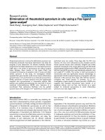
Figure 1
Proliferation capacity of rheumatoid arthritis synovial fibroblasts (RASFs)Proliferation capacity of rheumatoid arthritis synovial fibroblasts
(RASFs). (a) Early-cultured RASFs exhibit only a very low proliferation
rate in vitro (>2 n DNA equates S phase and G
2
/M phase, representa-
tive histogram). ~2 n DNA (arrow at 200) refers to the normal DNA
content of interphase (G
0
/G
1
phase) RASFs [32]. 4 n DNA (arrow at
G
2
/M peak at 400) refers to twice the amount of DNA in G
2
/M com-
pared with G
0
/G
1
phase. (b) Decrease in proliferation rate in confluent
RASFs. (c) Quantitative analysis. Values are mean ± standard deviation
as a percentage of early-cultured and confluent RASFs obtained from
11 or 6 individual patients with rheumatoid arthritis. **P < 0.01.
Available online />Page 5 of 10
(page number not for citation purposes)
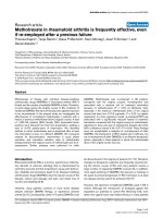
Figure 2
Effects of synchronisation and cell cycle arrest on proliferation of rheumatoid arthritis synovial fibroblasts (RASFs)Effects of synchronisation and cell cycle arrest on proliferation of rheumatoid arthritis synovial fibroblasts (RASFs). (a) The effect of nocodazol on the
cell cycle of early-cultured RASFs. Treatment of higher-proliferating RASFs with nocodazol (2.5 g/ml, 18 hours) resulted in a marginal increase of
RASFs arrested in G
2
/M phase (~4 n DNA at G
2
/M peak, black line, representative histogram). (b) Quantitative analysis. Values are mean ± stand-
ard deviation as a percentage of RASFs treated/untreated with nocodazol obtained from 3 individual patients with RA. (c) Effects of hydroxyurea
(HU) on cell cycle of RASFs were illustrated in a representative three-dimensional histogram with the y-axis (time in hours) pointing away from the
observer. RASFs treated with HU for 6 hours (time 0 hours) showed an accumulation of RASFs in G
0
/G
1
phase. Analysis of cell cycle 18, 24, and
30 hours after HU exposure showed a decrease of RASFs in G
0
/G
1
phase with a simultaneous increase of proliferating RASFs in S phase and G
2
/
M phase, indicating that the cell population remained highly synchronised. Cell cycle analysis after 42 and 48 hours confirmed an increase of RASFs
in G
0
/G
1
phase, indicating that cell division commenced between 30 and 48 hours. (d) Quantitative analysis as mean ± standard deviation. (e)
Early-cultured RASFs became arrested at G
0
/G
1
phase after 8 to 10 days of incubation with ITS medium. Subsequent incubation for another 1 or 2
days with complete Dulbecco's modified Eagle's medium (9/1, 9/2 days) resulted in an increase of proliferating RASFs. Bar graphs in frames on
right show quantitative analysis. Values are presented as mean ± standard deviation of percentages of RASFs obtained from at least three individual
patients with rheumatoid arthritis. Representative three-dimensional histogram. (f) Quantitative analysis.
Arthritis Research & Therapy Vol 11 No 1 Pundt et al.
Page 6 of 10
(page number not for citation purposes)
in Figure 3 are presented as OD/OD unstimulated RASFs.
Staurosporin-treated cells served as a positive control. We
hypothesise that the susceptibility of RASFs to receptor-medi-
ated apoptosis depends on the proliferation state of these
cells in vitro.
Expression of Fas and TRAIL receptors on rheumatoid
arthritis synovial fibroblasts
Finally, to investigate whether altered expression of death
receptors may provide an explanation for differences in the
susceptibility of RASFs to FasL- and TRAIL-induced apopto-
sis, the expression of Fas- and TRAIL-receptor changes during
cell cycle progression, synchronisation, or at cell cycle arrest
was examined. As shown by flow cytometry, TRAIL-R1 and
TRAIL-R2 were expressed constitutively on higher-proliferat-
ing RASFs in vitro, whereas TRAIL-R3 and TRAIL-R4 were not
detectable. The expression levels did not change in confluent
RASFs (Figure 4a, representative histogram, n = 3). In addi-
tion, expression of these receptors was unaltered when
RASFs were treated for 18 hours with 100 ng/ml TRAIL (data
not shown). Furthermore, cell surface expression of TRAIL
receptors on RASFs remained unchanged in RASFs synchro-
nised with HU (Figure 4b, representative histogram, n = 3) or
on RASFs arrested by using ITS medium (data not shown).
Fas (CD95) is a well-known apoptosis-signalling cell surface
receptor belonging to the TNF receptor family [36]. To investi-
gate the susceptibility of RASFs to FasL-mediated apoptosis,
cell surface expression of Fas on RASFs was determined by
flow cytometry in vitro. In agreement with data from Kobayashi
and colleagues [37], who showed surface expression of Fas
on RA synoviocytes, Fas was constitutively expressed on
higher-proliferating RASFs (data not shown). Cell surface
expression remained unchanged in confluent RASFs and
under all investigated conditions (data not shown).
Discussion
A decreased susceptibility to apoptosis and synovial prolifera-
tion has been described to contribute to RASF hyperplasia
[5,10,11,14,38,39]. In this context, the TRAIL receptor/TRAIL
system and the Fas/FasL system have raised much interest.
Increasing evidence suggests that RASFs are relatively resist-
ant to FasL-induced apoptosis in vitro [10,11,40]. Specifically,
several studies with cultured RASFs showed that synovio-
cytes from rheumatoid synovium tissue express functional Fas
[8,17,18] and that Fas activation induces apoptosis only in a
small population of cells, even though the Fas/FasL system
seems to be incapable of eliminating cells in proliferative RA
synovium [8,18,37,40,41]. The data concerning TRAIL appear
to be controversial [25,26,40]. Ichikawa and colleagues [26]
analysed the effect of TRAIL on RASFs and reported an
increased DR5 expression and an induction of DR5-mediated
apoptosis up to 80%, although varying levels of apoptosis
were induced by TRAIL using different RASF cultures. In
agreement with these findings, Miranda-Carus and colleagues
[38] analysed fibroblasts of 50 RA synovial fluid samples and
showed that these fibroblasts underwent apoptosis when
treated in vitro with an agonistic anti-DR5 antibody. In con-
trast, Morel and colleagues [25] proposed that TRAIL might
have two different effects on RASFs, namely an initial rapid
induction of apoptosis of up to 30% within the first 24 hours
followed by an increase in the proliferation [25]. In addition, it
is well documented that, depending on the cellular system,
TRAIL can promote both proliferation and apoptosis, as has
been established for other members of the TNF cytokine family
[42]. In the present study, we hypothesised that the suscepti-
bility of RASFs to receptor-mediated apoptosis depends on
the proliferation status of these cells and, therefore, analysed
the cell cycle dependency of FasL- and TRAIL-induced pro-
grammed cell death of RASFs in vitro.
Our results indicate that freshly prepared RASFs exhibit only a
very low proliferation rate in vitro. The proliferation rate
decreases further over time, particularly when RASFs become
confluent. Furthermore, we describe for the first time that up to
65% of RASFs exhibit a G
0
/G
1
-phase arrest in vitro. Moreo-
ver, our study shows that early-cultured RASFs are less sensi-
tive to TRAIL- and FasL-induced apoptosis than late-cultured
RASFs and far less sensitive than 100% confluent RASFs.
The difference in sensitivity to TRAIL- and FasL-mediated
apoptosis between early-cultured and confluent RASFs is not
due to differences in the surface expression of Fas and TRAIL
receptors. Rather, the susceptibility clearly depended on the
cell cycle of these cells as RASFs that were synchronised in S
phase or G
2
/M phase were less sensitive to TRAIL-induced
apoptosis than RASFs that were arrested in G
0
/G
1
phase.
These results suggest an inverse correlation between cell pro-
liferation and apoptosis. However, how the proliferation influ-
ences TRAIL- and FasL-mediated synovial cell death remains
unclear. Miyashita and colleagues [43] proposed that the ser-
ine/threonine protein kinase Akt, which affects several impor-
tant cellular functions (including cell growth, cell cycle entry,
migration, and cell survival), is an endogenous inhibitor of the
TRAIL-mediated synovial cell apoptotic pathway. Furthermore,
numerous data have shown that activation of Akt inhibits
TRAIL-mediated apoptosis in various cancer cells and Akt has
been shown to be overexpressed and activated in rheumatoid
synovial cells in situ [44-47]. Therefore, it might be speculated
that there is a correlation between cell proliferation and apop-
tosis, which may be regulated by the Akt pathway, but clearly
further studies are required to elaborate on these
observations.
Conclusion
In summary, we have shown that a relatively high number of
RASFs are arrested in G
0
/G
1
phase. Furthermore, our data
indicate that the sensitivity to TRAIL- or FasL-mediated apop-
tosis may be closely linked to synovial proliferation. These find-
ings will further enhance our understanding of the
pathophysiology of RA.
Available online />Page 7 of 10
(page number not for citation purposes)
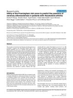
Figure 3
Susceptibility of proliferating rheumatoid arthritis synovial fibroblasts (RASFs) to Fas ligand (FasL)-induced and tumour necrosis factor-related apop-tosis-inducing ligand (TRAIL)-induced apoptosisSusceptibility of proliferating rheumatoid arthritis synovial fibroblasts (RASFs) to Fas ligand (FasL)-induced and tumour necrosis factor-related apop-
tosis-inducing ligand (TRAIL)-induced apoptosis. (a) As assessed by Cell Death Detection (ELISA
Plus
), higher-proliferating RASFs (50% of conflu-
ency) were less sensitive to TRAIL-induced apoptosis than lower-proliferating RASFs (80% of confluency) and significantly less sensitive than
confluent RASFs (100% confluent). (b) As revealed by the Apo-ONE
®
Homogeneous Caspase-3/7 Assay, higher-proliferating RASFs showed
lower activities of caspase-3 and caspase-7 after induction of apoptosis with FasL than less-proliferating RASFs and confluent RASFs. Highly syn-
chronised RASFs in S phase (HU/24 h) or G
2
/M phase (HU/30 h) were less sensitive to TRAIL-induced (c) and FasL-induced (d) apoptosis than
synchronised RASFs in G
0
/G
1
phase (HU/0 h), as measured by the Apo-ONE
®
Homogeneous Caspase-3/7 Assay. Moreover, RASFs arrested in
G
0
/G
1
phase through serum deprivation using insulin-transferrin-sodium selenite (ITS) medium (8 d) were more sensitive to TRAIL-induced (e) and
FasL-induced (f) apoptosis than proliferating RASFs in S phase (9/1 d) or G
2
/M phase (9/2 d). Staurosporin-induced apoptosis was measured as a
positive control. All values are mean ± standard deviation of fluorescence/fluorescence of unstimulated RASFs from at least three independent
patients with rheumatoid arthritis. *P < 0.05, **P < 0.01, ***P < 0.001.
Arthritis Research & Therapy Vol 11 No 1 Pundt et al.
Page 8 of 10
(page number not for citation purposes)
Figure 4
Surface expression of tumour necrosis factor-related apoptosis-inducing ligand (TRAIL) receptors on rheumatoid arthritis synovial fibroblasts (RASFs)Surface expression of tumour necrosis factor-related apoptosis-inducing ligand (TRAIL) receptors on rheumatoid arthritis synovial fibroblasts
(RASFs). (a) Staining of TRAIL receptors, as analysed by flow cytometry, showed constitutive surface expression of TRAIL-R1 and TRAIL-R2 on
RASFs in vitro. TRAIL-R3 and TRAIL-R4 were not detectable. The expression levels did not change in confluent RASFs. (b) Furthermore, cell sur-
face expression of TRAIL-R1 and TRAIL-R3 on RASFs remained unchanged in RASFs synchronised with hydroxyurea (HU). Representative histo-
grams of three separate experiments are shown.
Available online />Page 9 of 10
(page number not for citation purposes)
Competing interests
The authors declare that they have no competing interests.
Authors' contributions
NP helped to design research, to perform research, and to
analyse data and wrote the paper. MAP, CW, and TP helped
to design research, to perform research, and to analyse data.
IM helped to design research and to analyse data. SS, CF, KN,
and CS helped to perform research. FvV helped to perform
research and to analyse data. All authors read and approved
the final manuscript.
Acknowledgements
The authors thank Borna Truckenbrod and Vera Eckervogt for technical
assistance and Jennifer Gerding for observant reading of the manu-
script. This work was funded in part by the Deutsche Forschungsge-
meinschaft (DFG) (Pa689/2 and Pa689/3), the Assoziation für
Rheumatologische Orthopädie (ARO) of the German Society of Rheu-
matology (DGRh), and the Interdisciplinary Center for Clinical Research
(IZKF) of the University of Muenster.
References
1. Muller-Ladner U, Gay RE, Gay S: Cellular pathways of joint
destruction. Curr Opin Rheumatol 1997, 9:213-220.
2. Fassbender HG: Histomorphological basis of articular carti-
lage destruction in rheumatoid arthritis. Coll Relat Res 1983,
3:141-155.
3. Gay S, Gay RE, Koopman WJ: Molecular and cellular mecha-
nisms of joint destruction in rheumatoid arthritis: two cellular
mechanisms explain joint destruction? Ann Rheum Dis 1993,
52 Suppl 1:S39-S47.
4. Meinecke I, Rutkauskaite E, Gay S, Pap T: The role of synovial
fibroblasts in mediating joint destruction in rheumatoid
arthritis. Curr Pharm Des 2005, 11:563-568.
5. Pap T, Muller-Ladner U, Gay RE, Gay S: Fibroblast biology. Role
of synovial fibroblasts in the pathogenesis of rheumatoid
arthritis. Arthritis Res 2000, 2:361-367.
6. Perlman H, Pagliari LJ, Volin MV: Regulation of apoptosis and
cell cycle activity in rheumatoid arthritis. Curr Mol Med 2001,
1:597-608.
7. Riedl SJ, Shi Y: Molecular mechanisms of caspase regulation
during apoptosis. Nat Rev Mol Cell Biol 2004, 5:897-907.
8. Firestein GS, Yeo M, Zvaifler NJ: Apoptosis in rheumatoid arthri-
tis synovium. J Clin Invest 1995, 96:1631-1638.
9. Pap T: [Regulation of apoptosis in aggressive fibroblasts]. Z
Rheumatol 2007, 66:239-240. 242.
10. Hayashi S, Miura Y, Nishiyama T, Mitani M, Tateishi K, Sakai Y,
Hashiramoto A, Kurosaka M, Shiozawa S, Doita M: Decoy recep-
tor 3 expressed in rheumatoid synovial fibroblasts protects
the cells against Fas-induced apoptosis. Arthritis Rheum 2007,
56:1067-1075.
11. Meinecke I, Cinski A, Baier A, Peters MA, Dankbar B, Wille A,
Drynda A, Mendoza H, Gay RE, Hay RT, Ink B, Gay S, Pap T: Mod-
ification of nuclear PML protein by SUMO-1 regulates Fas-
induced apoptosis in rheumatoid arthritis synovial fibroblasts.
Proc Natl Acad Sci USA 2007, 104:5073-5078.
12. Kobayashi T, Okamoto K, Kobata T, Hasunuma T, Sumida T, Nish-
ioka K: Tumor necrosis factor alpha regulation of the FAS-
mediated apoptosis-signaling pathway in synovial cells.
Arthritis Rheum 1999, 42:519-526.
13. Wakisaka S, Suzuki N, Takeba Y, Shimoyama Y, Nagafuchi H, Tak-
eno M, Saito N, Yokoe T, Kaneko A, Asai T, Sakane T: Modulation
by proinflammatory cytokines of Fas/Fas ligand-mediated
apoptotic cell death of synovial cells in patients with rheuma-
toid arthritis (RA). Clin Exp Immunol 1998, 114:119-128.
14. Palao G, Santiago B, Galindo M, Paya M, Ramirez JC, Pablos JL:
Down-regulation of FLIP sensitizes rheumatoid synovial
fibroblasts to Fas-mediated apoptosis. Arthritis Rheum 2004,
50:2803-2810.
15. Baier A, Meineckel I, Gay S, Pap T: Apoptosis in rheumatoid
arthritis. Curr Opin Rheumatol 2003, 15:274-279.
16. Franz JK, Pap T, Hummel KM, Nawrath M, Aicher WK, Shigeyama
Y, Muller-Ladner U, Gay RE, Gay S: Expression of sentrin, a
novel antiapoptotic molecule, at sites of synovial invasion in
rheumatoid arthritis. Arthritis Rheum 2000, 43:599-607.
17. Nishioka K, Hasunuma T, Kato T, Sumida T, Kobata T: Apoptosis
in rheumatoid arthritis: a novel pathway in the regulation of
synovial tissue. Arthritis Rheum 1998, 41:1-9.
18. Nakajima T, Aono H, Hasunuma T, Yamamoto K, Shirai T, Hirohata
K, Nishioka K: Apoptosis and functional Fas antigen in rheuma-
toid arthritis synoviocytes. Arthritis Rheum 1995, 38:485-491.
19. Asahara H, Hasumuna T, Kobata T, Yagita H, Okumura K, Inoue H,
Gay S, Sumida T, Nishioka K: Expression of Fas antigen and Fas
ligand in the rheumatoid synovial tissue. Clin Immunol
Immunopathol 1996, 81:27-34.
20. Baetu TM, Hiscott J: On the TRAIL to apoptosis. Cytokine
Growth Factor Rev 2002, 13:199-207.
21. Emery JG, McDonnell P, Burke MB, Deen KC, Lyn S, Silverman C,
Dul E, Appelbaum ER, Eichman C, DiPrinzio R, Dodds RA, James
IE, Rosenberg M, Lee JC, Young PR: Osteoprotegerin is a recep-
tor for the cytotoxic ligand TRAIL. J Biol Chem 1998,
273:14363-14367.
22. Vitovski S, Phillips JS, Sayers J, Croucher PI: Investigating the
interaction between osteoprotegerin and receptor activator of
NF-kappaB or tumor necrosis factor-related apoptosis-induc-
ing ligand: evidence for a pivotal role for osteoprotegerin in
regulating two distinct pathways.
J Biol Chem 2007,
282:31601-31609.
23. Truneh A, Sharma S, Silverman C, Khandekar S, Reddy MP, Deen
KC, McLaughlin MM, Srinivasula SM, Livi GP, Marshall LA, Alnemri
ES, Williams WV, Doyle ML: Temperature-sensitive differential
affinity of TRAIL for its receptors. DR5 is the highest affinity
receptor. J Biol Chem 2000, 275:23319-23325.
24. Wang S, El-Deiry WS: TRAIL and apoptosis induction by TNF-
family death receptors. Oncogene 2003, 22:8628-8633.
25. Morel J, Audo R, Hahne M, Combe B: Tumor necrosis factor-
related apoptosis-inducing ligand (TRAIL) induces rheuma-
toid arthritis synovial fibroblast proliferation through mitogen-
activated protein kinases and phosphatidylinositol 3-kinase/
Akt. J Biol Chem 2005, 280:15709-15718.
26. Ichikawa K, Liu W, Fleck M, Zhang H, Zhao L, Ohtsuka T, Wang Z,
Liu D, Mountz JD, Ohtsuki M, Koopman WJ, Kimberly R, Zhou T:
TRAIL-R2 (DR5) mediates apoptosis of synovial fibroblasts in
rheumatoid arthritis. J Immunol 2003, 171:1061-1069.
27. Pap T, Shigeyama Y, Kuchen S, Fernihough JK, Simmen B, Gay
RE, Billingham M, Gay S: Differential expression pattern of
membrane-type matrix metalloproteinases in rheumatoid
arthritis. Arthritis Rheum 2000, 43:1226-1232.
28. Matsue H, Edelbaum D, Hartmann AC, Morita A, Bergstresser PR,
Yagita H, Okumura K, Takashima A: Dendritic cells undergo
rapid apoptosis in vitro during antigen-specific interaction with
CD4
+
T cells. J Immunol 1999, 162:5287-5298.
29. Straus DS: Growth-stimulatory actions of insulin in vitro and in
vivo. Endocr Rev 1984, 5:356-369.
30. Barros JC, Marshall CJ: Activation of either ERK1/2 or ERK5
MAP kinase pathways can lead to disruption of the actin
cytoskeleton. J Cell Sci 2005, 118:1663-1671.
31. Drynda A, Quax PH, Neumann M, Laan WH van der, Pap G,
Drynda S, Meinecke I, Kekow J, Neumann W, Huizinga TW, Nau-
mann M, Konig W, Pap T: Gene transfer of tissue inhibitor of
metalloproteinases-3 reverses the inhibitory effects of TNF-
alpha on Fas-induced apoptosis in rheumatoid arthritis syno-
vial fibroblasts. J Immunol 2005, 174:6524-6531.
32. Roumier T, Valent A, Perfettini JL, Metivier D, Castedo M, Kroemer
G: A cellular machine generating apoptosis-prone aneuploid
cells. Cell Death Differ 2005, 12:91-93.
33. Hsu SL, Yu CT, Yin SC, Tang MJ, Tien AC, Wu YM, Huang CY:
Caspase 3, periodically expressed and activated at G
2
/M tran-
sition, is required for nocodazole-induced mitotic checkpoint.
Apoptosis 2006, 11:765-771.
34. van Valen F, Fulda S, Truckenbrod B, Eckervogt V, Sonnemann J,
Hillmann A, Rodl R, Hoffmann C, Winkelmann W, Schafer L, Dock-
horn-Dworniczak B, Wessel T, Boos J, Debatin KM, Jurgens H:
Apoptotic responsiveness of the Ewing's sarcoma family of
tumours to tumour necrosis factor-related apoptosis-inducing
ligand (TRAIL). Int J Cancer 2000, 88:252-259.
Arthritis Research & Therapy Vol 11 No 1 Pundt et al.
Page 10 of 10
(page number not for citation purposes)
35. Plagemann PG, Richey DP, Zylka JM, Erbe J: Thymidine transport
by Novikoff rat hepatoma cells synchronized by double
hydroxyurea treatment. Exp Cell Res 1974, 83:303-310.
36. Suda T, Takahashi T, Golstein P, Nagata S: Molecular cloning
and expression of the Fas ligand, a novel member of the tumor
necrosis factor family. Cell 1993, 75:1169-1178.
37. Kobayashi T, Okamoto K, Kobata T, Hasunuma T, Kato T, Hamada
H, Nishioka K: Differential regulation of Fas-mediated apopto-
sis of rheumatoid synoviocytes by tumor necrosis factor alpha
and basic fibroblast growth factor is associated with the
expression of apoptosis-related molecules. Arthritis Rheum
2000, 43:1106-1114.
38. Miranda-Carus ME, Balsa A, ito-Miguel M, De Ayala CP, Martin-
Mola E: Rheumatoid arthritis synovial fluid fibroblasts express
TRAIL-R2 (DR5) that is functionally active. Arthritis Rheum
2004, 50:2786-2793.
39. Harris ED Jr: Rheumatoid arthritis. Pathophysiology and impli-
cations for therapy. N Engl J Med 1990, 322:1277-1289.
40. Itoh K, Hase H, Kojima H, Saotome K, Nishioka K, Kobata T: Cen-
tral role of mitochondria and p53 in Fas-mediated apoptosis of
rheumatoid synovial fibroblasts. Rheumatology (Oxford) 2004,
43:277-285.
41. Okamoto K, Fujisawa K, Hasunuma T, Kobata T, Sumida T, Nish-
ioka K: Selective activation of the JNK/AP-1 pathway in Fas-
mediated apoptosis of rheumatoid arthritis synoviocytes.
Arthritis Rheum 1997, 40:919-926.
42. Screaton G, Xu XN: T cell life and death signalling via TNF-
receptor family members. Curr Opin Immunol 2000,
12:316-322.
43. Miyashita T, Kawakami A, Tamai M, Izumi Y, Mingguo H, Tanaka F,
Abiru S, Nakashima K, Iwanaga N, Aratake K, Kamachi M, Arima K,
Ida H, Migita K, Origuchi T, Tagashira S, Nishikaku F, Eguchi K: Akt
is an endogenous inhibitor toward tumor necrosis factor-
related apoptosis inducing ligand-mediated apoptosis in
rheumatoid synovial cells. Biochem Biophys Res Commun
2003, 312:397-404.
44. Chen X, Thakkar H, Tyan F, Gim S, Robinson H, Lee C, Pandey SK,
Nwokorie C, Onwudiwe N, Srivastava RK: Constitutively active
Akt is an important regulator of TRAIL sensitivity in prostate
cancer. Oncogene 2001, 20:6073-6083.
45. Gibson EM, Henson ES, Haney N, Villanueva J, Gibson SB:
Epi-
dermal growth factor protects epithelial-derived cells from
tumor necrosis factor-related apoptosis-inducing ligand-
induced apoptosis by inhibiting cytochrome c release. Cancer
Res 2002, 62:488-496.
46. Thakkar H, Chen X, Tyan F, Gim S, Robinson H, Lee C, Pandey SK,
Nwokorie C, Onwudiwe N, Srivastava RK: Pro-survival function
of Akt/protein kinase B in prostate cancer cells. Relationship
with TRAIL resistance. J Biol Chem 2001, 276:38361-38369.
47. Zhang HG, Wang Y, Xie JF, Liang X, Liu D, Yang P, Hsu HC, Ray
RB, Mountz JD: Regulation of tumor necrosis factor alpha-
mediated apoptosis of rheumatoid arthritis synovial fibrob-
lasts by the protein kinase Akt. Arthritis Rheum 2001,
44:1555-1567.