Báo cáo y học: "Blockade of lymphotoxin-beta receptor signaling reduces aspects of Sjögren''''s syndro" pdf
Bạn đang xem bản rút gọn của tài liệu. Xem và tải ngay bản đầy đủ của tài liệu tại đây (4.58 MB, 12 trang )
Available online />
Research article
Vol 11 No 1
Open Access
Blockade of lymphotoxin-beta receptor signaling reduces aspects
of Sjögren's syndrome in salivary glands of non-obese diabetic
mice
Margaret K Gatumu1, Kathrine Skarstein1, Adrian Papandile2, Jeffrey L Browning2, Roy A Fava3,4
and Anne Isine Bolstad5
1Section
for Pathology, The Gade Institute, University of Bergen, Haukeland University Hospital, Jonas Lies vei 65, N-5021 Bergen, Norway
of Immunobiology, Biogen Idec, 12 Cambridge Center, Cambridge, MA 02142, USA
3Department of Veterans Affairs Medical Center, 215 North Main Street, White River Junction, VT 05009, USA
4Department of Micro/Immunology, Dartmouth Medical School, 1 Rope Ferry Road, Hanover, NH 03756, USA
5Department of Clinical Dentistry – Periodontics, University of Bergen, Aarstadveien 17, N-5009 Bergen, Norway
2Department
Corresponding author: Anne Isine Bolstad,
Received: 18 Dec 2008 Revisions requested: 13 Jan 2009 Revisions received: 30 Jan 2009 Accepted: 18 Feb 2009 Published: 18 Feb 2009
Arthritis Research & Therapy 2009, 11:R24 (doi:10.1186/ar2617)
This article is online at: />© 2009 Gatumu et al.; licensee BioMed Central Ltd.
This is an open access article distributed under the terms of the Creative Commons Attribution License ( />which permits unrestricted use, distribution, and reproduction in any medium, provided the original work is properly cited.
Abstract
Introduction The lymphotoxin-beta receptor (LTR) pathway is
important in the development and maintenance of lymphoid
structures. Blocking this pathway has proven beneficial in
murine models of autoimmune diseases such as diabetes and
rheumatoid arthritis. The aim of this study was to determine the
effects of LTR pathway blockade on Sjögren syndrome (SS)like salivary gland disease in non-obese diabetic (NOD) mice.
Methods The course of SS-like disease was followed in NOD
mice that were given lymphotoxin-beta receptor-immunoglobulin
fusion protein (LTR-Ig) starting at 9 weeks of age. Treatment
was given as a single weekly dose for 3, 7, or 10 weeks. Agematched NOD mice treated with mouse monoclonal IgG1, or
not treated at all, were used as controls. The severity of
inflammation, cellular composition, and lymphoid neogenesis in
the
submandibular
glands
were
determined
by
immunohistochemistry. Mandibular lymph nodes were also
studied. Saliva flow rates were measured, and saliva was
analyzed by a multiplex cytokine assay. The salivary glands were
Introduction
Sjögren syndrome (SS) is a chronic autoimmune disorder
characterized by lymphocytic infiltrates of the exocrine glands.
Patients present clinical symptoms of dry eyes and dry mouth.
analyzed for CXCL13, CCL19, and CCL21 gene expression by
quantitative polymerase chain reaction.
Results Treatment with LTR-Ig prevented the increase in size
and number of focal infiltrates normally observed in this SS-like
disease. Compared with the controls, the submandibular glands
of LTR-Ig-treated mice had fewer and smaller T- and B-cell
zones and fewer high endothelial venules per given salivary
gland area. Follicular dendritic cell networks were lost in LTRIg-treated mice. CCL19 expression was also dramatically
inhibited in the salivary gland infiltrates. Draining lymph nodes
showed more gradual changes after LTR-Ig treatment. Saliva
flow was partially restored in mice treated with 10 LTR-Ig
weekly injections, and the saliva cytokine profile of these mice
resembled that of mice in the pre-disease state.
Conclusions Our findings show that blocking the LTR
pathway results in ablation of the lymphoid organization in the
NOD salivary glands and thus an improvement in salivary gland
function.
The exocrinopathy may occur alone (primary SS) or in association with another autoimmune disorder (secondary SS) such
as rheumatoid arthritis or systemic lupus erythematosus [1].
Inflammatory reactions in the lacrimal and salivary glands with
ANOVA: analysis of variance; BSA: bovine serum albumin; CCL: C-C chemokine ligand; CXCL: C-X-C chemokine ligand; FDC: follicular dendritic
cell; HEV: high endothelial venule; IDDM: insulin-dependent diabetes mellitus; IL: interleukin; LIGHT: a cytokine homologous to lymphotoxins that
shows inducible expression and competes with herpes simplex virus glycoprotein D for herpes virus mediator (HVEM), a receptor induced on T cells;
LT: lymphotoxin; LTR: lymphotoxin-beta receptor; LTR-Ig: lymphotoxin-beta receptor-immunoglobulin fusion protein; MOPC 21: mouse monoclonal
IgG1; NOD: non-obese diabetic; NT: not treated; PBS: phosphate-buffered saline; PNAd: peripheral (lymph) node addressin; QPCR: quantitative
polymerase chain reaction; SS: Sjögren syndrome; TNF: tumor necrosis factor.
Page 1 of 12
(page number not for citation purposes)
Arthritis Research & Therapy
Vol 11 No 1
Gatumu et al.
lymphoid neogenesis, and specifically germinal center formation, are reported in SS patients [2-5].
The lymphotoxins (LT and LT) and LIGHT (a cytokine
homologous to LTs that shows inducible expression and competes with herpes simplex virus glycoprotein D for herpes virus
mediator [HVEM], a receptor induced on T cells) and their
receptors are part of the tumor necrosis factor (TNF) superfamily. LT exists in both secreted and membrane-bound
forms. The membrane-bound form is a complex of LT and
LT [6], forming in humans a predominant LT12 heteromer
[7]. This ligand binds the LT receptor (LTR) [8] along with
LIGHT [9]. For a detailed review of LT and LIGHT signaling,
see [10].
The LTR signaling pathway is important in the development
of organized lymphoid structures [10-18]. In the differentiated
tissue, the LTR signaling pathway is involved in the control of
expression of chemokines and adhesion molecules that aid in
the movement of lymphocytes and in their compartmentalization into T- and B-cell zones, high endothelial venule (HEV)
development, and follicular dendritic cell (FDC) network formation and in the positioning and numbers of dendritic cells
[19]. This signaling pathway is also crucial in lymphoid neogenesis at sites of inflammation [20]. Lymphoid neogenesis is
reported in human chronic inflammatory diseases (including
SS) and associated mouse models (reviewed in [21,22]).
The efficacy of LTR pathway blockade (that is, the blocking
of activation of LTR by either of its ligands via the pharmacological inhibitor LTR-immunoglobulin fusion protein [LTRIg]) has been studied in many murine disease models
(reviewed in [19]), and a human version was used in rheumatoid arthritis clinical trials [13].
In non-obese diabetic (NOD) mice, treatment with LTR-Ig
prevented insulin-dependent diabetes mellitus (IDDM) and
reversed insulitis [23,24]. In addition, soluble LTR-Ig transgene expression on the NOD background blocked diabetes
development [25]. In addition to spontaneously developing
IDDM, the NOD mouse spontaneously develops lymphocytic
infiltrates in the exocrine glands, with associated glandular
dysfunction [26]. Although the Idd3 and Idd5 susceptibility
loci for diabetes have a role in the development of sialadenitis,
the IDDM and sialadenitis are two independent autoimmune
events [27-29].
The aim of the present study was to analyze the effects of LT/
LIGHT axis blockade by the pharmacological inhibitor LTR-Ig
on SS-like salivary gland disease in female NOD mice, introducing the inhibitor prior to the development of the SS-like disease. The study confirmed that blocking the LTR pathway
results in ablation of the lymphoid organization in the NOD salivary glands, leading to an improvement in salivary gland function.
Page 2 of 12
(page number not for citation purposes)
Materials and methods
Animals and treatment protocols
Female NOD/LtJ mice were purchased from The Jackson Laboratory (Bar Harbor, ME, USA) and maintained in accordance
with standard animal housing and welfare guidelines. The protocol is shown in Figure 1a. In summary, eight mice that were
not treated (NT) were euthanized at 9 weeks (NT9 group) and
formed the baseline of the experiment. Beginning at 9 weeks
of age, groups of eight mice were given weekly intraperitoneal
injections of 100 g of either LTR-Ig (murine receptor fused
to mouse IgG1, which has been described previously [30]) or
mouse monoclonal IgG1 (MOPC 21) for 3, 7, and 10 weeks.
Mice were then euthanized 1 week after the last injection (that
is, at 12, 16, or 19 weeks). The LTR-Ig-treated mice are subsequently referred to as LT12, LT16, and LT19 and the MOPC
21-treated ones as MO12, MO16, and MO19, respectively.
Groups of age-matched control mice that were NT (with either
LTR-Ig or MOPC 21) were also investigated and are subsequently referred to as NT12, NT16, and NT19. The experimental protocol (research number 2006012BB) was approved by
the National Animal Research Authority of Norway.
Diabetes monitoring
The mice were monitored weekly for changes in weight. On
the day of euthanasia, blood glucose was measured after 2
hours of fasting (with water ad libitum) using the HemoCue
Glucose 201 RT test kit (HemoCue Norge, Oslo, Norway).
Salivary gland function analysis
On the day of euthanasia, a subcutaneous injection of a combination of ketamine and medetomidine hydrochloride (0.15
mL per 20 g of body weight) was used to anaesthetize the
mice. Saliva secretion was stimulated by intraperitoneal pilocarpine in saline (0.1 mL per 20 g of body weight), collected
for 10 minutes by a pipette, and transferred to pre-weighed
microtubes. The volume of the saliva sample was determined
and standardized against the weight of the individual mouse.
The saliva samples were then frozen at -80°C.
Multiplex cytokine analysis of saliva
Saliva samples were analyzed for cytokines using a mouse
cytokine twenty-plex kit (catalog number LMC006, BioSource;
Invitrogen Corporation, Carlsbad, CA, USA). The instructions
of the manufacturer were adhered to. The assay was measured by a Luminex 100™ instrument (Luminex Corporation,
Austin, TX, USA) and analyzed using StarStation 2.0 software
(Applied Cytometry Systems, Sacramento, CA, USA).
Salivary gland inflammation analysis
Submandibular and sublingual salivary glands were excised
and either snap-frozen in isopentane by liquid nitrogen and
stored at -80°C or placed in 4% formalin, left overnight, and
processed under standard techniques to obtain paraffin
blocks. The submandibular glands of all of the mice were
examined histologically after hematoxylin-and-eosin staining of
Available online />
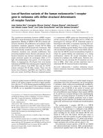
Figure 1
Experimental protocolinflammation of lymphotoxin-beta receptor-immunoglobulin fusion protein (LTR-Ig) treatment on blood glucose levels and on
submandibular gland and the effect
submandibular gland inflammation. (a) A weekly injection (i) of either LTR-Ig (LT) or mouse monoclonal IgG1 (MOPC 21) (MO). An isotypematched control Ig was administered. Age-matched mice that were not treated (NT) were also examined. Saliva flow rate measurements were performed, and the mice were then euthanized and salivary glands were harvested (†). (b) Blood glucose levels of individual mice in the experiment.
Horizontal lines indicate the median. At 19 weeks, the LTR-Ig-treated group blood glucose level was significantly lower than that of the agematched untreated mice (P < 0.001). Symbols refer to individual mice treated with LTR-Ig (ᮀ), MOPC 21 (❍), or no treatment (). Each symbol
indicates data from one mouse, and data were obtained on the day of euthanasia. The ratio index (c) and focus score (d) were used to analyze the
level of inflammation in the submandibular glands. Each group comprised seven or eight mice. Bars indicate the mean and the standard error of the
mean.
two to six sections at different levels in the gland and morphometrically analyzed using a Leica DMLB light microscope
(Leica, Wetzlar, Germany) connected to an Olympus Color
View III camera (Olympus, Tokyo, Japan) and AnalySIS software (Soft Imaging System, Münster, Germany). The focus
score [31] (number of foci made up of at least 50 mononuclear
cells per square millimeter of glandular tissue) and ratio index
[32] (ratio of the area of inflammation to the total area of the
gland) were determined.
Antibodies
The antibodies used were anti-B220, rat IgG2B, clone RA36B2 (R&D Systems, Minneapolis, MN, USA) (stains B cells);
anti-CD4, rat IgG2B, clone YTS191.1 (Dako Denmark A/S,
Glostrup, Denmark) (stains T cells); anti-CD21/CD35, clone
7G6 (BD Pharmingen, San Diego, CA, USA) (stains FDCs);
anti-peripheral (lymph) node addressin (anti-PNAd) carbohydrated epitope, clone MECA-79 (BD Pharmingen) (stains
HEVs); anti-CXCL13 goat IgG, lot numbers COZ025081 and
COZ026061 (R&D Systems); and anti-CCL19 goat IgG, lot
number BUL026021 (R&D Systems). The avidin-biotin complex technique was used for the immunohistochemistry analysis as described previously [32]. Frozen and paraffin sections
of salivary glands and mandibular lymph nodes (processed in
same manner as indicated above for salivary glands) were analyzed.
Analysis of T- and B-cell zones and high endothelial
venules
The submandibular glands were examined histologically by
authors MKG and KS in a blinded fashion after immunohistochemical single-staining with CD4 (stains T cells), B220
(stains B cells), and PNAd (stains HEVs) and morphometrically analyzed as stated above. The ratio of the zone of T-cell
aggregates or B-cell aggregates to the total area of the submandibular gland was determined. Isolated stained cells were
not included in this analysis. The number of HEVs per given
submandibular gland area was also analyzed.
Page 3 of 12
(page number not for citation purposes)
Arthritis Research & Therapy
Vol 11 No 1
Gatumu et al.
Quantitative polymerase chain reaction
Dissected submandibular glands from seven or eight mice
were placed in RNA later® (Ambion, Inc., Austin, TX, USA) at
4°C overnight before being frozen at -80°C until use. Organs
were disrupted in TRIzol® Reagent (Invitrogen Corporation).
RNA was extracted using RNeasy (Qiagen Inc., Valencia, CA,
USA). RNA was DNase-treated and cDNA was prepared
using a cDNA archive kit (Applied Biosystems, Foster City,
CA, USA). Quantitative polymerase chain reaction (QPCR)
was performed as described previously [33]. All RNA samples
were run in quadruplicate. The data were normalized to glyceraldehyde 3-phosphate dehydrogenase (GAPDH) as an
endogenous control. QPCR primers and probes were as follows: CXCL13: forward GTAAAACGCAGGCTTCCAAAA,
reverse GATGGCATTGCACCAGCTT, and FAM probe
AGTCTCCAGAAGGTTC; CCL19: forward CCTCGGCCTCTCAGATTCTTG, reverse GGCAGGCCACAGAGAGTGA, and FAM probe CACACAGTCTCTCAGGCT; and
CCL21: forward GGGCTGCAAGAGAACTGAACA, reverse
GGCGGGCTACTGGGCTAT,
and
FAM
probe
ACACAGCCCTCAAGAG.
Serum analysis for LTR-Ig levels by enzyme-linked
immunosorbent assay
A 96-well plate was coated with hamster anti-murine LTR
ACH6 (Biogen Idec, Cambridge, MA, USA) and incubated
overnight. The plate was blocked with 1% bovine serum albumin (BSA) in phosphate-buffered saline (PBS). Sera, at dilution 1:900 in 1% BSA in PBS, were added to the wells and
incubated for 1 hour. Peroxidase-conjugated donkey antimouse IgG (code number 715-036-150; Jackson ImmunoResearch Laboratories, Inc., West Grove, PA, USA) was then
incubated for 35 minutes. Samples were run in duplicate.
Serial dilutions of LTR-Ig were used to construct the standard curve. Plates were washed with PBS (pH 7.0) with 0.05%
Tween. Absorbance values were measured by a Multiskan®
microplate photometer (Labsystems Inc., Franklin, MA, USA)
and analyzed by Ascent software (Thermo Fisher Scientific
Inc., Waltham, MA, USA).
Statistics
Parametric data were analyzed using one-way betweengroups analysis of variance (ANOVA) with Tukey HSD (honestly significant difference) post hoc test and the Student t
test. Two-way between-groups ANOVA was undertaken for
specific comparisons of LTR-Ig and MOPC 21 treatment on
blood glucose levels, inflammation, and saliva flow rates. The
Mann-Whitney U test was used for non-parametric data. Pearson's correlation was used to describe linear relationships.
Saliva cytokines were Z-standardized. SPSS version 15.0
(SPSS Inc., Chicago, IL, USA) was used for these analyses.
The Kruskal-Wallis test and Dunn's multiple comparison test
were used for the LTR-Ig level analysis using GraphPad version 4 (GraphPad Software, Inc., San Diego, CA, USA). P values of less than 0.05 were considered significant.
Page 4 of 12
(page number not for citation purposes)
Results
Effect of LTR-Ig treatment on blood glucose levels
As highlighted in the Introduction, several studies have
reported successful prevention of diabetes development in
NOD mice upon treatment with LTR-Ig. We used the blood
glucose levels as an in-house control to determine the efficacy
of LTR-Ig treatment. Both the number of injections and the
type of treatment (either LTR-Ig or MOPC 21) played a role
in determining the glucose levels, as shown by the significant
interaction effect (P < 0.001). This finding was underscored
by the low glucose levels at 16 weeks in the LT16 group compared with age-matched controls, and at 19 weeks, the LT19
group glucose levels were not only lower than those of the
LT16 group, but also significantly different from those of agematched controls (Figure 1b).
LTR-Ig treatment inhibits submandibular gland
inflammation
Submandibular salivary glands were examined for inflammation. The ratio index and focus score were used to determine
the level of inflammation and the two methods showed a
strong correlation (r = 0.807, n = 75, P < 0.001). LTR-Igtreated groups had reduced levels of inflammation as they
aged and also the lowest inflammation levels compared with
the controls at the different ages at which the mice were examined (Figure 1c, d). Collectively, the levels of inflammation
increased as the NOD mice aged, with the oldest (19-weekold) untreated mice (NT19) recording the highest inflammation
in the study (Figure 1c, d). In marked contrast to MOPC 21treated and untreated mice, the inflammation levels (focus
score and ratio index) in LTR-Ig-treated mice never differed
significantly from levels of healthy 9-week-old pre-disease
mice. The inflammation levels of the LT19 group were significantly different from those of the NT19 group, but the comparison between LT19 and MO19 was not significant (Figure 1c,
d). However, the main effect of treatment (comparing all LTRIg-treated mice with all MOPC 21-treated groups using twoway between-groups ANOVA) was significant for both ratio
index (P = 0.005) and focus score (P = 0.007).
After establishing that inflammation was indeed inhibited, we
were interested in determining the specific components
affected by the treatment and in establishing a time line for disease development. For these studies, an average of four mice
per group were randomly selected to represent each treated
group and each age. Tissue sections were prepared and
appropriate antibodies were used to determine the specific
cell phenotypes and structures (Tables 1 and 2).
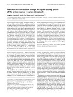
LTR-Ig treatment reduces the B- and T-cell zones in
submandibular glands
There were no B-cell zones at the beginning of the experiment
(NT9). These appeared in all of the groups at 12 weeks and
peaked at 16 weeks of age (Figure 2a, b). At 19 weeks, the
LTR-Ig-treated group B-cell zones were almost completely
Available online />
pole of the infiltrate. Scattered T and B cells were also
observed (Figure 2a).
Table 1
Absolute numbers of mice positive for high endothelial venules
and follicular dendritic cell networks
High endothelial venules
Treatment
LTR-Ig treatment inhibits high endothelial venules and
follicular dendritic cell networks in submandibular
glands
LTR-Ig treatment inhibited/reversed development of HEVs
(Table 1 and Figure 2a, d). In all of the LTR-Ig-treated mice
examined, only one HEV in one 12-week-old mouse was
observed. FDC networks were first noted at 12 weeks of age
in the untreated mice (NT12). Treatment with LTR-Ig blocked
development of FDC networks in the salivary glands examined
(Table 1 and Figure 3a).
Follicular dendritic cells
LT
MO
NT
LT
MO
NT
9
-
-
0/4
-
-
0/4
12
1/4
1/4
2/4
0/4
0/4
2/4
16
0/4
4/4
3/4
0/4
1/4
3/4
19
0/4
3/4
2/3
0/4
2/4
1/3
Age in weeks
The effect of LTR-Ig treatment on chemokine mRNA
expression and immunoreactive protein and the
profound reduction of CCL19 protein
CXCL13 was present in the inflammatory infiltrates of all of the
groups studied except the untreated 9-week-old mice (NT9)
(Table 2 and Figure 3a). Some cells showed membranous
staining but more commonly CXCL13 was noted as a dispersed substance within the infiltrates (Figure 3a). CCL19
was demonstrated in the salivary glands at sites of inflammation, more commonly as a dispersed substance within the infiltrate (Figure 3a). In some cells, CCL19 membranous staining
was also observed (Figure 3a). CCL19 expression was also
found on ductal and acinar epithelia (Table 2 and Figure 3a).
Unlike CXCL13, CCL19 was not expressed in the infiltrate of
any of the mice treated with LTR-Ig (Table 2). The mRNA
expression levels of CXCL13, CCL19, and CCL21 were also
analyzed, as shown in Figure 3b–d. When we compared the
chemokines in the LTR-Ig-treated mice with those of agematched controls or the baseline group (NT9), only CXCL13
was clearly downmodulated.
Treatment with either lymphotoxin-beta receptor-immunoglobulin
fusion protein (LTR-Ig) or mouse monoclonal IgG1 (MOPC 21) was
started at week 9. Euthanization was carried out 1 week after the last
injection. Mice receiving either LTR-Ig or MOPC 21 and euthanized
at 12 weeks of age had received 3 injections, those euthanized at 16
weeks of age had received 7 injections, and those euthanized at 19
weeks of age had received 10 injections. -, no mice in these groups;
LT, mice treated with lymphotoxin-beta receptor-immunoglobulin
fusion protein; MO, mice treated with mouse monoclonal IgG1; NT,
mice not treated.
inhibited (B-cell zones recorded in only one mouse in the LT19
group) (Figure 2b and data not shown). Statistical comparisons between MOPC 21 and LTR-Ig groups indicate that Bcell zone ratios were influenced mainly by treatment type (P =
0.011) and number of injections (P = 0.037). Although the
intergroup differences were not statistically significant, T-cell
zones increased with age in the MOPC 21 and untreated
groups but declined with age in the LTR-Ig group (Figure 2a,
c). The B-cell zones were smaller than T-cell zones (see y-axis
of Figure 2a, b). When B-cell zones or T-cell zones were
observed in LTR-Ig-treated mice, these structures were composed of groups of closely packed cells concentrated at one
Table 2
Absolute numbers of mice positive for chemokines
CXCL13
Treatment
LT
MO
CCL19
NT
Age in weeks
LT
Acini/duct
MO
Infiltrate
Acini/duct
NT
Infiltrate
Acini/duct
Infiltrate
9
-
-
0/4
-
-
-
-
4/4
0/4
12
2/4
1/4
4/4
4/4
0/4
4/4
1/4
4/4
2/4
16
4/4
4/4
4/4
4/4
0/4
4/4
4/4
4/4
3/4
19
2/4
4/4
3/3
3/4
0/4
2/4
2/4
2/3
1/3
Treatment with either lymphotoxin-beta receptor-immunoglobulin fusion protein (LTR-Ig) or mouse monoclonal IgG1 (MOPC 21) was started at
week 9. Euthanization was carried out 1 week after the last injection. Mice receiving either LTR-Ig or MOPC 21 and euthanized at 12 weeks of
age had received 3 injections, those euthanized at 16 weeks of age had received 7 injections, and those euthanized at 19 weeks of age had
received 10 injections. -, no mice in these groups; LT, mice treated with lymphotoxin-beta receptor-immunoglobulin fusion protein; MO, mice
treated with mouse monoclonal IgG1; NT, mice not treated.
Page 5 of 12
(page number not for citation purposes)
Arthritis Research & Therapy
Vol 11 No 1
Gatumu et al.
Figure 2
Effect of
(HEVs) lymphotoxin-beta receptor-immunoglobulin fusion protein (LTR-Ig) treatment on B-cell zones, T-cell zones, and high endothelial venules
(HEVs). (a) Photomicrographs of immunohistochemical staining for B cells, T cells, and HEVs in the submandibular glands. Three or four mice from
each of the 10 groups were randomly selected for immunohistochemistry. Data shown here are representative examples of control mice (19 weeks
old) that were not treated (NT19) or that were treated with 10 injections of mouse monoclonal IgG1 (MOPC 21) (MO19) and of mice treated with
10 injections of LTR-Ig (LT19). Bars = 100 m. (b) Proportion of B-cell zones to the total area of the gland (that is, the B-cell ratio index). (c) Proportion of T-cell zones to the total area of the gland (that is, the T-cell ratio index). (d) Number of HEVs to the total area of the gland. Each of the 10
groups had three or four randomly selected mice for each staining and analysis. Legend applies to frames (b-d). Bars indicate the mean and the
standard error of the mean.
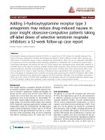
Effects of LTR-Ig treatment on lymph nodes
In contrast to the salivary glands where no LTR-Ig-treated
mice had FDC networks, the mandibular lymph nodes were
affected by LTR-Ig treatment in a more gradual manner:
Some disorganized or remnant networks were found in both
LT12 (Figure 4) and LT16, but none was found in LT19 group
(data not shown). The effect of LTR-Ig treatment on CCL19
(Figure 4) was also less dramatic in the mandibular lymph
nodes than in the salivary glands as this chemokine was
expressed in some but not all lymph nodes in the LT16 and
LT19 groups. Whereas HEVs in the salivary glands were very
sensitive to LTR-Ig treatment and were completely cleared in
the LT16 and LT19 groups, HEVs as revealed by MECA-79
staining were diminished in the mandibular lymph nodes
although some HEVs were still visible in the LT12 (Figure 4),
LT16, and LT19 groups. These histological changes to the
lymph nodes resemble those described previously following
Page 6 of 12
(page number not for citation purposes)
LTR-Ig treatment [33]. CXCL13 was expressed in all of the
draining lymph nodes from the LTR-Ig-treated mice. However, more intense staining was noted in the controls than in
the LTR-Ig-treated mice (Figure 4), indicating the likelihood of
a quantitative difference.
Salivary gland function is partially restored after
treatment with LTR-Ig
Generally, a subtle dichotomous distribution of saliva flow
rates within each group was observed (Figure 5a). To determine whether saliva flow rate was restored, all of the groups of
mice were compared with the pre-disease state (that is,
untreated mice euthanized at 9 weeks [NT9]). Overall, the
saliva flow rate declined in all of the groups of NOD mice that
were 9 to 12 weeks old but this decline was not statistically
significant (Figure 5a). At 16 weeks, all three groups had a significant decline in saliva flow (Figure 5a). At 19 weeks of age,
Available online />
Figure 3
Effect of lymphotoxin-beta receptor-immunoglobulin fusion protein (LTR-Ig) treatment on follicular dendritic cell (FDC) networks and chemokines
chemokines.
(a) Photomicrographs of immunohistochemical staining for FDC networks and CXCL13 and CCL19 in the submandibular glands. Three or four mice
from each of the 10 groups were randomly selected for immunohistochemistry. Data shown here are representative examples of control mice (19
weeks old) that were not treated (NT19) or that were treated with 10 injections of mouse monoclonal IgG1 (MOPC 21) (MO19) and of mice treated
with 10 injections of LTR-Ig. Arrowheads indicate sites of chemokine staining. Bars = 100 m. A comparison of relative mRNA expressions of
CXCL13 (b), CCL19 (c), and CCL21 (d) in salivary glands of the baseline group (NT9 group) and the oldest mice (19 weeks old) treated with either
LTR-Ig or MOPC 21 or untreated. Salivary glands from seven or eight mice per group were used in this analysis. Quantitative polymerase chain
reactions (Q PCRs) using RNA from each organ were run in quadruplicate, and the mean signal was normalized to the glyceraldehyde 3-phosphate
dehydrogenase (GAPDH). Data are presented as the mean of the values for the replicate organs (seven or eight organs per point) and the standard
deviation. The differences between LT19 and controls were not significant.
the saliva flow rate of the LT19 group was partially restored to
the levels recorded in the NT9 group (Figure 5a). This phenomenon did not occur in either the MO19 or the NT19 group.
There was a statistically significant main effect on the saliva
flow rate of the number of injections administered (P = 0.042).
The saliva flow rates of the MO19 and LT19 groups were significantly different (P = 0.049). To determine whether the
hyperglycemic state of the mice influenced the saliva flow
rates, we compared the saliva flow rates in mice within the
same group with either low or high glucose levels and found
no significant differences (data not shown).
Cytokines in saliva samples
Interleukin (IL)-1-beta was detected in all of the samples analyzed (from 55 individual mice), whereas vascular endothelial
growth factor was detected in all but one of the samples. For
macrophage inflammatory protein-1 alpha (MIP-1), IL-10, IL2, and IL-1, samples with a mean fluorescent intensity corresponding to a value lower than the least detectable concentration were assigned values of 0. Basic fibroblast growth factor
and CXCL10 were each detected in only one of the samples.
The other cytokines/chemokines tested (that is, granulocyte
macrophage-colony stimulating factor, interferon-gamma, IL-4,
IL-5, IL-12, IL-13, IL-17, CXCL1, CCL2, CXCL9, and TNF-)
were not detected in any of the samples. There was a striking
Page 7 of 12
(page number not for citation purposes)
Arthritis Research & Therapy
Vol 11 No 1
Gatumu et al.
Figure 4
Immunohistochemical analysis of lymph nodes Mandibular lymph nodes from two to six mice per group were examined (multiple lymph nodes in
nodes.
some mice). Representative photomicrographs of immunohistochemical staining of lymph nodes from mice from the LT12 group and NT12 group.
For the follicular dendritic cell (FDC) network panels, the LT12 lymph node has no FDC networks, but B cells are picked by the CD21 antibody, and
the NT12 lymph node has prominent FDC networks. Original magnifications: × 50 (insets), × 400 (CCL19), and × 200 (all other images).
similarity between the saliva cytokine profile of the LT19 and
NT9 groups. High levels of saliva cytokines (both pro- and antiinflammatory) were observed in the NT16 group. The results of
the analysis of the pro- and anti-inflammatory cytokines are
shown in Figure 5b.
Serum LTR-Ig levels decrease as the mice age
The serum levels of LTR-Ig were determined 1 week after the
final LTR-Ig injection in each group. A more-than-twofold
drop in the LTR-Ig levels was observed in older mice (88.19
± 12.04 and 40.17 ± 8.87 g/mL in 12-week-old and 19week-old mice, respectively).
Effect of mouse monoclonal IgG1
An effect of MOPC 21 was observed. However, except for
expression levels of MO19 and NT19 CCL21b mRNA (P =
0.042), the levels of the various parameters studied in MOPC
21 mice were not significantly different from those of the
untreated mice.
Page 8 of 12
(page number not for citation purposes)
Discussion
The results disclosed that blocking the LTR pathway in NOD
mice not only has an effect on the blood glucose levels, as
reported previously [23,25], but also has profound effects on
the salivary gland lesion, a hallmark of SS-like disease in NOD
mice. The loss of FDC networks on blocking the LTR pathway in adult mice has been described previously [19,34], but
the present study is the first to report that LTR-Ig treatment
results in FDC network loss in the salivary glands. FDC networks constitutively release a substantial amount of CXCL13
[35], although there are other sources of CXCL13, such as
macrophages, dendritic cells, and primed follicular helper T
cells [35]. CXCL13 recruits B cells and follicular helper T cells
to the B-cell zones. Immunohistochemistry disclosed that both
LTR-Ig-treated mice and controls expressed CXCL13 at the
protein level in the salivary glands. There was, however, a
depletion of B-cell zones by 19 weeks (LT19 group). This was
probably attributable to the reduction in the amount of
CXCL13, as indicated by the reduction in CXCL13 mRNA
expression levels in the LT19 group compared with controls.
Available online />
Figure 5
Salivary gland function and saliva cytokine analysis. (a) Saliva flow rates of the 10 groups. The saliva flow rate was expressed as microliters of saliva
analysis
secreted per minute per gram of mouse body weight. Data were analyzed by one-way analysis of variance followed by Tukey post hoc test. *P <
0.05, **P < 0.01, and ***P < 0.001 indicate statistically significant differences between the specific group and the NT9 group. Horizontal lines indicate the mean. Symbols refer to individual mice treated with lymphotoxin-beta receptor-immunoglobulin fusion protein (LTR-Ig) (ᮀ), mouse monoclonal IgG1 (MOPC 21) (❍), or no treatment (). Each symbol indicates data from one mouse. (b) Heat map of saliva cytokines showing fluctuations
in the levels of cytokines in the control mice groups and LTR-Ig-treated mice over time. The mean cytokine level of each mouse group was determined and Z-score standardization was computed for each cytokine using SPSS version 15.0. The heat map was drawn in Excel® 2003. IL, interleukin; MIP-1, macrophage inflammatory protein-1 alpha; VEGF, vascular endothelial growth factor.
The complete loss of FDC networks following LTR-Ig treatment may also have contributed to the lack of B-cell zones in
the LT19 group.
There was also an all-out loss of HEVs in the salivary glands in
the LT16 and LT19 groups. HEVs exhibit specific address signals that are recognized by circulating lymphocytes. PNAd is
one such signal (antigen) that was first defined by reactivity
with monoclonal antibody MECA-79 [36]. L-selectin on the circulating lymphocytes binds carbohydrate epitopes of PNAd,
facilitating homing of these naïve lymphocytes [37]. Previous
studies have reported a prominent role for the LTR pathway
in maintaining the HEV structures in a functional state in lymph
nodes of adult mice [13,33] and in sites of lymphoid neogenesis [38]. The LTR pathway is also important in de novo lymphangiogenesis in sites of lymphoid neogenesis [39].
Although the mRNA expression levels of CCL19 were not significantly reduced in the LTR-Ig-treated mice, there was a
complete loss of CCL19 protein in the salivary gland infiltrates
of LTR-Ig-treated mice. CCL19 and CCL21 are necessary
for the transmigration of naïve T cells and central memory T
cells across the HEVs and in the formation of T-cell zones [40].
Therefore, the combination of ablation of FDC networks, loss
of HEVs, and reduced chemokine expression would ultimately
result in a reduced influx of B and T cells to the salivary glands.
This may explain the reduced B- and T-cell zones in the LTRIg-treated mice.
Another interesting observation was the different response to
LTR-Ig treatment by the salivary gland lesion and the draining
lymph nodes. There is no consensus as to which of these sites
drives the immune response or whether there is a contribution
from both [13]. Our findings indicate that the LTR-Ig treatment had a more profound effect on the microarchitecture of
the salivary gland lymphoid tissue than on that of the lymph
node.
Our findings also suggest that the salivary gland lesion has a
prominent role in SS-like disease, as partial saliva flow resto-
Page 9 of 12
(page number not for citation purposes)
Arthritis Research & Therapy
Vol 11 No 1
Gatumu et al.
ration occurs in association with the phenotypic changes in
the salivary gland. The ectopic lymphoid tissue has a prominent role in several diseases/conditions [13]. In the NOD
mouse, pancreatic lymphoid neogenesis could initiate and
sustain the progression of IDDM in the absence of draining
lymph nodes [41]. Interestingly, it has been proposed that
LTR-Ig treatment would result in a quiescent state of the
lymph nodes, draining the sites of inflammation [13]. This is
partly supported by the present study, showing microarchitectural differences between LTR-Ig-treated mice and control
lymph nodes. However, the lymph node microarchitectural
changes in our experimental setup are unlike the grossly disturbed primary or secondary organ development observed in
mice deficient in components of the LTR pathway.
The present study was based on the hypothesis that LTR-Ig
treatment would preserve the integrity of the salivary gland by
preventing development and progression of inflammation and
lymphoid neogenesis, thus preventing the occurrence of salivary gland dysfunction. Our results, however, show that LTRIg treatment may reverse pathological events that have already
occurred in the salivary gland. This possibility is underscored
by the finding of HEVs in the salivary glands of 12-week-old
mice given three LTR-Ig injections (although in only one
mouse) but loss of HEVs in 16-week-old and 19-week-old
mice treated with LTR-Ig. Earlier analyses in mice suggested
that a period of at least 2 weeks of LTR-Ig treatment was
required to downmodulate HEVs and therefore any early HEVs
could still be present at week 12 [33]. Also, the saliva flow rate
appeared to fall (comparing LT12 and LT16 groups) but
recovered in the LT19 group, indicating that the salivary gland
dysfunction may have been prevented or reversed.
The dichotomous distribution of saliva flow rates in NOD mice
has been described previously [42]. The same study also used
saliva proteins to describe the efficacy of intervention agents
in SS-like disease of NOD mice. In our study, the striking similarity in the cytokine profiles of the pre-disease NT9 group and
the LT19 group may indicate a restoration of the pre-disease
quality of saliva in the LT19 group. However, analysis of proand anti-inflammatory cytokines and chemokines in saliva from
untreated mice across different ages disclosed no obvious
trend indicative of progressive disease status. However, 16
weeks may mark a time of potentially heightened cytokine production and activity in the NOD mouse.
Accelerated clearance of immunoglobulin-based agents is
commonly observed in many rodent autoimmune disease models, and our finding of a reduction in the serum levels of LTRIg as the mice age also points to a possible increased clearance of the fusion protein in these older NOD mice. It is postulated that, had the high levels of LTR-Ig observed in 12week-old mice been sustained throughout the experiment, the
protective/reversal effects of LTR-Ig would have been more
pronounced.
Page 10 of 12
(page number not for citation purposes)
There are severe technical limitations to finding proper controls for fusion proteins when faced with the need to use highlevel dosing in autoimmune disease models to maintain coverage. A protective effect of polyclonal human IgG used as a
control protein in experiments to determine the efficacy of
LTR-Ig in collagen-induced arthritis was reported [34]. The
mechanism behind the MOPC 21 effect may involve non-specific binding of MOPC 21 to targets in the pathways involved
in the disease process, resulting in a protective effect.
The immune dysregulation occurring in the female NOD background leads to autoreactivity and inflammation of the salivary
glands. This inflammation is accompanied by the formation of
lymphoid tissues in the salivary glands with HEVs and FDC
networks. Although the role of these tertiary lymphoid tissues
in the pathogenesis remains unclear, the present study clearly
demonstrates that LTR inhibition, whether via its effects on
trafficking or lymphoid architecture, results in a less destructive environment in the target organ.
Conclusion
This study shows that blocking the LTR pathway in NOD
mice ablates lymphoid neogenesis in the salivary glands and
this is accompanied by an improvement in salivary gland function. To our knowledge, the effect of interfering with the LTR
axis has not previously been explored in SS-like disease. It is
hoped that our results will contribute to the development of
appropriate methods for treatment of human SS.
Competing interests
JLB and AP are employees of Biogen Idec. The other authors
declare that they have no competing interests.
Authors' contributions
AIB designed the study and helped to carry out the animal
experiments, to analyze the results, and to write the paper.
MKG helped to carry out the animal experiments, performed
the immunohistochemistry, enzyme-linked immunosorbent
assay, multiplex analysis, and statistics, and helped to perform
the morphometric analysis, to analyze the results, and to write
the paper. KS helped to carry out the animal experiments, to
perform the morphometric analysis, to analyze the results, and
to write the paper. AP performed the QPCR and helped to
analyze the results. JLB and RAF helped to analyze the results
and to write the paper. All authors read and approved the final
manuscript.
Acknowledgements
We thank Edith Fick, Siren Hammer, Gunnvor Øijordsbakken, and Ewa
Szyszko, of the University of Bergen, and Norm Allaire and Tanushree
Phadke, of Biogen Idec, for technical assistance. The language editing
by Joan Bevenius is gratefully acknowledged. The study was funded by
Western Norway Regional Health Authority, The Meltzer Foundation,
The Research Council of Norway, and VA Merit Review.
Available online />
References
1.
2.
3.
4.
5.
6.
7.
8.
9.
10.
11.
12.
13.
14.
15.
16.
17.
18.
Fox RI: Sjögren's syndrome. Lancet 2005, 366:321-331.
Stott DI, Hiepe F, Hummel M, Steinhauser G, Berek C: Antigendriven clonal proliferation of B cells within the target tissue of
an autoimmune disease. The salivary glands of patients with
Sjögren's syndrome. J Clin Invest 1998, 102:938-946.
Salomonsson S, Jonsson MV, Skarstein K, Brokstad KA, Hjelmström P, Wahren-Herlenius M, Jonsson R: Cellular basis of
ectopic germinal center formation and autoantibody production in the target organ of patients with Sjögren's syndrome.
Arthritis Rheum 2003, 48:3187-3201.
Jonsson MV, Szodoray P, Jellestad S, Jonsson R, Skarstein K:
Association between circulating levels of the novel TNF family
members APRIL and BAFF and lymphoid organization in primary Sjögren's syndrome. J Clin Immunol 2005, 25:189-201.
Jonsson MV, Skarstein K, Jonsson R, Brun JG: Serological implications of germinal center-like structures in primary Sjögren's
syndrome. J Rheumatol 2007, 34:2044-2049.
Browning JL, Androlewicz MJ, Ware CF: Lymphotoxin and an
associated 33-kDa glycoprotein are expressed on the surface
of an activated human T cell hybridoma. J Immunol 1991,
147:1230-1237.
Browning JL, Dougas I, Ngam-ek A, Bourdon PR, Ehrenfels BN,
Miatkowski K, Zafari M, Yampaglia AM, Lawton P, Meier W: Characterization of surface lymphotoxin forms. Use of specific
monoclonal antibodies and soluble receptors. J Immunol
1995, 154:33-46.
Crowe PD, VanArsdale TL, Walter BN, Ware CF, Hession C,
Ehrenfels B, Browning JL, Din WS, Goodwin RG, Smith CA: A
lymphotoxin-beta-specific
receptor.
Science
1994,
264:707-710.
Mauri DN, Ebner R, Montgomery RI, Kochel KD, Cheung TC, Yu GL, Ruben S, Murphy M, Eisenberg RJ, Cohen GH, Spear PG, Ware
CF: LIGHT, a new member of the TNF superfamily, and lymphotoxin alpha are ligands for herpesvirus entry mediator.
Immunity 1998, 8:21-30.
Ware CF: Targeting lymphocyte activation through the lymphotoxin and LIGHT pathways.
Immunol Rev 2008,
223:186-201.
De Togni P, Goellner J, Ruddle NH, Streeter PR, Fick A, Mariathasan S, Smith SC, Carlson R, Shornick LP, Strauss-Schoenberger J: Abnormal development of peripheral lymphoid
organs in mice deficient in lymphotoxin. Science 1994,
264:703-707.
Ettinger R, Browning JL, Michie SA, van Ewijk W, McDevitt HO:
Disrupted splenic architecture, but normal lymph node development in mice expressing a soluble lymphotoxin-beta receptor-IgG1 fusion protein. Proc Natl Acad Sci USA 1996,
93:13102-13107.
Browning JL: Inhibition of the lymphotoxin pathway as a therapy for autoimmune disease.
Immunol Rev 2008,
223:202-220.
Koni PA, Sacca R, Lawton P, Browning JL, Ruddle NH, Flavell RA:
Distinct roles in lymphoid organogenesis for lymphotoxins
alpha and beta revealed in lymphotoxin beta-deficient mice.
Immunity 1997, 6:491-500.
Alimzhanov MB, Kuprash DV, Kosco-Vilbois MH, Luz A, Turetskaya
RL, Tarakhovsky A, Rajewsky K, Nedospasov SA, Pfeffer K: Abnormal development of secondary lymphoid tissues in lymphotoxin beta-deficient mice. Proc Natl Acad Sci USA 1997,
94:9302-9307.
Futterer A, Mink K, Luz A, Kosco-Vilbois MH, Pfeffer K: The lymphotoxin beta receptor controls organogenesis and affinity
maturation in peripheral lymphoid tissues. Immunity 1998,
9:59-70.
Rennert PD, Browning JL, Mebius R, Mackay F, Hochman PS: Surface lymphotoxin alpha/beta complex is required for the
development of peripheral lymphoid organs. J Exp Med 1996,
184:1999-2006.
Scheu S, Alferink J, Potzel T, Barchet W, Kalinke U, Pfeffer K: Targeted disruption of LIGHT causes defects in costimulatory T
cell activation and reveals cooperation with lymphotoxin beta
in mesenteric lymph node genesis. J Exp Med 2002,
195:1613-1624.
19. Gommerman JL, Browning JL: Lymphotoxin/LIGHT, lymphoid
microenvironments and autoimmune disease. Nat Rev Immunol 2003, 3:642-655.
20. Kratz A, Campos-Neto A, Hanson MS, Ruddle NH: Chronic
inflammation caused by lymphotoxin is lymphoid neogenesis.
J Exp Med 1996, 183:1461-1472.
21. Aloisi F, Pujol-Borrell R: Lymphoid neogenesis in chronic
inflammatory diseases. Nat Rev Immunol 2006, 6:205-217.
22. Drayton DL, Liao S, Mounzer RH, Ruddle NH: Lymphoid organ
development: from ontogeny to neogenesis. Nat Immunol
2006, 7:344-353.
23. Wu Q, Salomon B, Chen M, Wang Y, Hoffman LM, Bluestone JA,
Fu Y-X: Reversal of spontaneous autoimmune insulitis in nonobese diabetic mice by soluble lymphotoxin receptor. J Exp
Med 2001, 193:1327-1332.
24. Levisetti MG, Suri A, Frederick K, Unanue ER: Absence of lymph
nodes in NOD mice treated with lymphotoxin-{beta} receptor
immunoglobulin protects from diabetes. Diabetes 2004,
53:3115-3119.
25. Ettinger R, Munson SH, Chao C-C, Vadeboncoeur M, Toma J,
McDevitt HO: A critical role for lymphotoxin-beta receptor in
the development of diabetes in nonobese diabetic mice. J Exp
Med 2001, 193:1333-1340.
26. Hu Y, Nakagawa Y, Purushotham KR, Humphreys-Beher MG:
Functional changes in salivary glands of autoimmune diseaseprone NOD mice. Am J Physiol 1992, 263:E607-E614.
27. Cha S, Nagashima H, Brown VB, Peck AB, Humphreys-Beher MG:
Two NOD Idd-associated intervals contribute synergistically to
the development of autoimmune exocrinopathy (Sjögren's
syndrome) on a healthy murine background. Arthritis Rheum
2002, 46:1390-1398.
28. Johansson AC, Nakken B, Sundler M, Lindqvist AK, Johannesson
M, Alarcón-Riquelme M, Bolstad AI, Humphreys-Beher MG, Jonsson R, Skarstein K, Holmdahl R: The genetic control of sialadenitis versus arthritis in a NOD.Q × B10.Q F2 cross. Eur J
Immunol 2002, 32:243-250.
29. Hjelmervik T, Lindqvist A-K, Petersen K, Johannesson M, Stavrum
A-K, Johansson A, Jonsson R, Holmdahl R, Bolstad A: The influence of the NOD Nss1/Idd5 loci on sialadenitis and gene
expression in salivary glands of congenic mice. Arthritis Res
Ther 2007, 9:R99.
30. Gommerman JL, Giza K, Perper S, Sizing I, Ngam-ek A, NickersonNutter C, Browning JL: A role for surface lymphotoxin in experimental autoimmune encephalomyelitis independent of
LIGHT. J Clin Invest 2003, 112:755-767.
31. Jonsson R, Tarkowski A, Bäckman K, Holmdahl R, Klareskog L:
Sialadenitis in the MRL-l mouse: morphological and immunohistochemical characterization of resident and infiltrating
cells. Immunology 1987, 60:611-616.
32. Skarstein K, Johannessen AC, Holmdahl R, Jonsson R: Effects on
sialadenitis after cellular transfer in autoimmune MRL/lpr
mice. Clin Immunol Immunopathol 1997, 84:177-184.
33. Browning JL, Allaire N, Ngam-ek A, Notidis E, Hunt J, Perrin S, Fava
RA: Lymphotoxin-beta receptor signaling is required for the
homeostatic control of HEV differentiation and function.
Immunity 2005, 23:539-550.
34. Fava RA, Notidis E, Hunt J, Szanya V, Ratcliffe N, Ngam-ek A, de
Fougerolles AR, Sprague A, Browning JL: A role for the lymphotoxin/LIGHT axis in the pathogenesis of murine collageninduced arthritis. J Immunol 2003, 171:115-126.
35. Allen CD, Cyster JG: Follicular dendritic cell networks of primary follicles and germinal centers: phenotype and function.
Semin Immunol 2008, 20:14-25.
36. Streeter PR, Rouse BT, Butcher EC: Immunohistologic and
functional characterization of a vascular addressin involved in
lymphocyte homing into peripheral lymph nodes. J Cell Biol
1988, 107:1853-1862.
37. Berg EL, Robinson MK, Warnock RA, Butcher EC: The human
peripheral lymph node vascular addressin is a ligand for
LECAM-1, the peripheral lymph node homing receptor. J Cell
Biol 1991, 114:343-349.
38. Luther SA, Lopez T, Bai W, Hanahan D, Cyster JG: BLC expression in pancreatic islets causes B cell recruitment and lymphotoxin-dependent lymphoid neogenesis.
Immunity 2000,
12:471-481.
39. Furtado GC, Marinkovic T, Martin AP, Garin A, Hoch B, Hubner W,
Chen BK, Genden E, Skobe M, Lira SA: Lymphotoxin beta
Page 11 of 12
(page number not for citation purposes)
Arthritis Research & Therapy
Vol 11 No 1
Gatumu et al.
receptor signaling is required for inflammatory lymphangiogenesis in the thyroid. Proc Natl Acad Sci USA 2007,
104:5026-5031.
40. Ebert LM, Schaerli P, Moser B: Chemokine-mediated control of
T cell traffic in lymphoid and peripheral tissues. Mol Immunol
2005, 42:799-809.
41. Lee Y, Chin RK, Christiansen P, Sun Y, Tumanov AV, Wang J,
Chervonsky AV, Fu Y-X: Recruitment and activation of naive T
cells in the islets by lymphotoxin [beta] receptor-dependent
tertiary lymphoid structure. Immunity 2006, 25:499-509.
42. Delaleu N, Madureira AC, Immervoll H, Jonsson R: Inhibition of
experimental Sjögren's syndrome through immunization with
HSP60 and its peptide amino acids 437–460. Arthritis Rheum
2008, 58:2318-2328.
Page 12 of 12
(page number not for citation purposes)