Báo cáo y học: "In vitro model for the analysis of synovial fibroblast-mediated degradation of intact cartilage" pptx
Bạn đang xem bản rút gọn của tài liệu. Xem và tải ngay bản đầy đủ của tài liệu tại đây (5.53 MB, 20 trang )
Open Access
Available online />Page 1 of 20
(page number not for citation purposes)
Vol 11 No 1
Research article
In vitro model for the analysis of synovial fibroblast-mediated
degradation of intact cartilage
David Pretzel
1
, Dirk Pohlers
1
, Sönke Weinert
1,2
and Raimund W Kinne
1
1
Experimental Rheumatology Unit, Department of Orthopedics, University Hospital Jena, Friedrich Schiller University Jena, Klosterlausnitzer Strasse
81, Eisenberg, D-07607, Germany
2
Current address: Experimental Cardiology, Otto von Guericke University Magdeburg, Leipziger Strasse 44, Magdeburg, D-39120, Germany
Corresponding author: David Pretzel,
Received: 4 Jun 2008 Revisions requested: 24 Jul 2008 Revisions received: 20 Jan 2009 Accepted: 18 Feb 2009 Published: 18 Feb 2009
Arthritis Research & Therapy 2009, 11:R25 (doi:10.1186/ar2618)
This article is online at: />© 2009 Pretzel et al.; licensee BioMed Central Ltd.
This is an open access article distributed under the terms of the Creative Commons Attribution License ( />),
which permits unrestricted use, distribution, and reproduction in any medium, provided the original work is properly cited.
Abstract
Introduction Activated synovial fibroblasts are thought to play a
major role in the destruction of cartilage in chronic, inflammatory
rheumatoid arthritis (RA). However, profound insight into the
pathogenic mechanisms and the impact of synovial fibroblasts in
the initial early stages of cartilage destruction is limited. Hence,
the present study sought to establish a standardised in vitro
model for early cartilage destruction with native, intact cartilage
in order to analyse the matrix-degrading capacity of synovial
fibroblasts and their influence on cartilage metabolism.
Methods A standardised model was established by co-culturing
bovine cartilage discs with early-passage human synovial
fibroblasts for 14 days under continuous stimulation with TNF-
α, IL-1β or a combination of TNF-α/IL-1β. To assess cartilage
destruction, the co-cultures were analysed by histology,
immunohistochemistry, electron microscopy and laser scanning
microscopy. In addition, content and/or neosynthesis of the
matrix molecules cartilage oligomeric matrix protein (COMP)
and collagen II was quantified. Finally, gene and protein
expression of matrix-degrading enzymes and pro-inflammatory
cytokines were profiled in both synovial fibroblasts and cartilage.
Results Histological and immunohistological analyses revealed
that non-stimulated synovial fibroblasts are capable of
demasking/degrading cartilage matrix components
(proteoglycans, COMP, collagen) and stimulated synovial
fibroblasts clearly augment chondrocyte-mediated, cytokine-
induced cartilage destruction. Cytokine stimulation led to an
upregulation of tissue-degrading enzymes (aggrecanases I/II,
matrix-metalloproteinase (MMP) 1, MMP-3) and pro-
inflammatory cytokines (IL-6 and IL-8) in both cartilage and
synovial fibroblasts. In general, the activity of tissue-degrading
enzymes was consistently higher in co-cultures with synovial
fibroblasts than in cartilage monocultures. In addition, stimulated
synovial fibroblasts suppressed the synthesis of collagen type II
mRNA in cartilage.
Conclusions The results demonstrate for the first time the
capacity of synovial fibroblasts to degrade intact cartilage matrix
by disturbing the homeostasis of cartilage via the production of
catabolic enzymes/pro-inflammatory cytokines and suppression
of anabolic matrix synthesis (i.e., collagen type II). This new in
vitro model may closely reflect the complex process of early
stage in vivo destruction in RA and help to elucidate the role of
synovial fibroblasts and other synovial cells in this process, and
the molecular mechanisms involved in cartilage degradation.
Introduction
Rheumatoid arthritis (RA) is a chronic disorder primarily affect-
ing the joints and leading to destruction of the articular carti-
lage with subsequent severe morbidity and disability. It is
characterised by a chronic infiltration of inflammatory cells into
the synovial membrane and the development of a hyperplastic
pannus tissue [1].
This pannus tissue, consisting of both inflammatory and resi-
dent mesenchymal cells, invades and destroys the underlying
cartilage and bone. Therefore, the role of macrophages [2], T-
APMA: aminophenylmercuric acetate; CFSE: carboxyfluoroscein succinimidyl ester; COMP: cartilage oligomeric matrix protein; DMEM: Dulbecco's
modified eagle medium; ELISA: enzyme-linked immunosorbent assay; FCS: fetal calf serum; H&E: haematoxylin and eosin; HRP: horseradish perox-
idase; Ig: immunoglobulin; IL: interleukin; OA: osteoarthritis; PBS: phosphate buffered saline; qPCR: quantitative polymerase chain reaction; RA: rheu-
matoid arthritis; SDS: sodium dodecyl sulfate; SFB: synovial fibroblast; TNF: tumour necrosis factor.
Arthritis Research & Therapy Vol 11 No 1 Pretzel et al.
Page 2 of 20
(page number not for citation purposes)
and B-cells [3] and synovial fibroblasts (SFB) [4] in the patho-
genesis of RA, including their multilateral interactions, has
been intensely investigated. Due to their aggressive features
and over-expression of matrix-degrading enzymes, activated
SFB seem to play a major role in the invasion and proteolytic
degradation of the cartilage matrix [5]. In addition, they can
indirectly induce a catabolic metabolism in chondrocytes via
soluble mediators [6]. The destructive properties of SFB have
been analysed in several in vivo and in vitro models. Despite
their unquestionable advantages, established animal models
of arthritis, including co-implantation models in immunodefi-
cient mice (reviewed in [7,8]), also have disadvantages. They
reflect a very complex cellular network rather than the particu-
lar influence of a certain cell type, are time-consuming and
expensive, and can be ethically problematic.
In an attempt to replace, or at least reduce, the number of ani-
mal experiments, several co-culture models of cartilage
destruction have been established to date. Besides differ-
ences in the co-cultured cell types and their purity (whole syn-
ovial membranes, pools of synovial macrophages, fibroblasts,
T- and B-cells, or polymorphic neutrophilic leucocytes), most
notably the type of cartilage (-like) matrix varied widely. The
types of cartilage ranged from artificial, cell-free matrix substi-
tutes based on collagen/peptide matrices [9] or extracted car-
tilage components (reconstituted from milled cartilage) [10] to
in vitro generated, cell-containing matrices (derived from the
three-dimensional (3D) culture of chondrocytes) [11]. In artifi-
cial matrices, however, the matrix structure barely resembles
the natural structure and properties of native cartilage con-
cerning zonal architecture, density, rigidity and composition of
matrix constituents. In the case of in vitro models with isolated
chondrocytes, on the other hand, cells may de-differentiate
from their chondrogenic phenotype (even in 3D culture) and a
re-differentiation of the expanded chondrocytes may be diffi-
cult to achieve, especially in long-term cultures.
Therefore, some research groups have used native cartilage
explants (mostly human) for studies on the matrix-degrading
capacities of synovial cells [12,13]. However, the human car-
tilage available via joint replacement surgery is from patients
with severe osteoarthritis (OA) or RA and is mostly of poor
quality and shows a high heterogeneity of the pre-existing car-
tilage erosions, so standardisation for in vitro models is diffi-
cult.
The objective of the present study, therefore, was to establish
a standardised in vitro model of RA-related early cartilage
destruction with native, intact cartilage in order to analyse the
matrix-degrading capacity of SFB and their influence on the
cartilage metabolism. Purified, early-passage SFB were used
in co-culture with cartilage to reduce the complex cellular net-
work to the main elements of interest. The focus of the model
was the representation of initial cartilage destruction, thereby
reflecting the main features of early matrix degradation in RA
under well-defined and reproducible conditions.
For this purpose, a 48-well plate in vitro system was estab-
lished, consisting of an interactive co-culture of bovine carti-
lage discs with isolated RA SFB. In addition, the system was
stimulated with TNF-α and IL-1β (two pro-inflammatory
cytokines centrally involved in the pathogenic process of RA)
in order to simulate the influence of macrophage (leukocyte)-
derived pro-inflammatory cytokines on both chondrocytes and
SFB in vivo.
Cartilage destruction was monitored by histological and immu-
nohistological methods and tissue-degrading enzymes, as well
as pro-inflammatory cytokines in both SFB and chondrocytes
were studied on a transcriptional and protein level.
Materials and methods
Isolation and culture of synovial fibroblasts
Synovial tissue was obtained during synovectomy from
patients with RA in the Orthopedic Clinic, Waldkrankenhaus
'Rudolf Elle' Eisenberg, Germany. All patients fulfilled the
American Rheumatism Association criteria for RA [14] and
had given their informed consent (for additional patient infor-
mation see Table 1). The study was approved by the Ethics
Committee of the Friedrich Schiller University. Negative purifi-
cation of SFB from primary culture synovial cells was carried
out as previously described (purity ≥ 98%) [15].
Frozen and subsequently thawed SFB (first passage) were
cultured to 80 to 100% confluency in SFB-medium (Dul-
becco's modified eagle media (DMEM) containing 100 μg/ml
gentamycin, 100 μg/ml penicillin/streptomycin, 20 mM 4-(2-
Table 1
Clinical characteristics of the patients at the time of synovectomy
Patients (total) Gender
(male/female)
Age (years) Disease
duration
(years)
RF (titre) ESR
(mm/hour)
CRP (mg/l) Number of
ARA criteria
Concomitant
medication
5 3/2 6.18 ± 4.4 9.6 ± 2.6 100 ± 33.7 49.4 ± 11.8 47.6 ± 12.3 5.6 ± 0.5 MTX (2)
NSAID (4)
For the parameters age, disease duration, erythrocyte sedimentation rate (ESR), C-reactive protein (CRP; normal range, < 5 mg/l) and number of
American Rheumatism Association (ARA) criteria, means ± standard error of the mean are given. MTX = methotrexate; NSAID = non-steroidal
anti-inflammatory drugs; RF = rheumatoid factor.
Available online />Page 3 of 20
(page number not for citation purposes)
hydroxyethyl)-1-piperazineethanesulfonic acid and 10% FCS).
As a preparation for the co-culture experiments, SFB were cul-
tured for 24 hours before co-culture with a medium mixture
containing equal parts of SFB medium and co-culture medium
(DMEM and F12 Nutmix; ratio 1:1 (Invitrogen, Karlsruhe, Ger-
many), containing 100 μg/ml gentamycin, 5% FCS, and ITS-
culture supplement (1:1000; final concentrations: 5 μg/ml
insulin and transferrin, 5 ng/ml selenic acid; BD Biosciences,
Heidelberg, Germany)).
Preparation and embedding of bovine cartilage
Cartilage was obtained on the day of slaughter from bovine
knee joints (German Holstein Friesian Cattle; average age 24
months). Cartilage discs were aseptically dissected from the
lateral sites of the facies articularis of the bovine femur using a
biopsy punch (inner diameter 3 mm) and a scalpel (resulting
height of the discs 1.3 ± 0.3 mm). The cartilage discs were
directly transferred into a dish containing co-culture medium.
The cartilage discs obtained from different locations were ran-
domly distributed to the different experimental groups. For
embedding of the discs, a total of 450 μl hot, liquid, 2% agar-
ose (normal melting point; Invitrogen) was filled into the wells
of a 48-well plate. Cylinders of a defined size were created by
inserting a self-manufactured metal-pin plate into the hot aga-
rose (Figures 1a, b; upper panel). The cartilage discs were
then embedded in the preformed cylinders with the intact sur-
face orientated upside (Figure 1c; upper panel). Afterwards,
the wells were filled with 300 μl co-culture medium and kept
in an atmosphere of 37°C, 5% carbon dioxide for 48 hours
(Figure 1d; upper panel).
This was performed to ensure the reliable fixation of the carti-
lage discs on the bottom of the pre-formed cylinders, to create
a defined space above the discs for subsequent application
and seeding of the SFB exclusively on the cartilage surface
and to reduce the shear forces acting on the co-culture system
during media exchange (Figures 1e to 1h; upper panel). The
use of agarose, on the other hand, allowed sufficient diffusion
of nutrients from the medium into the embedded cartilage
matrix.
Cartilage co-culture with synovial fibroblasts
Co-culture medium was removed from the cartilage pre-cul-
ture and 25 μl of the trypsin-treated SFB suspensions (n = 5
separate RA-SFB cultures; 2 × 10
4
cells each) in the 1:1
medium mixture were carefully added drop-wise onto the car-
tilage surface. After three hours of co-culture, 550 μl co-cul-
ture medium with/without TNF-α (10 ng/ml), IL-1β (5 ng/ml) or
a combination of TNF-α/IL-1β (PeproTech, Hamburg, Ger-
many) were added to the well. These cytokine concentrations
represent the dose of each cytokine with the maximum effect
in monocultures of stimulated SFB (concerning the induction
of several matrix-metalloproteinase (MMP), as determined in
initial experiments, data not shown). The co-culture was then
continued for 14 days at 37°C and 5% (v/v) carbon dioxide.
Every two to three days, 550 μl of the culture supernatants
were carefully removed for analysis and replaced with fresh
co-culture medium with/without cytokines. Supernatants were
pooled over two weeks and stored at -20°C for further analy-
ses (Figure 1; central panel).
In each experimental group, six replicates were cultured in par-
allel, four were analysed histologically and two were proc-
essed for mRNA analysis of the SFB layer and cartilage. For
each experimental parameter, patient SFB were analysed sep-
arately for each donor.
After 14 days of in vitro co-culture, multiple layers of SFB were
observed exclusively on the intact cartilage surface (but not on
the adjacent cutting edges; Figure 1a, lower panel). SFB and
chondrocytes remained vital (except for the chondrocytes
close to the lateral edges, probably as a result of the compres-
sion by the biopsy punch), as shown by positive staining with
prolyl-4-hydroxylase (Figure 1b, lower panel) and mRNA pro-
duction for several molecules. To ensure that the cell layers on
top of the cartilage surface were formed by SFB and not by
migrated chondrocytes, immunohistochemistry for the human-
specific fibroblast marker Thy-1 (CD90) was used. According
to this marker, human SFB formed a distinct layer on the carti-
lage, whereas chondrocytes in the cartilage matrix were not
stained at all (Figure 1c, lower panel).
Labelling of synovial fibroblasts for analysis by laser
scanning microscopy
Twenty-four hours before co-culture, SFB were labelled with 5
μM 5-(and-6)-carboxyfluorescein diacetate, succinimidyl ester
(CFSE; Molecular Probes, Karlsruhe, Germany) according to
the supplier's instructions. This fluorescent dye becomes
impermeable to cell membranes after cellular intake and
remains trapped intracellularly for the whole co-culture period
of two weeks. Invasion of SFB into cartilage matrix was ana-
lysed in a wet state after two weeks of co-culture using a laser
scanning microscope LSM 510 Meta (Carl Zeiss, Jena, Ger-
many). Filters were chosen according to the emission wave-
length of the CFSE dye (λ
ex
= 488 nm and λ
em
= 530 nm). In
addition, the reflection signal of the unlabelled cartilage was
measured in a second detector channel.
Histology and immunohistochemistry
Fresh, non-cultured cartilage discs or cultured cartilage discs
with/without SFB were embedded in tissue freezing medium
(Leica, Nussloch, Germany) and immediately frozen in 2-
methyl-butane cooled with liquid nitrogen. Cryosections (8
μm) were mounted on aminoalkyl-silane-coated slides. Carti-
lage and SFB morphology was analysed after conventional
H&E staining (Hollborn, Leipzig, Germany). Proteoglycan loss
from cartilage was quantified after staining with safranin-O and
counterstaining with light green at low magnification (40×)
using the image analysing software DatInfMeasure (DatInf
Arthritis Research & Therapy Vol 11 No 1 Pretzel et al.
Page 4 of 20
(page number not for citation purposes)
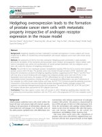
Figure 1
Experimental setup of the in vitro model and histological characterization of the cartilage co-cultured with SFBExperimental setup of the in vitro model and histological characterization of the cartilage co-cultured with SFB. Upper panel: Embedding of cartilage
and subsequent co-culture with synovial fibroblasts (SFB). (a) Hot 2% agarose was filled in each well of a 48-well plate and (b) a cylinder was cre-
ated in the agarose by inserting a metal pin plate and removing the plate after polymerisation. (c-d) Subsequently cartilage disc were embedded in
the preformed cylinder and pre-cultured for two days. (e) The SFB suspension was then applied and (f) left for three hours for settling and initial
adherence of the SFB on the cartilage surface. Finally, (g-h) co-culture medium was carefully added into the upper well compartment. Middle panel:
Experimental setup. Cultures were maintained for two weeks, medium was replaced every two to three days, and supernatants were collected and
subjected to protein analysis. Cultured constructs were either further processed for histological evaluation and quantification of cartilage oligomeric
matrix protein (COMP) content in cartilage or used for gene expression analysis of SFB and cartilage. Lower panel: Histological and immunohisto-
chemical staining of cartilage co-cultures with SFB after 14 days of in vitro culture. (a, b) H&E staining demonstrates the formation of a SFB multi-
layer on the cartilage surface. (c) Immunostaining for prolyl-4-hydroxylase verifies the viability of SFB and chondrocytes and (d) immunostaining for
human Thy-1 proves the fibroblast origin of the co-cultivated cells. Magnification (a) 40×, (b) 630× and (c, d) 400×.
Available online />Page 5 of 20
(page number not for citation purposes)
GmbH, Tübingen, Germany) and by measuring the total area
and the safranin-O positive/negative areas.
For immunohistochemistry, frozen sections were dried over-
night and fixed for 10 minutes either in acetone (anti-prolyl-4-
hydroxylase and anti-Thy-1 monoclonal antibodies) or in 4%
paraformaldhyde in PBS. Endogenous peroxidase activity was
blocked with 0.5% hydrogen peroxide in ethanol. Demasking
of epitopes (cartilage oligomeric matrix protein (COMP) and
COL2-3/4-C (short)) was performed by incubation with chon-
droitinase ABC (Sigma-Aldrich, Deisenhofen, Germany). After
blocking nonspecific binding sites with 10% rabbit or goat
serum (same species as the source of the secondary antibody)
in PBS, sections were incubated for one hour with primary
antibodies against prolyl-4-hydroxylase (Biomeda, Foster City,
CA, USA), Thy-1 (CD90; Dianova, Hamburg, Germany),
COMP (rabbit polyclonal antibody directed against human
and bovine COMP; Kamiya Biomedicals, Seattle, WA, USA)
or the collagen cleavage epitope Col2-3/4C-C (short) (immu-
noreactive with human and bovine epitopes, TECO Medical,
Sissach, Switzerland) and, subsequently, with horseradish
peroxidase (HRP)-conjugated rabbit anti-mouse immunoglob-
ulin (Ig) G/or goat anti-rabbit IgG (Santa Cruz Biotechnology,
Santa Cruz, CA, USA). The peroxidase was revealed using
diaminobenzidine or 3-amino-9-ethylcarbazole (both Sigma-
Aldrich). Slides were counterstained with haematoxylin and
covered with Aquatex (Merck, Darmstadt, Germany). Mouse
IgG
1
/IgG
2a
(DAK-GO1/DAK-GO5; both from Dako, Glostrup,
Denmark) or affinity-purified rabbit IgG (Sigma-Aldrich) served
as isotype controls and always yielded negative results.
Transmission electron microscopy
Cartilage discs were chemically pre-fixed for 24 hours at room
temperature (4% glutaraldehyde; 0.1 M sodium cacodylate
buffer; pH 7.2; Roth, Karlsruhe, Germany), post-fixed for 24
hours (1% osmium tetroxide; 0.1 M sodium cacodylate buffer,
pH 7.4), rinsed three times in isotonic buffer solution (0.1 M
sodium cacodylate buffer, pH 7.2), and finally transferred to
100% acetone by dehydration through a graded series of ace-
tone. Discs were then incubated with 2% uranyl acetate for
one hour, washed with propylene oxide and embedded in
araldite by polymerisation at 60°C. Vertical, semi-thin sections
were cut on a Leica Ultracut E ultramicrotome and stained for
15 minutes in 1% Richardson solution (Hollborn). Subse-
quently, thin sections were cut (about 60 nm thick), mounted
on copper grids, stained for five minutes with a mixture of 80
mM sodium citrate, 40 mM lead nitrate and 40 mM sodium
hydroxide, and examined on a Philips CM 10 transmission
electron microscope (Philips, Hamburg, Germany). Transmis-
sion electron microscopy of cartilage is known to illustrate the
collagen network structure, whereas proteoglycans are col-
lapsed and not visible due to the fixation method.
RNA isolation
The SFB layer was carefully detached from the cartilage disc
by incubating the cartilage/SFB composite for 10 seconds in
75 μl RLT-lysis buffer (RNeasy
®
Micro kit; Qiagen, Hilden,
Germany) containing 15 ng carrier RNA. This procedure com-
pletely removed the SFB from the cartilage surface, but left the
chondrocytes in the cartilage intact as assessed by histologi-
cal analysis (Figure 2) and quantitative PCR (qPCR) using
species-specific primers (data not shown). Total RNA was
then isolated using the RNeasy
®
Micro kit according to the
supplier's instructions including a DNase digestion step.
Following removal of the SFB (in the case of co-culture), the
shock-frozen cartilage was pulverised in a microdismembrator
(Braun, Melsungen, Germany) by milling it for 30 seconds with
an agitated grinding ball in a liquid nitrogen-cooled, stainless
steel vessel (shaking rate of 2000 per minute and amplitude of
16 mm). Subsequently, RNA was extracted by resuspension
of the powder in 400 μl RLT-lysis buffer containing carrier
RNA and centrifugation. After addition of 800 μl RNase-free
water, interfering matrix components were removed by digest-
ing the supernatant for 10 minutes at 55°C with proteinase K
(20 mg/ml; Qiagen). Total RNA was then isolated as above.
Figure 2
Histological analysis of cartilage co-cultures with synovial fibroblasts (SFB) (a) before and (b) after detachment of SFB by short incubation with lysis bufferHistological analysis of cartilage co-cultures with synovial fibroblasts (SFB) (a) before and (b) after detachment of SFB by short incubation with lysis
buffer. SFB were completely removed from the cartilage surface, whereas cartilage matrix and chondrocytes remained intact. H&E staining, magnifi-
cation 200×.
Arthritis Research & Therapy Vol 11 No 1 Pretzel et al.
Page 6 of 20
(page number not for citation purposes)
This method enabled us to isolate intact RNA from small carti-
lage samples (about 5 mg per preparation) with a good yield.
The integrity of the RNA samples was demonstrated by detec-
tion of distinct 28S and 18S rRNA bands without smear by
agarose gelelectrophoresis in selected samples (data not
shown).
Reverse transcription and quantitative PCR
Total RNA eluate (10 μl) was primed with oligo(d)T and
reverse-transcribed for one hour at 42°C using SuperScript-II
reverse transcriptase (Invitrogen).
qPCR reactions were performed as previously described [16]
with cloned standards for the quantitation of human MMP-1,
MMP-3, IL-6, IL-8, and the housekeeping gene aldolase and a
batch preparation of bovine cDNA for the cartilage samples.
qPCR was performed on a mastercycler 'realplex2' (Eppen-
dorf, Hamburg, Germany) with HotMaster Taq (Eppendorf)
and the primer pairs and PCR conditions presented in Table
2. The relative concentrations of cDNA present in each sample
were calculated by the software using the standard curves. In
order to normalise the amount of cDNA in each sample and to
guarantee the comparability of the calculated mRNA expres-
sion in all analysed samples, the housekeeping gene aldolase
was amplified. Product specificity was confirmed by melting
curve analysis and initial cycle sequencing of the PCR prod-
ucts.
Table 2
Primers, product length and specific amplification conditions for qPCR
Gene Primer forward Primer reverse Accession number T annealing Melting T product
Human/bovine
Aldolase A
5'-
TCATCCTCTTCCATGAG
ACACTCTA-3'
5'ATTCTGCTGGCAGAT
ACTGGCATAA-3'
[GenBank:
NM_000034]
58°C 88°C
Human MMP-1 5'-
GACCTGGAGGAAATCT
TGC-3'
5'-
GTTAGCTTACTGTCACA
CGC-3'
[GenBank:
NM_002421
]
58°C 86°C
Human MMP-3 5'-
CTCACAGACCTGACTC
GGTT-3'
5'-
CACGCCTGAAGGAAG
AGATG-3'
[GenBank:
NM_002422
]
58°C 81°C
Human IL-6 5'-
ATGAACTCCTTCTCCAC
AAGCG-3'
5'-
CTCCTTTCTCAGGGCT
GAG-3'
[GenBank:
NM_000600
]
60°C 86°C
Human IL-8 5'-
GCCAAGAGAATATCCG
AACT-3'
5'-
AGGCACAGTGGAACAA
GGACTTGT-3'
[GenBank:
NM_000584
]
60°C 78°C
Bovine MMP-1 5'-
CAAGAGCAGATGTGGA
CCAA-3'
5'-
CTGGTTGAAAAGCATG
AGCA-3'
[GenBank:
NM_174112
]
61°C 83°C
Bovine MMP-3 5'-
CTGGTGTCCAGAAGGT
GGAT-3'
5'-
TAGGCGCCCTTGAAGA
AGTA-3'
[GenBank: AB043995
] 61°C 83°C
Bovine IL-6 5'-
ATGAACTCCCGCTTCA
CAAG-3'
5'-
CCTTGCTGCTTTCACA
CTCA-3'
[GenBank:
NM_173923]
61°C 83°C
Bovine IL-8 5'-
TGCTCTCTGCAGCTCT
GTGT-3'
5'-
CAGACCTCGTTTCCATT
GGT-3'
[GenBank:
NM_173925
]
64°C 81°C
Bovine Col II (α 1
chain)
5'-
CATCTGGTTTGGAGAA
ACCATC-3'
5'-
GCCCAGTTCAGGTCTC
TTAG-3'
[GenBank:
NM_001001135
]
61°C 83°C
Bovine COMP 5'-
ATGCGGACAAGGTGG
TAGAC-3'
5'-
TCTCCATACCCTGGTT
GAGC-3'
[GenBank: X74326
] 61°C 87°C
General amplification protocol (40 cycles): initial denaturation for two minutes at 95°C; denaturation for 15 seconds at 95°C, specific primer
annealing temperature (see table) for 15 seconds, amplification at 68°C for 20 seconds, additional heating step to 5°C below the melting
temperature of the PCR product (see table). General melting curve protocol (one cycle): denaturation for one second at 95°C; cooling to 5°C
above the primer annealing temperature (holding for 10 seconds); heating to 95°C (0.1°C/second); final cooling for five minutes at 40°C.
Col = collagen; COMP = cartilage oligomeric matrix protein; IL = interleukin; MMP = matrix metalloproteinase; qPCR = quantitative polymerase
chain reaction.
Available online />Page 7 of 20
(page number not for citation purposes)
MMP-activity assay
The synthetic peptide Mca-Pro-Leu-Gly-Leu-Dap(Dnp)-Ala-
Arg-NH
2
(Bachem, Heidelberg, Germany) was used to quan-
tify the sum activity of bovine and human MMP in pooled
supernatants (two weeks of culture). This fluorogenic sub-
strate peptide is a very sensitive substrate for the in situ deter-
mination of the MMP activity. Cleavage at the Gly-Leu bond
separates the highly fluorescent Mca group from the efficient
2,4-dinitrophenyl quencher, resulting in an increase of fluores-
cence intensity. The substrate peptide can be cleaved by
numerous MMP, with MMP-2, MMP-9 and, to a lesser extent,
MMP-1, MMP-3 and MMP-13 showing the highest rates of
turnover [17]. To estimate the potential total activity of latent
and active MMP, latent MMP were activated by incubation
with 2 mM aminophenylmercuric acetate (APMA; Sigma-
Aldrich); without APMA activation, none of the samples
showed any MMP activity.
For the assay, 10 μl culture-supernatant were incubated for
two hours at 37°C with 20 μl of 25 μM MCA-Pro-Leu-Gly-Leu-
DAP(DNP)-Ala-Arg-NH
2
in 70 μl incubation buffer (100 mM
Tris/HCl, 30 mM calcium chloride, 1 μM zinc chloride
,
2 mM
APMA, 0.05% Brij, pH 7.6) and the increase of the fluores-
cence intensity was measured at 390 nm. Fresh, co-culture
medium containing FCS was analysed as an internal control
for MMP activity. Although the values in the medium control
were only marginally higher than those in the buffer control, the
values in the co-culture medium were nevertheless subtracted
from the values in the experimental samples in order to correct
for background MMP activity.
Casein zymography
Caseinolytic activity in pooled supernatants was assayed by
electrophoresis in polyacrylamide gels containing sodium
dodecyl sulfate (SDS) and casein (Sigma-Aldrich) using a
batch of HT1080-conditioned media as a standard [18]. Fresh
co-culture medium served as an internal control for the casei-
nolytic activity derived from the supplemented FCS. The MMP
suggested on the basis of their known caseinolytic activity and
the molecular weight of their latent and active forms were then
identified by western blot analysis of the same pooled super-
natants.
Western Blot for bovine/human MMP-1 and MMP-3
Pooled culture supernatants (20 μl) were resolved by native
SDS-PAGE. MMP-1 was detected by immunoblotting using a
primary antibody against active/latent MMP-1 (MAB901,
R&DSystems, Wiesbaden, Germany) and goat-anti-mouse
IgG HRP as a secondary antibody (Sigma-Aldrich). The blots
were stripped and re-probed with primary antibody against
active/latent MMP-3 (MAB 513, R&DSystems).
Enzyme-linked immunosorbent assay
In the supernatants of cartilage cultures with SFB, levels of
SFB-derived active/latent MMP-1 were measured using a
mouse-anti-human MMP-1 monoclonal antibody (MAB901,
R&DSystems) as a capture antibody (1 μg/ml), biotinylated
goat-anti-human MMP-1 (BAF901, R&DSystems) as a detec-
tor antibody (200 ng/ml) and recombinant human MMP-1
(901-MP-010, R&D Systems) as a standard (39 to 5000 pg/
ml). SFB-derived active/latent MMP-3 levels were determined
using the anti-human MMP-3 Total Duo Set (R&D Systems),
and the levels of SFB-derived IL-6 and IL-8 were analysed
using anti-human OptEIA-ELISA Sets (BD Biosciences).
Combined aggrecanase I/II activity (reflecting both SFB-
derived human and cartilage-derived bovine activity) in the
supernatants of cartilage cultures with/without SFB was
measured according to the manufacturer's instructions using
a commercially available ELISA-Kit (Invitek, Berlin, Germany).
For all enzyme-linked immunosorbent assay (ELISA) measure-
ments, fresh co-culture medium was also analysed for the con-
tent of the corresponding molecule in the supplemented
serum. Although the values in the medium control were only
marginally higher than those in the buffer control, the values in
the co-culture medium were nevertheless subtracted from the
values in the experimental samples.
Extraction and quantification of COMP from bovine
cartilage
COMP was isolated from cartilage according to the method of
DiCesare et al. [19] with minor modifications. Briefly, 20 mg of
shock-frozen cartilage from monoculture/co-culture with SFB
was pulverised according to the procedure described above
for RNA isolation; in the case of samples from co-culture
experiments, a step with lysis of the SFB layer and subsequent
PBS washing of the remaining cartilage was included. The pul-
ver was transferred to a tube with 500 μl ice-cold neutral salt
buffer (10 mM Tris/hydrochloric acid, 0.15 M sodium chloride,
pH 7.4, containing 1 mM phenylmethylsulfonyl fluoride, 0.025
mg/ml leupeptin, 0.025 mg/ml aprotinin and 0.025 mg/ml
pepstatin). After centrifugation, the supernatant was decanted
and the tissue was re-suspended in the same buffer. The
extraction procedure was completed by two cycles of centrif-
ugation and addition of neutral salt buffer containing 10 mM
EDTA. Aliquots (10 μl) of all extracts were analysed by non-
reducing and reducing SDS/PAGE. Western blots were
developed using a polyclonal rabbit antibody against COMP
(same antibody as used for immunohistochemistry) and an
HRP-conjugated anti-(rabbit IgG) as a secondary antibody.
The major proportion of COMP (pentameric, oligomeric and
monomeric, as well as degraded COMP) was enriched in the
first neutral salt buffer extract, although only small amounts
were detected in the second neutral salt buffer extract and the
subsequent two extracts with EDTA-containing buffer (data
not shown). The content of cartilage-derived COMP was ana-
lysed in pooled extracts using a bovine-specific ELISA-Kit
(Anamar Medical, Gothenburg Sweden) according to manu-
facturer's instructions. The polyclonal antibody used in this
assay detected the same COMP species as the polyclonal
Arthritis Research & Therapy Vol 11 No 1 Pretzel et al.
Page 8 of 20
(page number not for citation purposes)
antibody employed for western blots (personal communica-
tion; Anders Sjödin, Anamar Medical) and, therefore, the
results represent the overall COMP content in the cartilage
matrix expressed as units/mg cartilage.
Statistics
Analyses were performed using the Mann-Whitney U test and
the statistical software SPSS/Win version 10.0 (SPSS, Chi-
cago, USA); differences with p ≤ 0.05 were considered to be
statistically significant.
Results
Proteoglycan release from cartilage
Strong safranin O staining was observed in sections of freshly
isolated cartilage or in non-stimulated cartilage monocultures,
demonstrating minimal loss of proteoglycan after two weeks of
in vitro culture (about 1%; Figure 3a, left panel).
TNF-α stimulated cartilage was characterised by a slight, but
significant proteoglycan loss (10%; Figure 3a, left panel)
exclusively at the cartilage surface. This was significantly
enhanced in IL-1β stimulated samples, in which a drastic pro-
teoglycan release (50%) occurred in the upper half of the car-
tilage matrix. In TNF-α/IL-1β stimulated cartilage the
Figure 3
Analysis of proteoglycan loss from cartilage monocultures and co-cultures with SFBAnalysis of proteoglycan loss from cartilage monocultures and co-cultures with SFB. Cartilage monocultures (n = 5, with four replicates each) and
co-cultures with SFB (n = 5 patients with four replicates for each patient) with or without stimulation with TNF-α, IL-1β or TNF-α/IL-1β (14 days), as
detected by safranin-O staining. (a) The upper panel shows representative histological samples, in which red colour indicates the presence and
green colour the absence of proteoglycans in the cartilage matrix. Fresh, non-cultured cartilage serves as a positive control. The lower chart depicts
the results of quantitative image analysis of the stained sections. (b) Aggrecanase I/II activity in culture supernatants of cartilage monocultures and
co-cultures with SFB (n = 5 with four replicates for each patient). Mean ± standard error of the mean (SEM) are plotted. § p ≤ 0.05 Mann-Whitney
U Test compared with non-stimulated control; * p ≤ 0.05 Mann-Whitney U Test compared with stimulation with TNF-α; # p ≤ 0.05 Mann-Whitney U
Test compared with stimulation with IL-1β; + p ≤ 0.05 Mann-Whitney U Test compared with cartilage-monoculture.
Available online />Page 9 of 20
(page number not for citation purposes)
proteoglycan loss was higher than the sum of the individual
effects (80%; p ≤ 0.05 versus TNF-α), indicating a synergistic
effect of the two cytokines.
In comparison to cartilage monocultures, strikingly, non-stimu-
lated cartilage co-cultures with RA-SFB showed a significantly
stronger depletion of proteoglycan from the cartilage matrix
(15% versus 1%; Figure 3a, right panel). As in the case of
monocultures, also the proteoglycan depletion in co-cultures
was augmented by stimulation with TNF-α and further
enhanced by IL-1β or the combination of TNF-α/IL-1β (both p
≤ 0.05 versus TNF-α; Figure 3a, right panel).
A considerable contribution of the SFB, whether direct or indi-
rect, was demonstrated by the fact that all co-cultures showed
a significantly higher proteoglycan depletion than the respec-
tive monocultures (Figure 3a, compare left and right panel).
Aggrecanase activity in the supernatant
There was no aggrecanase activity in non-stimulated cartilage
monocultures. Stimulation with TNF-α, IL-1β or the combina-
tion of TNF-α/IL-1β led to a similar, significant induction of
aggrecanase activity (0.21 to 0.36 nM/15 minutes; Figure 3b,
left panel).
As in the case of monocultures, there was no aggrecanase
activity in non-stimulated cartilage co-cultures. Again, stimula-
tion with TNF-α and, in particular, IL-1β led to a significant
induction of aggrecanase activity (0.52 and 0.82 nM/15 min-
utes, respectively; Figure 3b, right panel). The aggrecanase
activity in the supernatants of double-stimulated co-cultures
was significantly higher compared with that after stimulation
with either TNF-α or IL-1β.
Interestingly, the aggrecanase activity in cartilage co-culture
with SFB was either numerically (for TNF-α) or significantly
higher (for IL-1β and TNF-α/IL-1β) than in the corresponding
monoculture (Figure 3b, compare left and right panel), again
pointing to a contribution of SFB.
COMP detection in cartilage
COMP was barely detectable in fresh, non-cultured cartilage
and undetectable in non-stimulated cartilage monocultures. In
contrast, faint COMP staining throughout the whole matrix
was observed in TNF-α and, in particular, in IL-1β or TNF-α/IL-
1β stimulated cartilage monocultures (Figures 4a and c1 to
c4).
In contrast, already non-stimulated co-cultures with SFB
showed a noticeable staining in the cartilage matrix and SFB
(visually even stronger than in cytokine-stimulated monocul-
tures). This staining was further increased by stimulation with
TNF-α or, in a more pronounced fashion, with IL-1β and TNF-
α/IL-1β (Figures 4d1 to d4). Interestingly, fresh human OA
cartilage with its known loss of matrix integrity also exhibited a
considerable COMP staining (Figure 4b).
Detection of collagen cleavage
In fresh, non-cultured cartilage or non-stimulated cartilage
monocultures, no staining for cleaved collagen was observed.
In contrast, stimulation with TNF-α and IL-1β led to a clear
appearance of the collagen cleavage epitope in the extracellu-
lar matrix. Collagen cleavage was even more pronounced in
TNF-α/IL-1β stimulated cartilage samples (Figures 4e and g1
to g4).
Interestingly, collagen cleavage was already observed in non-
stimulated cartilage co-cultured with SFB, indicating the
capacity of non-stimulated SFB to degrade cartilage collagen
(Figure 4h1). The staining intensity for the collagen cleavage
epitope was further increased after stimulation with TNF-α
and, in particular, with IL-1β or TNF-α/IL-1β (Figures 4h2 to
h4). Fresh human OA cartilage also exhibited a considerable
degree of collagen cleavage (Figure 4f).
Morphological destruction of the cartilage matrix
Transmission electron microscopy showed an organized colla-
gen network with sharp and distinct collagen fibers (rich in
contrast) in freshly isolated cartilage or in non-stimulated
monocultures (Figure 4i to j1). In contrast, TNF-α, and espe-
cially IL-1β or TNF-α/IL-1β stimulated monocultures, showed
a clear loss of fibril structure, distinguishable as a decreased
contrast of the collagen fibrils (Figure 4j2 to j4).
Even more pronounced destruction was observed in co-cul-
tures with SFB (both non-stimulated and stimulated with TNF-
α, IL-1β or TNF-α/IL-1β), in all cases showing a massively
reduced optical contrast of the collagen structures in areas
near the cartilage surface (Figure 4k1 to k4).
Invasion of synovial fibroblasts into the cartilage
Using light microscopy, an invasive behaviour of co-cultured
SFB was not observed in any samples after two weeks (not
shown). In contrast, after co-culture for six weeks an initial inva-
sion of SFB into superficial cartilage areas was observed in
TNF-α/IL-1β co-stimulated samples (Figure 5b), but not in the
case of non-stimulated samples (Figure 5a) and samples stim-
ulated with TNF-α or IL-1β alone (data not shown).
The attachment of SFB to the cartilage surface and the ero-
sion of cartilage matrix was also analyzed by laser scanning
microscopy using SFB fluorescence-labelled before co-cul-
ture. Although the SFB layer on top of the cartilage only shows
the fluorescence signal of labelled SFB (Figure 6a) and deep
cartilage regions only exhibit the reflection signal of the unla-
belled cartilage (Figure 6c), the signal in the superficial carti-
lage consists of both components and therefore indicates an
initial invasion of labelled SFB into the cartilage matrix (Figure
6b). This effect was already present in non-stimulated co-cul-
Arthritis Research & Therapy Vol 11 No 1 Pretzel et al.
Page 10 of 20
(page number not for citation purposes)
Figure 4
Immunohistochemical staining and electron microscopyImmunohistochemical staining and electron microscopy. (a, e, i) Fresh, non-cultured bovine cartilage and (b, f) human osteoarthritis (OA) cartilage,
as well as (c1 to c4, g1 to g4, j1 to j4) bovine cartilage from monocultures or (d1 to d4, h1 to h4, k1 to k4) co-cultures with synovial fibroblast (SFB)
after 14 days are shown. Immunostaining for cartilage oligomeric matrix protein (COMP) clearly reveals a (c1 to c4) strong correlation between the
appearance/detection of COMP within the cartilage matrix and the stimulation with TNF-α, IL-1β and TNF-α/IL-1β, (d1 to d4) which is dramatically
augmented by the co-culture with SFB. (a) Fresh, non-cultured bovine cartilage and (c1) non-stimulated cartilage monocultures do not stain for
COMP; in contrast, (b) human OA cartilage shows a positive staining for COMP. (g1 to h4) Immunostaining for the collagen cleavage neo-epitope
Col2-3/4C-(short) demonstrates the matrix-degrading capacity of SFB and the amplifying impact of TNF-α, IL-1β and TNF-α/IL-1β on this process.
(e) Whereas fresh, non-cultured bovine cartilage lacks signs of collagen cleavage, (f) human OA cartilage exhibits positive staining for the
neoepitope. (i to k4) Transmission electron microscopy confirmed the immunohistologically detected collagen breakdown by a decreased optical
density of collagen fibres (the dotted line indicates the cartilage surface or the interface between the cartilage and the co-cultured SFB). Magnifica-
tions in (a to h4) 200×; inserts 630×; (i to k4) 39,000×.
Available online />Page 11 of 20
(page number not for citation purposes)
tures and not enhanced by cytokine stimulation (data not
shown).
mRNA synthesis and protein content of COMP in
cartilage
Stimulation of cartilage monocultures with IL-1β or with TNF-
α/IL-1β, but not with TNF-α, significantly reduced the mRNA
for COMP compared with the respective non-stimulated con-
trol (13- and 10-fold, respectively). Interestingly, the co-culture
with SFB had no further influence on the mRNA expression of
COMP in cartilage (Figure 7; upper panel).
The reduction of COMP mRNA could be confirmed at the pro-
tein level in cartilage monocultures, in which a significant
decrease of COMP was detected after IL-1β and TNF-α/IL-1β
stimulation (both about three-fold compared with non-stimu-
lated cartilage). Strikingly, a significantly reduced COMP con-
tent was observed in all cartilage co-cultures with SFB as
compared with the respective monocultures (about three- to
nine-fold reduction; Figure 7; lower panel). This further under-
lines the ability of the co-cultured SFB to fundamentally disturb
the cartilage matrix homeostasis. In this case, additional
cytokine stimulation seems to play a minor role.
Figure 5
Invasion of cartilage by synovial fibroblasts (SFB) after six weeks of co-culture (Light microscopy)Invasion of cartilage by synovial fibroblasts (SFB) after six weeks of co-culture (Light microscopy). An initial invasion of SFB into superficial cartilage
areas was observed in (b) TNF-α/IL-1β co-stimulated samples, but not in the case of (a) non-stimulated samples and samples stimulated with TNF-
α or IL-1β alone (data not shown). Magnifications in (a) and (b) 400×; inserts 100×.
Figure 6
Invasion of cartilage by synovial fibroblasts (SFB) after two weeks of co-culture (Laser scanning microscopy)Invasion of cartilage by synovial fibroblasts (SFB) after two weeks of co-culture (Laser scanning microscopy). Erosion of cartilage matrix by fluores-
cence-labelled SFB was examined in an aqueous setting. Micrographs represent the view from above on the co-culture of cartilage with SFB in dif-
ferent sectional planes (indicated by red arrows) and the corresponding cross-sections. (a) plane within the SFB layer on top of the cartilage, (b)
plane within superficial cartilage and (c) plane within deep cartilage zone. Magnifications in 200×.
Arthritis Research & Therapy Vol 11 No 1 Pretzel et al.
Page 12 of 20
(page number not for citation purposes)
Analysis of collagen synthesis
Stimulation of cartilage monocultures with IL-1β (or TNF-α/IL-
1β), but not with TNF-α, significantly reduced the mRNA for
the α
1
chain of collagen II (Figure 8). This was also observed
in cartilage co-cultures with SFB, in which only IL-1β signifi-
cantly reduced collagen II expression compared with the non-
stimulated co-culture (Figure 8). Notably, both TNF-α and IL-
1β stimulated cartilage co-cultures revealed a significantly
lower collagen II expression in comparison to the respective
monoculture (Figure 8). This indicates that SFB disturb the
cartilage homeostasis by both degrading cartilage and sup-
pressing the neosynthesis of collagen II.
Matrix-metalloproteinase activity
Following activation of latent bovine and human MMP by incu-
bation with APMA, the MMP activity in both cartilage monoc-
ultures and co-cultures was significantly increased by
stimulation with TNF-α, IL-1β or TNF-α/IL-1β (Figure 9). In
addition, all co-cultures with SFB showed a significantly higher
MMP activity than the respective monocultures (Figure 9),
demonstrating a major contribution of the co-cultured SFB to
the secretion of matrix-degrading MMP.
Caseinolytic activity
In TNF-α, IL-1β or TNF-α/IL-1β stimulated, but not in non-stim-
ulated, monocultures or co-cultures with SFB, protease bands
with caseinolytic activity were detected at a molecular weight
of about 45 kD (presumably containing the active forms of
MMP-1 and/or MMP-3; Figure 10, lower panel). In TNF-α, IL-
1β or TNF-α/IL-1β stimulated co-cultures with SFB, interest-
ingly, additional bands were observed at a molecular weight of
about 57 kD, possibly representing the latent form of MMP-1
and/or MMP-3. This was confirmed by immunological detec-
tion of both MMP-1 (Figure 10, upper panel) and MMP-3 (Fig-
ure 10, middle panel). Successful inhibition of the caseinolytic
activity in zymography by EDTA and lack of inhibition by the
serine protease inhibitor phenylmethylsulfonyl fluoride further
confirmed the MMP character of the bands (data not shown).
Figure 7
mRNA expression (upper panel) and protein content (lower panel) of bovine cartilage oligomeric matrix protein (COMP)mRNA expression (upper panel) and protein content (lower panel) of bovine cartilage oligomeric matrix protein (COMP). Cartilage from monocul-
tures (n = 5, with two replicates each) and co-cultures with synovial fibroblasts (SFB) (n = 5, with two replicates each) with/without stimulation with
TNF-α, IL-1β or TNF-α/IL-1β (14 days) are shown. Gene expression values (means ± standard error of the mean (SEM)), as determined by quantiti-
ative PCR, are expressed as percentage of the values in non-stimulated cartilage monocultures (100%), protein values (means ± SEM) are
expressed as units/mg cartilage. § p ≤ 0.05 Mann-Whitney U Test compared with non-stimulated control; + p ≤ 0.05 Mann-Whitney U Test com-
pared with cartilage monoculture.
Available online />Page 13 of 20
(page number not for citation purposes)
In comparison with the respective non-stimulated cultures, the
caseinolytic activity in both monocultures and co-cultures was
significantly increased by stimulation with TNF-α, IL-1β or
TNF-α/IL-1β (Figure 10, lower panel). As in the case of MMP
activity, all stimulated co-cultures with SFB showed a signifi-
cantly higher caseinolytic activity than the respective monocul-
tures (Figure 10, lower panel), further underlining the
contribution of the co-cultured SFB.
Expression of matrix-metalloproteinases
On the basis of the MMP detected by casein zymography and
western blot, the gene expression of MMP-1 and -3 was ana-
lysed separately in cartilage derived from monocultures or co-
cultures and in SFB obtained from co-cultures. In cartilage
monocultures, the level of bovine MMP-1 mRNA was signifi-
cantly increased by TNF-α stimulation (3.6-fold; Figure 11a),
that of bovine MMP-3 mRNA was numerically increased (1.9-
fold; Figures 11b). This effect was significantly more pro-
nounced after IL-1β stimulation (36- and 35-fold) or TNF-α/IL-
1β stimulation (53- and 58-fold; Figures 11a, b). Notably, there
were no significant differences for the gene expression of
bovine MMP-1 and MMP-3 between cartilage derived from
monocultures or co-cultures (data not shown).
In SFB co-cultured with cartilage, the level of human MMP-1
and MMP-3 mRNA was significantly increased by TNF-α, IL-
1β or TNF-α/IL-1β stimulation (12-, 13- and 21-fold, respec-
tively, for MMP-1; 49-, 69- and 78-fold for MMP-3; Figures
Figure 8
mRNA expression of bovine collagen type II (α1 chain)mRNA expression of bovine collagen type II (α1 chain). Cartilage from monocultures (n = 5, with two replicates each) and co-cultures with SFB (n =
5, with two replicates each) with/without stimulation with TNF-α, IL-1β or TNF-α/IL-1β (14 days) were used. Gene expression values (means ±
standard error of the mean), as determined by qPCR, are expressed as percent of the values in non-stimulated cartilage monocultures (100%). § p
≤ 0.05 Mann-Whitney U Test compared with non-stimulated control; * p ≤ 0.05 Mann-Whitney U Test compared with stimulation with TNF-α; + p ≤
0.05 Mann-Whitney U Test compared with cartilage monoculture.
Figure 9
Bovine/human MMP-activityBovine/human MMP-activity. Supernatants of cartilage monocultures (n = 5, with six replicates each) and co-cultures with synovial fibroblasts (SFB)
(n = 5, with six replicates each) with/without stimulation with TNF-α, IL-1β or TNF-α/IL-1β (14 days) were used. Means +/- standard error of the
mean are plotted. § p ≤ 0.05 Mann-Whitney U Test compared with non-stimulated control; * p ≤ 0.05 Mann-Whitney U Test compared with stimula-
tion with TNF-α; + p ≤ 0.05 Mann-Whitney U Test compared with cartilage monoculture.
Arthritis Research & Therapy Vol 11 No 1 Pretzel et al.
Page 14 of 20
(page number not for citation purposes)
11c, d). These results were confirmed at the protein level (by
ELISA); the co-cultured SFB secreted significantly more
MMP-1 and MMP-3 after stimulation with TNF-α, IL-1β or TNF-
α/IL-1β (9-, 6- and 10-fold, respectively, for MMP-1; 11-, 21-
and 24-fold for MMP-3; Figures 11e, f).
Expression of pro-inflammatory cytokines
The influence of the pro-inflammatory cytokines TNF-α and IL-
1β on the gene expression of IL-6 and IL-8 was also assessed
in cartilage derived from monocultures or co-cultures and in
SFB obtained from co-cultures.
In cartilage monocultures, the levels of bovine IL-6 and IL-8
mRNA were exclusively augmented in a significant fashion by
IL-1β or TNF-α/IL-1β stimulation (170- and 510-fold for IL-6;
83- and 189-fold for IL-6; Figures 12a, b). As described above
for bovine MMP-1 and MMP-3, there were no significant differ-
ences for the gene expression of bovine IL-6 and IL-8 between
cartilage derived from monocultures or co-cultures (data not
shown).
In SFB co-cultured with cartilage, the levels of human IL-6 and
IL-8 mRNA were significantly increased by stimulation with
TNF-α (14- and 500-fold, respectively), IL-1β (38- and 958-
fold, respectively) or TNF-α/IL-1β (25- and 1712-fold, respec-
tively; Figures 12c, d). These results were again confirmed at
the protein level; the co-cultured SFB secreted significantly
more IL-6 and IL-8 after stimulation with TNF-α (10- and 14-
fold, respectively), IL-1β (28- and 19-fold, respectively) or
TNF-α/IL-1β (45- and 37-fold, respectively; Figures 12e, f).
Discussion
Suitability of the new model
Based on the experimental results described above, our
newly-developed in vitro destruction model offers several new
features in comparison with published in vitro models, in that
the model uses initially intact cartilage matrix and co-cultured,
early-passage SFB with properties close to their in vivo fea-
tures. In contrast, previous models of cartilage destruction
mostly worked with either artificial, in vitro generated, dam-
aged or devitalised cartilage matrices and were therefore not
suitable for examining the process of cartilage destruction in
'healthy' intact cartilage [9-12,20-22]. The mature bovine
joints employed in the present study turned out to be a suitable
cartilage source, because they are regularly available and are
able to harvest up to 80 cartilage discs per joint with standard-
ised, highly homogenous quality. These discs show a com-
pletely intact cartilage matrix and surface (no superficial
Figure 10
Caseinolytic activityCaseinolytic activity. Supernatants of cartilage monocultures (n = 5, with two replicates each) and co-cultures with synovial fibroblasts (SFB) (n = 5,
with two replicates each) with/without stimulation with TNF-α, IL-1β or TNF-α/IL-1β (14 days) were used. In order to assess the total caseinolytic
activity (lower panel), the bands for both the active and the latent forms were used for quantification. Means +/- standard error of the mean are plot-
ted. § p ≤ 0.05 Mann-Whitney U Test compared with non-stimulated control; + p ≤ 0.05 Mann-Whitney U Test compared with cartilage monocul-
ture. Parallel analysis of the supernatants by western blot revealed that bovine/human matrix metalloproteases (MMP) 1 and MMP-3 (upper and
middle panel) are responsible for the caseinolytic enzyme activity in culture supernatants.
Available online />Page 15 of 20
(page number not for citation purposes)
Figure 11
Expression of bovine MMP-1 and MMP-3 mRNAExpression of bovine MMP-1 and MMP-3 mRNA. Cartilage from (a, b) monoculture (n = 5, with two replicates each) and (c, d) human mRNA/protein
in synovial fibroblasts (SFB) after co-culture with cartilage (n = 5, with two replicates each) with/without stimulation with TNF-α, IL-1β or TNF-α/IL-
1β (14 days) were used.gene expression values (means ± standard error of the mean (SEM)), as determined by quantitiative PCR, are expressed as
percentage of the values in non-stimulated samples (100%). In addition, (e, f) the values for human MMP-1 and MMP-3 protein secreted by SFB into
the supernatant of co-cultures are shown. The protein levels, as measured in the supernatant by ELISA, are expressed as means +/- SEM. § p ≤
0.05 Mann-Whitney U Test compared with non-stimulated control; * p ≤ 0.05 Mann-Whitney U Test compared with stimulation with TNF-α.
Arthritis Research & Therapy Vol 11 No 1 Pretzel et al.
Page 16 of 20
(page number not for citation purposes)
Figure 12
Expression of bovine IL-6 and IL-8 mRNAExpression of bovine IL-6 and IL-8 mRNA. Cartilage from (a, b) monoculture (n = 5, with two replicates each) and (c, d) human mRNA/protein in syn-
ovial fibroblasts (SFB) (n = 5, with two replicates each) after co-culture with cartilage with/without stimulation with TNF-α, IL-1β or TNF-α/IL-1β (14
days) were used.gene expression values (means ± standard error of the mean (SEM)), as determined by quantitiative PCR, are expressed as per-
centage of the values in non-stimulated samples (100%). In addition, (e, f) the values for human IL-6 and IL-8 protein secreted by SFB into the
supernatant of co-cultures are shown. The protein levels, as measured in the supernatant by ELISA, are expressed as means ± SEM. § p ≤ 0.05
Mann-Whitney U Test compared with non-stimulated control; * p ≤ 0.05 Mann-Whitney U Test compared with stimulation with TNF-α; # p ≤ 0.05
Mann-Whitney U Test compared with stimulation with IL-1β.
Available online />Page 17 of 20
(page number not for citation purposes)
fissures or other OA abnormalities) without primary loss of pro-
teoglycan, both prerequisites for the unequivocal determina-
tion of early cartilage alterations. These features are difficult to
achieve with human samples because normal human cartilage
is usually not available and cartilage from OA or RA patients
normally shows drastic signs of matrix destruction/alteration,
which often extend from the surface throughout the cartilage
matrix. Therefore, models using shaved or cut cartilage sur-
faces or cartilage with a damaged surface and exposed fibrillar
matrix may investigate the progression of pre-existing erosions
rather than initial damage [11,13].
The only possible disadvantage of the present system, that is
the use of a xenogenic bovine cartilage matrix, may be of minor
importance since important matrix molecules (for example col-
lagen II and aggrecan) share a high sequence homology as a
result of the close evolutionary relationship between bovine
and human; and since human cytokines/chemokines and pro-
teases can also act on bovine chondrocytes or proteins and
vice versa (see below; [23]). In addition, limited access to nor-
mal human cartilage may allow the validation of selected data
in this newly-established in vitro cartilage model [24].
Regarding the characteristics of the co-cultured SFB, previ-
ous studies have worked with: complete synovial tissue or het-
erogeneous cell-mixtures (comprised of macrophages, B-
cells, T-cells and SFB) [11,13], fibroblasts cell lines or their
conditioned supernatants [20,25], and either non-purified,
early-passage SFB (possibly still contaminated with macro-
phages) [9] or late-passage SFB (fourth and higher) [10],
known to differ largely from early-passage SFB (first to fourth)
[15,26]. The present model, in contrast, uses purified, early-
passage SFB with a phenotype similar to their in vivo status in
the synovial membrane [15], allowing the exact assignment of
the observed effects to SFB. Taken together, the present sys-
tem allows for the first time to simulate the initial cartilage
destruction in rheumatoid joints mediated by aggressive SFB.
A partial/complete reconstitution of the mixture of inflamma-
tory and mesenchymal cells in the synovial tissue is also pos-
sible by adding some or all of the adherent and non-adherent
cells obtained during the isolation procedure of SFB [15].
Destructive processes in the cartilage are induced by co-
culture with synovial fibroblasts and are further
potentiated by cytokine stimulation
This study shows that non-stimulated RA SFB are capable of
rapidly degrading intact undamaged cartilage by inducing a
loss of matrix proteoglycan and a cleavage of collagen. The
degree of SFB-mediated matrix degradation was further
enhanced by stimulation with TNF-α and, in particular, IL-1β or
even more pronounced with the combination of TNF-α and IL-
1β. Matrix-degrading proteases (MMP and aggrecanases) and
pro-inflammatory cytokines (IL-6 and IL-8) in SFB and cartilage
were identified as potential direct or indirect mediators for this
cartilage destruction. In addition, a suppression of collagen
synthesis in chondrocytes by stimulated SFB appears to con-
tribute to the breakdown of cartilage homeostasis.
Synovial fibroblasts promote cartilage destruction by
degradation of extracellular matrix and suppression of
matrix synthesis
Proteoglycan loss
The limited/absent proteoglycan loss from the cartilage in non-
stimulated cartilage monocultures shows that the basic in vitro
conditions preserve/stabilise the normal cartilage metabolism
and render this monoculture a suitable control for cytokine-
stimulated monocultures and the respective co-cultures with
SFB.
Strong induction of proteoglycan loss in cytokine-stimulated
cartilage monocultures points to a major contribution of
chondrocytes to cytokine-induced matrix degradation. IL-1β
was a stronger inductor of proteoglycan loss than TNF-α, con-
firming previous results [27-29]. The combination of both
cytokines amplified the effect of the respective cytokines
alone. This is of particular interest, because the inflamed joint
(synovium) is characterised in vivo by the concomitant appear-
ance of these pro-inflammatory factors.
Significant enhancement of the proteoglycan loss in non-stim-
ulated or stimulated co-cultures with SFB demonstrates an
important role for SFB, whether directly or indirectly via stimu-
lation of chondrocytes. Interestingly, there was a gradient of
proteoglycan loss from the cartilage surface to deeper matrix
zones, most likely because of the specific zonal properties of
cartilage concerning their proteoglycan content [30] or the dif-
ferential zonal inhibition of matrix/aggrecan neosynthesis by IL-
1 (α/β) (data not shown [31]).
COMP
COMP was barely or not at all detectable in fresh, non-cul-
tured cartilage or non-stimulated cartilage monocultures, sug-
gesting that COMP, although clearly measurable [32], is
masked by other matrix molecules in normal cartilage. Similar
results were obtained in fetal human cartilage, which also
showed weak pericellular immunoreactivity for COMP [33]. In
contrast, COMP staining throughout the whole matrix was
observed in TNF-α or IL-1β stimulated monocultures and, in
particular, in non-stimulated or stimulated co-cultures with
SFB. Increased COMP detection could therefore either reflect
a futile, regenerative attempt of chondrocytes [34] or an
enhanced demasking/degradation of matrix-bound COMP,
although an exclusive connection with the loss of proteogly-
cans is unlikely because of the incongruent histological results
for the two molecules (present study; [34]). Steady-state anal-
ysis of the cartilage from monocultures and co-cultures after
two weeks showed a substantial reduction of COMP mRNA
and protein, at least excluding a successful net reconstitution
of COMP. Although the reduction of COMP mRNA was not
influenced by co-culture with SFB, the loss of immunoreactive
Arthritis Research & Therapy Vol 11 No 1 Pretzel et al.
Page 18 of 20
(page number not for citation purposes)
COMP protein from the cartilage matrix was strongly aug-
mented by the SFB, further underlining the contribution of SFB
to the disruption of the cartilage matrix homeostasis. There-
fore, the COMP appearance in histological sections seems to
be a consequence of the initial cartilage destruction and sub-
sequent demasking of this cartilage component. Independent
of the precise molecular mechanism, the present study dem-
onstrates for the first time that co-culture of cartilage with RA
SFB induces an increased appearance of matrix-bound
COMP.
Collagen breakdown
Although the above described loss of protective proteogly-
cans [35] may lead to an enhanced accessibility and cleavage
of collagen fibres, the primary collagen cleavage site was not
detectable in the supernatant of any experimental group (data
not shown). This is most probably based on the fact that
cleaved collagen initially remains in the cartilage matrix unless
further matrix disaggregation or secondary collagen cleavage
occurs [28,36,37]. Indeed, the primary collagen cleavage site
was detected immunohistochemically in stimulated (but not
non-stimulated) cartilage monocultures, indicating a strong
involvement of chondrocytes in the degradation process under
pathological conditions. On the other hand, the increased
staining intensity in cartilage samples co-cultured with SFB
shows that the collagen breakdown was further augmented by
SFB, for example via secreted soluble proteinases and/or
cytokines.
Electron microscopy confirmed the immunohistochemical
results and revealed that severe damage of the collagen net-
work appeared exclusively in stimulated monocultures, as well
as in all co-cultures with SFB. Strikingly, all co-cultures
showed higher amount of damage than the respective mono-
cultures.
Collagen synthesis
In view of an amplification of the potential net loss of collagen
from the cartilage matrix, the synthesis of collagen type II
(mRNA) was also significantly suppressed in TNF-α and IL-1β
stimulated co-cultures. To our knowledge, these are the first
data demonstrating a suppressive effect of SFB on collagen
type II gene expression in chondrocytes.
Synovial fibroblasts produce or induce the mediators to
destroy cartilage extracellular matrix
Aggrecanase activity
The absence of aggrecanase activity (the enzyme predomi-
nantly responsible for proteoglycan degradation/depletion
[35,38]) in non-stimulated monocultures and co-cultures is
consistent with previously reported data [29]. TNF-α and IL-1β
are potent inductors of aggrecanase activity in both cases,
underlining the key role of these cytokines in proteoglycan
depletion. On the other hand, the clearly increased aggrecan
activity in co-cultures as compared with monocultures sug-
gests an impact of activated SFB. However, this effect
appears to be mediated by the induction of aggrecanase
expression in chondrocytes rather than by increased aggreca-
nase expression in SFB, as indicated by qPCR experiments
(data not shown). Alternatively, an activation of matrix-bound
aggrecanases by SFB-derived MMP could contribute to the
augmented aggrecanase activity in co-culture [39].
MMP activity
The present results show a slight, but significant induction of
MMP-activity in cartilage monocultures after cytokine treat-
ment, which is further enhanced in co-culture samples with
non-stimulated or stimulated SFB. Although the MMP sub-
strate employed in this study can be cleaved by all known
MMP, MMP-2 and MMP-9 have the highest rate of turnover for
the substrate peptide [17,40], in agreement with their clear
detection in the supernatant of all samples by gelatine zymog-
raphy (data not shown). This may be of pathogenic relevance
in RA, because MMP-2 and MMP-9, among other MMP, can
further degrade cleaved collagen and thereby support its
release from cartilage [41].
MMP-1 and MMP-3 expression/activity
Casein zymography, western blots, qPCR and ELISA results
indicated that MMP-1 and MMP-3 are detectable at the
mRNA, protein and/or activity level (the latter only in stimulated
samples) in both cartilage and SFB and the expression of
these MMP is further enhanced by either co-culture and/or
stimulation with TNF-α, IL-1β or TNF-α/IL-1β. These results
are in good agreement with previous reports describing the
induction of MMP-1 and MMP-3 in cartilage and SFB by TNF-
α and/or IL-1β [42,43]. Therefore, they support the validity of
the new model for the analysis of the mechanisms of cartilage
destruction by SFB. Both MMP-1, capable of cleaving intact
collagen [44], and MMP-3, responsible for the cleavage of
other extracellular matrix components [45,46] and the subse-
quent increase of accessibility of collagen fibrils to other colla-
genases like MMP-1 [35,42,47], are presumed to be of major
importance in the initial joint destruction in the pathogenesis of
RA. In addition, MMP-1 is proteolytically activated by MMP-3
[46,48,49], indicating a concerted action of matrix destruction
by different MMP.
IL-6 and IL-8 expression
Whereas the exposition of SFB to TNF-α and particularly IL-1β
or TNF-α/IL-1β led to an enhanced production of the pro-
inflammatory cytokines IL-6 and IL-8 (mRNA and protein level),
only IL-1β and the combination of IL-1β with TNF-α were capa-
ble of inducing IL-6 and IL-8 mRNA in cartilage. As in the case
of MMP-1 and MMP-3, these results are in good agreement
with previous reports [50,51] and underline the potential
importance of these cytokines in the pathogenesis of RA. Con-
cerning IL-6, this is further supported by studies showing that
the serum levels of IL-6 correlate with those of C-reactive pro-
tein and rheumatoid factors, as well as the degree of joint
Available online />Page 19 of 20
(page number not for citation purposes)
destruction [52] and that the disruption of IL-6 signalling by
receptor-blocking antibodies shows clinical efficacy in RA in
phase II clinical trials [2,53,54]. In addition, IL-8 promotes the
invasive activity of SFB in co-culture with cartilage slices [22],
pointing to a possible connection between IL-8 and cartilage
degradation.
Invasion of SFB into cartilage
An invasive growth of non-stimulated and stimulated SFB into
the superficial cartilage zone was observed after two weeks of
co-culture when samples were analysed by laser scanning
micoscopy (LSM) (but not by histology), showing that LSM is
a suitable and sensitive tool for the analysis of initial stages of
cartilage erosion. After co-culture for six weeks the cartilage
damage induced by TNF-α/IL-1β stimulated SFB was already
detectable by conventional histology, suggesting a somewhat
more pronounced superficial cartilage erosion. Thus, SFB (in
this case RA SFB) appear capable of invading cartilage within
a relatively short time period, in particular if stimulated by pro-
inflammatory cytokines such as TNF-α and IL-1β.
Conclusion
The new in vitro model consisting of xenogenic, undamaged
bovine cartilage in an interactive culture with human SFB may
prove an effective instrument to study the impact of SFB in the
initial, early destruction in 'healthy' intact cartilage. This system
may be suitable to validate or even partially replace complex
animal studies and, in particular, address the importance of
isolated, specific synovial cell types in an experimental setting
which reflects prominent features of joint destruction in RA. In
the long run, the system may allow the testing/screening of the
molecular basis and efficacy of new therapeutic strategies and
thereby contribute to the improvement of anti-rheumatic ther-
apy.
Competing interests
The authors declare that they have no competing interests.
Authors' contributions
D Pretzel established the modified, present form of the model,
performed the real-time PCR, the immunohistochemistry and
the respective data analyses and wrote the manuscript. D Poh-
lers performed some of the experiments and participated in
writing the manuscript. SW established the initial form of the
in vitro destruction model and described it in his diploma the-
sis. RWK contributed to the design of the study and partici-
pated in the layout, writing and finalisation of the manuscript.
Acknowledgements
We thank Mrs Cordula Müller and Bianca Lanick for excellent technical
assistance. We are grateful to Dr Andreas Roth, Dr Rando Winter, Dr
Katrin Diener and Dr Renée Fuhrmann (Clinic of Orthopedics, FSU Jena,
Waldkrankenhaus 'Rudolf Elle', Eisenberg) for providing patient material
and to Dr Ernesta Palombo-Kinne for critical reading of the manuscript.
This work was supported by grants from the Deutsche Arthrose-Hilfe
e.V. (P88-A79-Furmann-EP2-kinn1-knorpel, P88-A79-Furmann-EP3-
kinn2-fuß-op), the Deutsche Forschungsgemeinschaft (Ki 439/6, Ki
439/7), the Interdisciplinary Center of Clinical Research Jena (FKZ
01ZZ9602, 01ZZ0105, and 01ZZ0405) and the Jena Centre for Bioin-
formatics (FKZ 0312704B).
References
1. Goronzy JJ, Weyand CM: Rheumatoid arthritis: epidemiology,
pathology, pathogenesis. In Primer on the rheumatic diseases
Edited by: Klippel JH, Weyand CM, Wortmann RL. Atlanta: Arthri-
tis Foundation; 1997:155-161.
2. Kinne RW, Stuhlmuller B, Burmester GR: Cells of the synovium
in rheumatoid arthritis. Macrophages. Arthritis Res Ther 2007,
9:224.
3. Goronzy JJ, Weyand CM: T and B cell-dependent pathways in
rheumatoid arthritis. Curr Opin Rheumatol 1995, 7:214-221.
4. Pap T, Muller-Ladner U, Gay RE, Gay S: Fibroblast biology. Role
of synovial fibroblasts in the pathogenesis of rheumatoid
arthritis. Arthritis Res 2000, 2:361-367.
5. Huber LC, Distler O, Tarner I, Gay RE, Gay S, Pap T: Synovial
fibroblasts: key players in rheumatoid arthritis. Rheumatology
(Oxford) 2006, 45:669-675.
6. Goldring SR: Pathogenesis of bone and cartilage destruction
in rheumatoid arthritis. Rheumatology (Oxford) 2003, 42(Suppl
2):ii11-ii16.
7. Bendele AM: Animal models of rheumatoid arthritis. J Muscu-
loskelet Neuronal Interact 2001, 1:377-385.
8. Muller-Ladner U, Kriegsmann J, Franklin BN, Matsumoto S, Geiler
T, Gay RE, Gay S: Synovial fibroblasts of patients with rheuma-
toid arthritis attach to and invade normal human cartilage
when engrafted into SCID mice. Am J Pathol 1996,
149:1607-1615.
9. Tolboom TC, Pieterman E, Laan WH van der, Toes RE, Huidekoper
AL, Nelissen RG, Breedveld FC, Huizinga TW: Invasive proper-
ties of fibroblast-like synoviocytes: correlation with growth
characteristics and expression of MMP-1, MMP-3, and MMP-
10. Ann Rheum Dis 2002, 61:975-980.
10. Neidhart M, Seemayer CA, Hummel KM, Michel BA, Gay RE, Gay
S: Functional characterization of adherent synovial fluid cells
in rheumatoid arthritis: destructive potential in vitro and in
vivo. Arthritis Rheum 2003, 48:1873-1880.
11. Schultz O, Keyszer G, Zacher J, Sittinger M, Burmester GR:
Development of in vitro model systems for destructive joint
diseases: novel strategies for establishing inflammatory pan-
nus. Arthritis Rheum
1997, 40:1420-1428.
12. Scott BB, Weisbrot LM, Greenwood JD, Bogoch ER, Paige CJ,
Keystone EC: Rheumatoid arthritis synovial fibroblast and
U937 macrophage/monocyte cell line interaction in cartilage
degradation. Arthritis Rheum 1997, 40:490-498.
13. Ermis A, Muller B, Hopf T, Hopf C, Remberger K, Justen HP,
Welter C, Hanselmann R: Invasion of human cartilage by cul-
tured multicellular spheroids of rheumatoid synovial cells – a
novel in vitro model system for rheumatoid arthritis. J Rheu-
matol 1998, 25:208-213.
14. Arnett FC, Edworthy SM, Bloch DA, McShane DJ, Fries JF, Cooper
NS, Healey LA, Kaplan SR, Liang MH, Luthra HS: The American
Rheumatism Association 1987 revised criteria for the classifi-
cation of rheumatoid arthritis. Arthritis Rheum 1988,
31:315-324.
15. Zimmermann T, Kunisch E, Pfeiffer R, Hirth A, Stahl HD, Sack U,
Laube A, Liesaus E, Roth A, Palombo-Kinne E, Emmrich F, Kinne
RW: Isolation and characterization of rheumatoid arthritis syn-
ovial fibroblasts from primary culture – primary culture cells
markedly differ from fourth-passage cells. Arthritis Res 2001,
3:72-76.
16. Pohlers D, Beyer A, Koczan D, Wilhelm T, Thiesen HJ, Kinne RW:
Constitutive upregulation of the transforming growth factor-
beta pathway in rheumatoid arthritis synovial fibroblasts.
Arthritis Res Ther 2007, 9:R59.
17. Fields G: Using fluorogenic peptide substrates to assay matrix
metalloproteinases. In Methods in Molecular Biology Edited by:
Clark I. Totowa, NJ: Humana Press; 2000:495-518.
18. Nissler K, Pohlers D, Huckel M, Simon J, Brauer R, Kinne RW:
Anti-CD4 monoclonal antibody treatment in acute and early
chronic antigen induced arthritis: influence on macrophage
activation. Ann Rheum Dis 2004, 63:1470-1477.
Arthritis Research & Therapy Vol 11 No 1 Pretzel et al.
Page 20 of 20
(page number not for citation purposes)
19. DiCesare PE, Morgelin M, Mann K, Paulsson M: Cartilage oligo-
meric matrix protein and thrombospondin 1. Purification from
articular cartilage, electron microscopic structure, and
chondrocyte binding. Eur J Biochem 1994, 223:927-937.
20. Ibold Y, Frauenschuh S, Kaps C, Sittinger M, Ringe J, Goetz PM:
Development of a high-throughput screening assay based on
the 3-dimensional pannus model for rheumatoid arthritis. J
Biomol Screen 2007, 12:956-965.
21. Neidhart M, Gay RE, Gay S: Anti-interleukin-1 and anti-CD44
interventions producing significant inhibition of cartilage
destruction in an in vitro model of cartilage invasion by rheu-
matoid arthritis synovial fibroblasts. Arthritis Rheum 2000,
43:1719-1728.
22. Wang AZ, Wang JC, Fisher GW, Diamond HS: Interleukin-
1beta-stimulated invasion of articular cartilage by rheumatoid
synovial fibroblasts is inhibited by antibodies to specific
integrin receptors and by collagenase inhibitors. Arthritis
Rheum 1997, 40:1298-1307.
23. Schuerwegh AJ, Dombrecht EJ, Stevens WJ, Van Offel JF, Bridts
CH, De Clerck LS: Influence of pro-inflammatory (IL-1 alpha,
IL-6, TNF-alpha, IFN-gamma) and anti-inflammatory (IL-4)
cytokines on chondrocyte function. Osteoarthritis Cartilage
2003, 11:681-687.
24. Aurich M, Mwale F, Reiner A, Mollenhauer JA, Anders JO, Fuhr-
mann RA, Kuettner KE, Poole AR, Cole AA: Collagen and prote-
oglycan turnover in focally damaged human ankle cartilage:
evidence for a generalized response and active matrix remod-
eling across the entire joint surface. Arthritis Rheum 2006,
54:244-252.
25. Andreas K, Lubke C, Haupl T, Dehne T, Morawietz L, Ringe J, Kaps
C, Sittinger M: Key regulatory molecules of cartilage destruc-
tion in rheumatoid arthritis: an in vitro study. Arthritis Res Ther
2008, 10:R9.
26. Neumann E, Judex M, Grifka J, Kullmann F, Masuda K, Schölmerich
J, Gay S, Müller-Ladner U: Veränderte Genexpression in synovi-
alen Fibroblasten von Patienten mit Rheumatoider Arthritis
nach wenigen Zellkulturpassagen. Z Rheumatol 2001,
60(Suppl 1):55.
27. Sandy JD: A contentious issue finds some clarity: on the inde-
pendent and complementary roles of aggrecanase activity and
MMP activity in human joint aggrecanolysis. Osteoarthritis Car-
tilage 2006, 14:95-100.
28. Sondergaard BC, Henriksen K, Wulf H, Oestergaard S, Schurigt
U, Brauer R, Danielsen I, Christiansen C, Qvist P, Karsdal MA: Rel-
ative contribution of matrix metalloprotease and cysteine pro-
tease activities to cytokine-stimulated articular cartilage
degradation. Osteoarthritis Cartilage 2006, 14:738-748.
29. Stevens AL, Wheeler CA, Tannenbaum SR, Grodzinsky AJ: Nitric
oxide enhances aggrecan degradation by aggrecanase in
response to TNF-alpha but not IL-1beta treatment at a post-
transcriptional level in bovine cartilage explants. Osteoarthritis
Cartilage 2007, 16:489-497.
30. Poole CA: The structure and function of articular cartilage
matrices. In Joint Cartilage Degradation. Basic and Clinical
Aspects Edited by: Woessner JF, Howell DS. New York: Marcel
Dekker; 1993:1-33.
31. Häuselmann HJ, Flechtenmacher J, Michal L, Thonar EJ, Shinmei
M, Kuettner KE, Aydelotte MB: The superficial layer of human
articular cartilage is more susceptible to interleukin-1-induced
damage than the deeper layers. Arthritis Rheum 1996,
39:478-488.
32. Hedbom E, Antonsson P, Hjerpe A, Aeschlimann D, Paulsson M,
Rosa-Pimentel E, Sommarin Y, Wendel M, Oldberg A, Heinegard
D: Cartilage matrix proteins. An acidic oligomeric protein
(COMP) detected only in cartilage. J Biol Chem 1992,
267:6132-6136.
33. Delot E, Brodie SG, King LM, Wilcox WR, Cohn DH: Physiologi-
cal and pathological secretion of cartilage oligomeric matrix
protein by cells in culture. J Biol Chem 1998,
273:26692-26697.
34. Wagner S, Hofstetter W, Chiquet M, Mainil-Varlet P, Stauffer E,
Ganz R, Siebenrock K: Early osteoarthritic changes of human
femoral head cartilage subsequent to femoro-acetabular
impingement. Osteoarthritis Cartilage 2003, 11:508-518.
35. Pratta MA, Yao W, Decicco C, Tortorella MD, Liu RQ, Copeland
RA, Magolda R, Newton RC, Trzaskos JM, Arner EC: Aggrecan
protects cartilage collagen from proteolytic cleavage. J Biol
Chem 2003, 278:45539-45545.
36. Dickinson SC, Vankemmelbeke MN, Buttle DJ, Rosenberg K, Hein-
egard D, Hollander AP: Cleavage of cartilage oligomeric matrix
protein (thrombospondin-5) by matrix metalloproteinases and
a disintegrin and metalloproteinase with thrombospondin
motifs. Matrix Biol 2003, 22:267-278.
37. Kozaci LD, Buttle DJ, Hollander AP: Degradation of type II colla-
gen, but not proteoglycan, correlates with matrix metallopro-
teinase activity in cartilage explant cultures.
Arthritis Rheum
1997, 40:164-174.
38. Little CB, Flannery CR, Hughes CE, Mort JS, Roughley PJ, Dent C,
Caterson B: Aggrecanase versus matrix metalloproteinases in
the catabolism of the interglobular domain of aggrecan in
vitro. Biochem J 1999, 344(Pt 1):61-68.
39. Patwari P, Gao G, Lee JH, Grodzinsky AJ, Sandy JD: Analysis of
ADAMTS4 and MT4-MMP indicates that both are involved in
aggrecanolysis in interleukin-1-treated bovine cartilage. Oste-
oarthritis Cartilage 2005, 13:269-277.
40. Fasciglione GF, Marini S, D'Alessio S, Politi V, Coletta M: pH- and
temperature-dependence of functional modulation in metallo-
proteinases. A comparison between neutrophil collagenase
and gelatinases A and B. Biophys J 2000, 79:2138-2149.
41. Mackay AR, Hartzler JL, Pelina MD, Thorgeirsson UP: Studies on
the ability of 65-kDa and 92-kDa tumor cell gelatinases to
degrade type IV collagen. J Biol Chem 1990,
265:21929-21934.
42. Ainola MM, Mandelin JA, Liljestrom MP, Li TF, Hukkanen MV, Kont-
tinen YT: Pannus invasion and cartilage degradation in rheu-
matoid arthritis: involvement of MMP-3 and interleukin-1beta.
Clin Exp Rheumatol 2005, 23:644-650.
43. Burrage PS, Mix KS, Brinckerhoff CE: Matrix metalloproteinases:
role in arthritis. Front Biosci 2006, 11:529-543.
44. Cunnane G, Fitzgerald O, Hummel KM, Youssef PP, Gay RE, Gay
S, Bresnihan B: Synovial tissue protease gene expression and
joint erosions in early rheumatoid arthritis. Arthritis Rheum
2001, 44:1744-1753.
45. Matrisian LM: The matrix-degrading metalloproteinases.
Bioessays 1992, 14:455-463.
46. Murphy G, Cockett MI, Stephens PE, Smith BJ, Docherty AJ:
Stromelysin is an activator of procollagenase: a study with nat-
ural and recombinant enzymes. Biochem J 1987, 248:265-268.
47. Hasty KA, Reife RA, Kang AH, Stuart JM: The role of stromelysin
in the cartilage destruction that accompanies inflammatory
arthritis. Arthritis Rheum 1990, 33:388-397.
48. Knauper V, Wilhelm S, Seperack PK, De Clerck YA, Langley KE,
Osthues A: Direct activation of human neutrophil procolla-
genase by recombinant stromelysin. Biochem J 1993,
295:581-586.
49. van Meurs J, van Lent P, Stoop R, Holthuysen A, Singer I, Bayne E,
Mudgett J, Poole R, Billinghurst C, van der KP, Buma P, van den
BW: Cleavage of aggrecan at the Asn341-Phe342 site coin-
cides with the initiation of collagen damage in murine antigen-
induced arthritis: a pivotal role for stromelysin 1 in matrix met-
alloproteinase activity. Arthritis Rheum 1999, 42:2074-2084.
50. Bondeson J, Wainwright SD, Lauder S, Amos N, Hughes CE: The
role of synovial macrophages and macrophage-produced
cytokines in driving aggrecanases, matrix metalloproteinases,
and other destructive and inflammatory responses in osteoar-
thritis. Arthritis Res Ther 2006, 8:R187.
51. Aida Y, Maeno M, Suzuki N, Namba A, Motohashi M, Matsumoto
M, Makimura M, Matsumura H: The effect of IL-1beta on the
expression of inflammatory cytokines and their receptors in
human chondrocytes. Life Sci 2006, 79:764-771.
52. Dasgupta B, Corkill M, Kirkham B, Gibson T, Panayi G: Serial esti-
mation of interleukin 6 as a measure of systemic disease in
rheumatoid arthritis. J Rheumatol 1992, 19:22-25.
53. Kavanaugh A: Interleukin-6 inhibition and clinical efficacy in
rheumatoid arthritis treatment – data from randomized clinical
trials. Bull NYU Hosp Jt Dis 2007, 65(Suppl 1):S16-S20.
54. Smolen JS, Maini RN: Interleukin-6: a new therapeutic target.
Arthritis Res Ther 2006, 8(Suppl 2):S5.