Báo cáo y học: "β Susceptibility to collagen-induced arthritis is modulated by TGFβ responsiveness of T cells" ppt
Bạn đang xem bản rút gọn của tài liệu. Xem và tải ngay bản đầy đủ của tài liệu tại đây (747.41 KB, 6 trang )
R114
Introduction
Collagen-induced arthritis (CIA) is an experimental model
sharing several clinical and pathological features with
rheumatoid arthritis (RA). CIA has been used to study the
pathogenesis of RA [1]. The importance of T cells in the
pathogenesis of CIA and RA has been established [2] and
numerous studies have been performed to determine the
cytokines and susceptibility factors involved in arthritis
development [3]. However, little is known about the regu-
lation of T cells that leads to the maintenance of immune
homeostasis within the joint.
Transforming growth factor beta (TGFβ) family members
are pleiotropic factors produced by a variety of cells and
with actions depending on the context of their production
[4]. Besides having effects on cell proliferation and differ-
entiation and on matrix regulation and tissue repair,
TGFβ1 is a major immunoregulatory factor [4]. TGFβ has
been detected in RA synovial tissue, and suppressive
effects of synovial fluid have been attributed to its actions
[5]. In line with its site- and context-specific action, con-
flicting results have emerged from the use of exogenous
TGFβ1 systemically or locally in joints and from the use of
CII = chicken collagen type II; CIA = collagen-induced arthritis; cpm = counts per minute; ELISA = enzyme-linked immunosorbent assay; IFNγ = inter-
feron γ; IL = interleukin; PBS = phosphate-buffered saline; PCR = polymerase chain reaction; RA = rheumatoid arthritis; RPMI = Roswell Park Memo-
rial Institute; SEM = standard error of the mean; TGFβ = transforming growth factor β; Th1 = T helper type 1; TNFα = tumor necrosis factor α.
Arthritis Research & Therapy Vol 6 No 2 Schramm et al.
Research article
Susceptibility to collagen-induced arthritis is modulated by TGF
ββ
responsiveness of T cells
Christoph Schramm
1
, Jörg Kriegsmann
2
, Martina Protschka
3
, Samuel Huber
1
, Torsten Hansen
4
,
Edgar Schmitt
5
, Peter Robert Galle
1
and Manfred Blessing
2,6
1
I. Medizinische Klinik, Johannes Gutenberg-Universität Mainz, Mainz, Germany
2
Gemeinschaftspraxis für Pathologie, Trier, Germany
3
I. Medizinische Klinik, Abteilung Pathophysiologie, Johannes Gutenberg-Universität Mainz, Mainz, Germany
4
Institut für Pathologie, Johannes Gutenberg-Universität Mainz, Mainz, Germany
5
Institut für Immunologie, Mainz, Germany
6
Biotechnologisch-Biomedizinisches Zentrum, Leipzig, Germany
Correspondence: Christoph Schramm (e-mail: )
Received: 7 Nov 2002 Revisions requested: 29 Nov 2002 Revisions received: 12 Dec 2003 Accepted: 17 Dec 2003 Published: 8 January 2004
Arthritis Res Ther 2004, 6:R114-R119 (DOI 10.1186/ar1039)
© 2004 Schramm et al., licensee BioMed Central Ltd (Print ISSN 1478-6354; Online ISSN 1478-6362). This is an Open Access article: verbatim
copying and redistribution of this article are permitted in all media for any purpose, provided this notice is preserved along with the article's original
URL.
Abstract
The objective of our study was to determine the regulatory
effects that endogenous transforming growth factor β (TGFβ)
exerts on T cells in the pathogenesis of collagen-induced arthritis
(CIA). CIA was induced in transgenic mice expressing a
dominant negative TGFβ type II receptor in T cells under the
control of the human CD2 promoter. Clinical and histological
arthritis scores were determined and experiments on disease
induction and the healing phase of disease were performed. The
proliferation and cytokine production of draining lymph node cells
in vitro were analyzed. Transgenic mice were more susceptible
to induction of CIA. The overall incidence was higher in
transgenic mice than in wild-type mice (57% vs 35%, P < 0.05).
Affected transgenic animals displayed a significantly higher
clinical (4.5 ± 0.6 vs 1.67± 0.19, P = 0.001) and histological
arthritis score (8.01 ± 0.9 vs 4.06 ±1.1, P< 0.05). Draining
lymph node cells of transgenic mice secreted more tumor
necrosis factor α and IFNγ and proliferated more vigorously in
response to collagen type II and upon CD3/CD28 costimulation
in vitro. Therefore, the regulation of T cells by endogenous TGFβ
is important for the maintenance of joint integrity after arthritis
induction. Defects in TGFβ-signalling as a susceptibility factor for
rheumatoid arthritis may warrant further investigation.
Keywords: dominant negative TGFβ type II receptor, IFNγ, transgenic mice
Open Access
Available online />R115
anti-TGFβ antibodies. The systemic administration of
TGFβ to mice ameliorated CIA [6], whereas its local
administration to foot pads and joints in rats induced syn-
ovitis and aggravated their disease [4,7]. Similarly, block-
ing endogenous TGFβ by the systemic injection of
anti-TGFβ antibody aggravated CIA in mice [6], whereas it
ameliorated the ongoing inflammation when injected into
the joints of rats [8]. TGFβ also has important functions in
tissue repair and fibrosis and chondrocyte differentiation
[9]. These conflicting results underline the need for a
better understanding of the role of endogenous TGFβ in
the maintenance of joint integrity.
The immunoregulatory effects of TGFβ have been clearly
demonstrated in TGFβ-null mice, which die by four weeks
of age because of multifocal inflammatory lesions, mainly
in the lung and heart [10]. No joint lesions have been
reported in these mice, but probably their life span was
too short for the development of arthritis. In addition, it is
difficult to delineate the effects of TGFβ to a specific cell
type in this model. We have therefore used transgenic
FVB/N mice with an impaired TGFβ-signalling pathway in
T cells to delineate the regulatory effects of TGFβ on
T cells in the maintenance of joint homeostasis in CIA
[11]. The transgenic mice express a dominant negative
TGFβ type II receptor under the control of the human CD2
promoter in T cells. This receptor lacks the intracellular
kinase domain that is responsible for the phosphorylation
of the type I receptor and the subsequent activation of the
signalling cascade [12]. The truncated receptor competes
with the endogenous type II receptor on the cell surface,
thereby blocking TGFβ signal transduction.
We found a higher incidence of CIA in transgenic mice
and a higher clinical and histological arthritis score with an
increased production of Th1 cytokines by draining lymph
node cells of transgenic mice. These findings indicate the
importance of regulatory effects of endogenous TGFβ on
T cells in the maintenance of joint integrity.
Materials and methods
Animals
The generation and characterization of transgenic hCD2-
∆kTβRII mice is described elsewhere [11]. In these mice,
impaired TGFβ-signalling in T cells was shown to be similar
to that in other models reported [13,14]. All transgenic lines
were established and maintained as heterozygotes on an
FVB/N background. FVB/N mice are naturally resistant to
the induction of CIA [15]. Therefore, hCD2-∆kTβRII mice
were crossed with DBA/1 mice (Charles River, Sulzfeld,
Germany). The male F
1
generation was genotyped using
PCR as described elsewhere [11] and included in the
experiments at 6 to 12 weeks of age. Nontransgenic male
littermates were used as controls. In four separate experi-
ments, 49 transgenic and 29 wild-type F
1
mice were
included in the analysis of acute arthritis. An additional
14 transgenic and 17 wild-type mice were included in the
analysis of the chronic phase of disease. Animal care was in
accordance with governmental and institutional guidelines.
Induction of CIA
Chicken collagen type II (CII) (Sigma, Deisenhofen,
Germany) was dissolved and stored in 0.01
M acetic acid
at 4 mg/ml. Wild-type and transgenic F
1
mice were
injected intradermally with 100 µg of CII emulsified in com-
plete Freund’s adjuvant (charge H37Ra) (Difco, Detroit,
MI, USA) in both ears (25 µg each) and the base of the tail
(50 µg). A booster injection of 100 µg CII in 100 µl PBS
was given intraperitoneally 21 days later. Arthritis usually
developed within the first week after the booster injection.
Clinical arthritis scoring
Mice were scored every two to three days in the acute
phase and once a week in the chronic phase of arthritis,
and grades ranging from 0 to 4 were allotted to each limb:
grade 0, no visible abnormalities; grade 1, mild redness or
swelling of wrist or up to three inflamed digits; grade 2,
more than three inflamed digits or moderate redness and
swelling of ankle or wrist; grade 3, severe ankle and wrist
inflammation; grade 4, extensive ankle and wrist inflamma-
tion including all digits, or new bone formation with
reduced motion. A maximum score of 16 could be
achieved for each mouse.
Histological assessment
For the analysis of acute arthritis, anesthetized mice were
killed by cervical dislocation when no further clinical dete-
rioration occurred, which was within the first six weeks
after the onset of arthritis. For the analysis of the healing
phase of arthritis, mice were observed up to 24 weeks
after arthritis induction. After removal of draining lymph
nodes, all four limbs of mice with a clinical arthritis score
of at least grade 1 were removed. Specimens were fixed in
formalin and decalcified in 10% Tris-buffered EDTA
(pH 7.3) for 24 to 72 hours using standard methods. Sec-
tions 5 µm thick were cut and stained with hematoxylin
and eosin.
The histological arthritis score was determined in a
blinded fashion for inflammatory and degenerative
changes and graded from 0 and 3 for each limb as
follows:
Synovial lining — grade 0, no changes; grade 1, localized
monolayer cubical transformation; grade 2, localized multi-
layer cubical transformation; grade 3, multilayer synovial
lining with extensive necrosis
Cellular infiltrate — grade 0, no changes; grade 1, few
focal infiltrates; grade 2, extensive focal infiltrates; grade 3,
extensive infiltrates invading the capsule with aggregate
formation
Cartilage — grade 0, no changes; grade 1, superficial,
localized cartilage degradation in more than one region;
Arthritis Research & Therapy Vol 6 No 2 Schramm et al.
R116
grade 2, localized deep cartilage degradation; grade 3,
extensive deep cartilage degradation at several locations
Pannus — grade 0, no changes; grade 1, pannus formation
at up to two sites; grade 2, pannus formation at up to four
sites, with infiltration or flat overgrowth of joint surface;
grade 3, pannus formation at more than four sites or exten-
sive pannus formation at two sites.
Of the four limbs analyzed per animal, the maximum score
for each category was used. Therefore, a maximum score
of 12 could be reached per animal.
Cell culture and cell proliferation assay
Popliteal and axillary draining lymph nodes were removed
and ground through a 40-µm nylon mesh. Cells were culti-
vated in RPMI 1640 medium (Biochrom, Berlin, Germany)
containing 5% fetal calf serum supplemented with penicillin
(100 U/ml) and streptomycin (100µg/ml) (Life Technolo-
gies, Eggenstein, Germany). 2 ×10
6
cells/ml were plated
and incubated with 50 µg/ml of CII or costimulated with
anti-CD3/CD28 antibodies at 37°C in a water-saturated
atmosphere with 5% CO
2
in air. For costimulation, plates
were precoated with 10 µg/ml antimouse CD3 monoclonal
antibodies (BD Pharmingen, Heidelberg, Germany) in 0.1
M
sodium phosphate buffer, pH 8.5, overnight at 4°C, and
10 µg/ml antimouse CD28 monoclonal antibodies (BD
Pharmingen) was then added to the medium. Supernatants
were collected after 48 hours and frozen in liquid nitrogen.
For proliferation assays, cells were seeded at 5 ×10
5
cells
per well in 96-well flat-bottomed plates (Greiner Bio-One,
Frickenhausen, Germany) in RPMI medium. Cells were
incubated for 48 hours and pulsed with 0.25 µCi/well
3
H-thymidine (37 MBq/ml) for the last 16 hours of culture.
Samples were harvested and counted in a Betaplate liquid
scintillation counter (Wallac, Freiburg, Germany).
ELISA
Cytokine levels of IL-2, IL-4, IL-5, IL-6, IL-10, tumour
necrosis factor α (TNFα), and IFNγ in supernatants were
measured using Mouse BD OptEIA ELISA Sets (BD
Pharmingen) in accordance with the manufacturer’s
instructions.
Statistical analysis
Means ±SEM are given. For comparison of groups, the
two-sided Mann–Whitney rank sum test was applied. A
value of P <0.05 was considered significant.
Results
Clinical and histological severity of arthritis
Mice with signs of inflammation at any time point during the
observation period were included in the analysis of the
severity of arthritis. In four separate experiments analysing
the acute phase of arthritis, the overall arthritis incidence in
transgenic mice was 57% (28/49), compared with only
35% (12/34) in wild-type littermates (P < 0.05). Arthritis
usually developed within the first 10 days after the booster
injection of CII and lasted for at least four weeks, when a
steady state was reached and the mice were killed. The
clinical arthritis score was significantly higher in transgenic
than in wild-type mice (4.5 ±0.6 vs 1.67 ±0.19, P = 0.001;
Fig. 1b). No significant joint inflammation was observed in
wild-type or transgenic FVB/N mice (data not shown). In
long-term experiments analysing the healing phase of the
disease, a plateau of disease activity in transgenic F
1
mice
was reached after the initial flare had subsided after about
12 weeks. Thereafter chronic arthritis developed, which
remained stable over the next 10–12 weeks without a ten-
dency to heal. Only minor changes in disease activity were
observed in wild-type mice. The time course of arthritis
development for one representative long-term experiment
out of two is shown in Fig. 1a.
The histological arthritis score was determined in all limbs
of mice with a clinical score of at least grade 1 during the
observation period. Inflammatory and degenerative
changes were more severe in mice with impaired TGFβ-
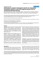
Figure 1
Increased clinical and histological severity of arthritis in transgenic
mice with impaired TGFβ-signalling in T cells. (a) The time course of
the severity of arthritis after the booster injection (day 0) of one long-
term experiment is shown. This experiment involved six wild-type and
eight transgenic mice. Means ± SEM are shown. (b) The maximum
clinical arthritis score of mice with a clinical score of at least grade 1
during the observation period of four separate short-term experiments
was significantly higher in transgenic mice than in wild-type mice
(mean ± SEM; **P =0.001). (c) Mice with a clinical score of at least
grade 1 were analyzed histologically for inflammatory and degenerative
changes. Transgenic mice had significantly higher histological scores
than wild-type mice (mean ± SEM; *P <0.05). TG, tg, transgenic; WT,
wt, wild-type.
0
1
2
3
4
5
6
7
8
456789101112131415161718192021
weeks
clinical score
TG
WT
(a)
0
1
2
3
4
5
6
wt tg
clinical score
(b)
**
0
2
4
6
8
10
wt tg
histological score
(c)
*
signalling in T cells than in wild-type mice (histological
score 8.01 ±0.9 vs 4.06 ± 1.1, P < 0.05; Fig.1c). In wild-
type mice, only minor inflammatory changes and a smooth
cartilage surface without significant cartilage or bone
destruction were observed. In transgenic mice, however,
severe inflammatory changes, also involving the periarticu-
lar soft tissue, and numerous neutrophils within the articu-
lar space were observed in small and large joints. Heavy
proliferation of fibrocellular tissue leading to pannus for-
mation with joint destruction was seen. Representative
sections of small and large joints from wild-type and trans-
genic mice are shown in Fig. 2.
Increased cell proliferation and Th1 cytokine production
in lymph node cells from transgenic mice
The draining axillary and popliteal lymph nodes of affected
animals were removed and the cells cultured in the pres-
ence of CII. Increased cell proliferation was found in the
lymph node cells of transgenic as compared with wild-
type mice five weeks after arthritis induction (779 ±85 vs
186 ±27 cpm; Fig. 3a). In addition, a marked difference
was noted in the production of TNFα and IFNγ in the
supernatants of CII-stimulated cultures of draining lymph
node cells (data not shown). Such cells from mice with
long-standing arthritis were not significantly stimulated by
CII in vitro, maybe because of an epitope spreading after
the long period of joint inflammation (data not shown).
However, after costimulation of these cells with
CD3/CD28, a markedly increased proliferative capacity
was observed in transgenic as compared with wild-type
mice 20 weeks after arthritis induction (23,603 ±2125 vs
3554 ±194 cpm; Fig. 3b). Th1 cytokines such as IFNγ
and TNFα were highly up-regulated after costimulation as
well as after stimulation with CII (Fig. 3c,d). In addition, it
appeared that IL-5 was down-regulated in transgenic
lymph node cells after stimulation with CII (Fig. 3d).
Discussion
Our results demonstrate the importance of endogenous
TGFβ in regulating T cells in order to maintain joint integrity
in vivo. Results of studies of the role of endogenous TGFβ
in the development of joint lesions have been contradictory
[6,8]. TGFβ is a pleiotropic cytokine, produced by a variety
of cells and known to exert its effects depending on the
effector cell and the context of production [4].
TGFβ has been detected in the synovium and effusions of
arthritic joints, and an immunosuppressive role has been
postulated from results of in vitro experiments [5]. The
importance of TGFβ in maintaining immune homeostasis
has been demonstrated in TGFβ knockout mice, which die
within the first weeks of life as a result of multifocal inflam-
matory lesions, especially in the heart and lungs [10].
Because it is difficult to delineate the effects of the lack of
TGFβ on a specific cell type in these mice, various
methods have been used to impair TGFβ-signalling in spe-
cific cell types using cell-specific promoters. A dominant
negative TGFβ type II receptor has been overexpressed in
T cells using the CD4 and the CD2 promoter [13,14]. In
addition, Smad7, an inhibitory Smad protein, has been
expressed in T cells [16]. The phenotypes of these trans-
genic mice have turned out to be different from each
other, probably because of strain differences and as yet
unknown mechanisms.
Although T cells have been shown in several models to be
important for the development of arthritis [2], in none of
these mice has the spontaneous development of arthritis
been described, indicating a tight regulation of immune
homeostasis within the joint. The transgenic mice used in
this study did not develop spontaneous arthritis even after
an observation period of more than nine months [11].
Moreover, hCD2-∆kTβRII mice developed only minimal
Available online />R117
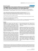
Figure 2
Increased inflammatory and degenerative changes in transgenic hCD2-
∆kTβRII mice after the induction of CIA. Representative sagittal
histological sections stained with hematoxylin and eosin are shown.
(a–c) A small joint of the extremities (a) of a wild-type mouse, and a
larger joint (b), show a smooth cartilage surface without any cartilage
or bone destruction. (c) The synovial lining layer is composed of flat
synovial cells or is mildly hyperplastic. (d–f) Joints of transgenic mice
with severe inflammatory changes also affecting the periarticular soft
tissue are shown. (d) Destruction was seen in small joints, with
fibroproliferative tissue (lower portion) and numerous neutrophils within
the articular space (upper portion and f). Bone destruction has
resulted in bone modulation. (e) In larger joints of the extremities, also,
there is heavy proliferation of fibrocellular tissue (pannus formation)
with joint destruction. Scale bars represent 100 µm.
inflammatory lesions on distal joints after immunization
with CII. These mice were generated on an FVB/N back-
ground. FVB/N mice have been reported to be resistant to
CIA, although they express the same MHC haplotype as
DBA/1 mice, a major susceptibility factor for the develop-
ment of CIA. Still, antigen recognition might be impaired in
FVB/N mice, resulting in resistance to the induction of CIA
due to deletions in the T-cell receptor Vβ or mutations in
the T-cell receptor Vα genes [15]. Therefore, the F
1
gener-
ation of crossings with DBA/1 mice was used for the
experiments. Wild-type F
1
mice still had a rather low inci-
dence and severity of CIA. In contrast, transgenic F
1
mice
showed a marked increase in the incidence and severity of
arthritis, demonstrating that the susceptibility of wild-type
F
1
mice was greatly enhanced by the impairment of TGFβ-
signalling in T cells. In addition, in long-term experiments,
no resolution of arthritis was observed after an initial flare
of disease. Clearly, these results indicate that impairment
of TGFβ-signalling in T cells alone is not sufficient to over-
come the resistance of FVB/N mice. However, in the
setting of T-cell activation through efficient antigen pre-
sentation, impairment of TGFβ-signalling seems to be an
additional susceptibility factor, a finding that underlines
the importance of T cells in the regulation of joint home-
ostasis.
We also demonstrated an increased production of the
Th1 cytokines TNFα and IFNγ in cultures of transgenic
draining lymph node cells after arthritis induction and after
long-standing arthritis. These cytokines are involved in the
pathogenesis of arthritis and could be elevated either
because of more severe inflammation in transgenic mice
or because of the spontaneous differentiation and
cytokine shift observed in T cells with impaired TGFβ-sig-
nalling [11,13].
In addition to its immunoregulatory effects, TGFβ has
been shown to play an important role in matrix regulation
and chondrocyte differentiation. As has been mentioned
elsewhere, the injection of TGFβ1 into joints results in
osteophyte formation and synovitis [7]. Moreover, the
impairment of TGFβ-signalling in skeletal tissue of trans-
genic mice expressing a dominant negative TGFβ type II
receptor under the control of a metallothionein-like pro-
moter has resulted in degenerative changes and bone
malformation, the changes in joints resembling those seen
in osteoarthritis [9]. TGFβ therefore seems to have benefi-
cial effects in the promotion of tissue repair and down-reg-
ulation of inflammation, but when these regulatory effects
are not sufficient to control disease, negative effects such
as fibrosis and bone remodelling could predominate in the
long term.
Conclusion
A significantly higher incidence and severity of CIA were
observed in transgenic mice with impaired TGFβ-sig-
nalling in T cells than in wild-type littermates. These results
demonstrate that endogenous TGFβ acts on T cells to
maintain joint integrity after the induction of arthritis and
during the healing phase of disease. Several studies have
been performed on the susceptibility factors contributing
to the development of arthritis. Our data suggest assess-
ment of the TGFβ-signalling cascade as an as yet
unknown susceptibility factor.
Competing interests
None declared.
Arthritis Research & Therapy Vol 6 No 2 Schramm et al.
R118
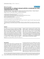
Figure 3
Increased proliferation and Th1 cytokine production in cultures of
draining lymph node cells from transgenic mice. (a) Cell proliferation
five weeks after arthritis induction. Cells were stimulated in vitro with
50 µg/ml CII and cultivated for 48 hours (mean± SEM). This
experiment involved 10 transgenic and 11 wild-type mice. (b) Draining
lymph node cell proliferation 20 weeks after arthritis induction. Cells
were stimulated with anti-CD3/CD28 antibody. Eight transgenic and
six wild-type mice were included. (c,d) Cytokine levels were
determined in the culture supernatants of draining lymph node cells
after 20 weeks of arthritis using ELISA, in (c) cells stimulated with CII
and (d) cells costimulated with anti-CD3/CD28 antibody. CII, chicken
collagen type II; cpm, counts per minute; TG, transgenic; TNF, tumor
necrosis factor; WT, wild-type.
0
5
10
15
20
25
30
WT TG
cpm × 10
3
(b)
0
200
400
600
800
1000
WT TG
cpm
(a)
0
1000
2000
3000
4000
5000
6000
7000
IL-2 IL-4 IL-5 IL-6 IL-10 IFNγ TNFα
cytokine level (pg/ml)
TG
WT
(d)
0
50
100
150
200
250
300
350
400
450
IL-2 IL-4 IL-5 IL-6 IL-10 IFN
γ TNFα
cytokine level (pg/ml)
TG
WT
(c)
Acknowledgements
This work was supported by the DFG, SFB 548 and MAIFOR, Faculty
of Medicine, University of Mainz. The authors thank Marina Snetkova for
excellent technical assistance.
References
1. Anthony DD, Haqqi TM: Collagen-induced arthritis in mice: an
animal model to study the pathogenesis of rheumatoid arthri-
tis. Clin Exp Rheumatol 1999, 17:240-244.
2. Taneja V, Taneja N, Paisansinsup T, Behrens M, Griffiths M, Luthra
H, David CS: CD4 and CD8 T cells in susceptibility/protection
to collagen-induced arthritis in HLA-DQ8-transgenic mice:
implications for rheumatoid arthritis. J Immunol 2002, 168:
5867-5875.
3. Feldmann M, Brennan FM, Maini RN: Role of cytokines in
rheumatoid arthritis. Annu Rev Immunol 1996, 14:397-440.
4. Letterio JJ, Roberts AB: Regulation of immune responses by
TGF-beta. Annu Rev Immunol 1998, 16:137-161.
5. Lotz M, Kekow J, Carson DA: Transforming growth factor-beta
and cellular immune responses in synovial fluids. J Immunol
1990, 144:4189-4194.
6. Thorbecke GJ, Shah R, Leu CH, Kuruvilla AP, Hardison AM, Pal-
ladino MA: Involvement of endogenous tumor necrosis factor
alpha and transforming growth factor beta during induction of
collagen type II arthritis in mice. Proc Natl Acad Sci USA 1992,
89:7375-7379.
7. Allen JB, Manthey CL, Hand AR, Ohura K, Ellingsworth L, Wahl
SM: Rapid onset synovial inflammation and hyperplasia
induced by transforming growth factor beta. J Exp Med 1990,
171:231-247.
8. Wahl SM, Allen JB, Costa GL, Wong HL, Dasch JR: Reversal of
acute and chronic synovial inflammation by anti-transforming
growth factor beta. J Exp Med 1993, 177:225-230.
9. Serra R, Johnson M, Filvaroff EH, LaBorde J, Sheehan DM,
Derynck R, Moses HL: Expression of a truncated, kinase-
defective TGF-beta type II receptor in mouse skeletal tissue
promotes terminal chondrocyte differentiation and
osteoarthritis. J Cell Biol 1997, 139:541-552.
10. Kulkarni AB, Huh CG, Becker D, Geiser A, Lyght M, Flanders KC,
Roberts AB, Sporn MB, Ward JM, Karlsson S: Transforming
growth factor beta 1 null mutation in mice causes excessive
inflammatory response and early death. Proc Natl Acad Sci
USA 1993, 90:770-774.
11. Schramm C, Protschka M, Köhler H, Podlech J, Reddehase MJ,
Schirmacher P, Galle PR, Lohse AW, Blessing M: Impairment of
TGF-beta signaling in T-cells increases susceptibility to
experimental autoimmune hepatitis in mice. Am J Physiol
2003, 284:G525-G535.
12. Brand T, MacLellan WR, Schneider MD: A dominant-negative
receptor for type beta transforming growth factors created by
deletion of the kinase domain. J Biol Chem 1993, 268:11500-
11503.
13. Gorelik L, Flavell RA: Abrogation of TGFbeta signaling in T cells
leads to spontaneous T cell differentiation and autoimmune
disease. Immunity 2000, 12:171-181.
14. Lucas PJ, Kim SJ, Melby SJ, Gress RE: Disruption of T cell
homeostasis in mice expressing a T cell-specific dominant
negative transforming growth factor beta II receptor. J Exp
Med 2000, 191:1187-1196.
15. Osman GE, Hannibal MC, Anderson JP, Lasky SR, Ladiges WC,
Hood L: FVB/N (H2(q)) mouse is resistant to arthritis induc-
tion and exhibits a genomic deletion of T-cell receptor V beta
gene segments. Immunogenetics 1999, 49:851-859.
16. Nakao A, Miike S, Hatano M, Okumura K, Tokuhisa T, Ra C,
Iwamoto I: Blockade of transforming growth factor beta/Smad
signaling in T cells by overexpression of Smad7 enhances
antigen-induced airway inflammation and airway reactivity. J
Exp Med 2000, 192:151-158.
Correspondence
Dr Christoph Schramm, I. Department of Medicine, Johannes
Gutenberg-University, Langenbeckstr. 1, 55101 Mainz, Germany;
Tel: +49 6131 3933359; fax: +49 6131 3933364; e-mail:
Available online />R119