Báo cáo y học: "Antibodies against the CB10 fragment of type II collagen in rheumatoid arthritis" pdf
Bạn đang xem bản rút gọn của tài liệu. Xem và tải ngay bản đầy đủ của tài liệu tại đây (328.11 KB, 7 trang )
Open Access
Available online />R477
Vol 6 No 5
Research article
Antibodies against the CB10 fragment of type II collagen in
rheumatoid arthritis
Andrew D Cook
1
, Robyn Gray
1
, John Ramshaw
2
, Ian R Mackay
1
and Merrill J Rowley
1
1
Department of Biochemistry and Molecular Biology, Monash University, Clayton, Victoria, Australia
2
CSIRO Molecular Science, Parkville, Victoria, Australia
Corresponding author: Merrill J Rowley,
Received: 2 Apr 2004 Revisions requested: 14 May 2004 Revisions received: 11 Jun 2004 Accepted: 18 Jun 2004 Published: 11 Aug 2004
Arthritis Res Ther 2004, 6:R477-R483 (DOI 10.1186/ar1213)
http://arthr itis-research.com/conte nt/6/5/R477
© 2004 Cook et al.; licensee BioMed Central Ltd. This is an Open Access article: verbatim copying and redistribution of this article are permitted in
all media for any purpose, provided this notice is preserved along with the article's original URL.
Abstract
Antibodies against intact type II collagen (CII) are a feature of
rheumatoid arthritis (RA) but have limited diagnostic value. Here
we assess whether either of the two major cyanogen bromide
fragments of CII, namely CB10 or CB11, are more sensitive
substrates for the detection of antibodies in RA. Cleavage of
bovine CII with cyanogen bromide yielded CB10 and CB11;
these were purified by column chromatography for use in an
enzyme-linked immunosorbent assay. Serum antibodies were
measured in patients with RA, psoriatic arthritis (PsA),
osteoarthritis (OA) and blood donors. Results were compared
with those using intact CII. Antibodies against CB10 were found
in as many as 88% of 96 patients with long-standing RA, but
only 12% of 33 patients with PsA, 6% of 34 patients with OA
and 3% of 93 control sera. Lower frequencies for these
diseases were obtained on testing for antibodies against CB11:
50%, 6%, 21% and 2%, respectively. The sensitivity of
detection in RA of antibodies against CB10 compared with
antibodies against intact CII (88% versus 24%) was not at the
expense of specificity, which remained high at 94%. The much
higher frequency of antibodies against CB10 in RA than in other
rheumatic diseases or control sera indicates that CB10 is
clearly a more sensitive substrate than the intact collagen
molecule and, combined with other assays (rheumatoid factor,
anti-cyclic citrullinated peptide [anti-CCP]), might comprise a
panel with a highly reliable predictive value. Moreover, our
findings should encourage renewed interest in the role of
collagen autoimmunity in the pathogenesis of RA.
Keywords: antibodies, cyanogen bromide peptides, rheumatoid arthritis, type II collagen
Introduction
Rheumatoid arthritis (RA) is a systemic autoimmune dis-
ease characterized by chronic articular inflammation and
progressive joint destruction. Autoimmunity to type II colla-
gen (CII), an abundant molecule in articular cartilage, is
implicated in pathogenesis because the immunization of
susceptible strains of rats, mice and nonhuman primates
using native CII in adjuvant results in collagen-induced
arthritis (CIA), which has close immunologic and patho-
logic similarities to RA [1]. CIA is dependent on immune
responses to CII by both T and B lymphocytes, with arthritis
being initiated by complement-fixing autoantibodies that
bind to CII in the cartilage matrix [2]. In humans, IgG-pro-
ducing B cells specific for CII are present in rheumatoid
synovium and synovial fluid [3,4], suggesting an intra-artic-
ular antigen-driven immune process. Serum autoantibodies
against native and denatured type II collagen (anti-NCII and
DCII) are detected in 24–30% of patients with RA, as
shown by cross-sectional studies on patients with well-
established disease [5-8]. However, this relatively low fre-
quency in RA of anti-CII, together with seropositivity (albeit
at even lower frequency) in other rheumatological and
inflammatory diseases [7] including psoriatic arthritis (PsA)
[7,9], osteoarthritis (OA) [7,10,11], juvenile arthritis [12],
Paget's disease [10,11], and systemic lupus erythemato-
sus [6,7,10,13], has negated the diagnostic utility and
pathogenetic significance of anti-CII in RA. However, sev-
eral studies have reported that the frequency of anti-NCII
might be as high as 60–75% in patients with RA when
tested very early in the disease [14-16], and thereafter fre-
quencies tend to fall to those ascertained in the earlier
cross-sectional studies [17], suggesting that their pres-
ence might provide a sensitive predictive marker for cases
of early RA.
CIA = collagen-induced arthritis; CII = type II collagen; CNBr = cyanogen bromide; ELISA = enzyme-linked immunosorbent assay; mAb = monoclonal
antibody; NCII = native type II collagen; NHS = normal human serum; OA = osteoarthritis; PsA = psoriatic arthritis; RA = rheumatoid arthritis; SD =
standard deviation.
Arthritis Research & Therapy Vol 6 No 5 Cook et al.
R478
Our interest in the reactivity of RA sera with various cleav-
age products of CII led to the present study, in which we
assess whether purified preparations of the two major cya-
nogen bromide (CNBr) fragments of CII, namely CB10 and
CB11, would serve as superior substrates for the detection
of antibodies in RA. It was found that the frequency of anti-
bodies against CB10 in cases of well-established RA was
markedly increased, from 24% to 88%, comparing the fre-
quency using the intact CII molecule; this was not at the
expense of the specificity, which remained at the high level
of 94%.
Materials and methods
Patients
Sera were studied from 96 of 114 consecutively selected
patients with the diagnosis of RA, according to the 1987
criteria of the American College of Rheumatology [18], who
were attending the rheumatology clinic at the Royal Mel-
bourne Hospital, Melbourne; clinical and immunogenetic
data have been reported on these patients [8,19,20]. Of
the 96 patients, 70% were female, 87% had been serop-
ositive for rheumatoid factor at some stage, 86% had the
RA HVR3 susceptibility motif, and 86% had erosive dis-
ease. At entry to the study, 78% had active disease, and
the median Ritchie Articular Index was 5 (range 0–25). The
median age at onset was 45 years (range 15–77), and the
median duration of disease was 13 years (range 3–51).
Patients were receiving various drugs including corticoster-
oids, non-steroidal anti-inflammatory drugs, or disease-
modifying anti-rheumatic drugs, including methotrexate.
Sera were also obtained from 33 patients with PsA and
from 34 patients with OA attending rheumatology clinics at
the Royal Melbourne Hospital and Monash Medical Centre,
Melbourne. Sera from 93 blood donors attending the Red
Cross Blood Bank in Melbourne were used as normal con-
trols. The use of these sera for the present study was
approved by the Standing Committee on Ethics in
Research involving Humans of Monash University.
Preparation of type II collagen
CII was prepared from human rib cartilage and bovine nasal
septum cartilage by digestion with pepsin and by differen-
tial salt precipitation [12], and the preparations appeared
pure according to an overloaded 5% SDS-PAGE gel
stained with 0.2% (w/v) Coomassie blue (Bio-Rad, Her-
cules, CA, USA); no contamination with other collagen
types or with proteoglycans was evident (data not shown).
CNBr digestion of bovine CII and purification of CB10
and CB11 peptides
CNBr digestion of bovine CII was performed with 3 mg of
purified bovine CII. This was dissolved in 1 ml of 70% (v/v)
formic acid, containing 50 mg of CNBr (Sigma, St Louis,
MO, USA) and cleavage was achieved by the method of
Scott and Veis [21].
CB10 and CB11 were then purified as described by Miller
and Lunde [22], with slight modifications. The CB peptides
were initially resolved into two major fractions by chroma-
tography on a Bio-Gel P-2 column (100–200 mesh, exclu-
sion limit 2600 Da, globular proteins; Bio-Rad),
equilibrated with 0.1 M acetic acid. The larger peptides that
were eluted in the excluded volume were pooled, freeze-
dried and re-chromatographed on a cation-exchange col-
umn of carboxymethylcellulose (Whatman CM-52), equili-
brated with 0.02 M sodium citrate, pH 3.6, containing 0.01
M NaCl (start buffer). Chromatography was performed at
42°C for 10 hours with a linear salt gradient obtained by the
addition of 1% 0.16 M NaCl in 0.02 M sodium citrate, pH
3.6, to the start buffer every 6 min, with a flow rate of 1.75
ml/min, using the Bio-Rad Econo system (Bio-Rad). Frac-
tions were collected at 10 min intervals, run on 15% Tricine
gels and stained with 0.2% (w/v) Coomassie blue. Frac-
tions containing CB10 or CB11 were pooled separately,
desalted on the Bio-Gel P-2 column and freeze-dried. To
purify CB10 from CB8 and CB9,7, and CB11 from CB12,
each fraction was applied to a P-30 column (100–200
mesh, exclusion limit 40 kDa, globular proteins; Bio-Rad),
equilibrated in 0.1 M acetic acid. To determine the purity of
the peptides after each column run, fractions were run on
15% Tricine gels and stained with 0.2% (w/v) Coomassie
blue. Purified CB10 and CB11 were renatured by stepwise
cooling, as described by Terato and colleagues [23], from
20 to 0.2°C.
Circular dichroism
We confirmed that renaturation of the CB peptides had
occurred by performing circular dichroism spectroscopy on
a Jasco J-810 spectropolarimeter equipped with a PFD
423S/L Peltier-type temperature controller, with constant
flushing with N
2
. CII and CB peptides were dissolved at 1
mg/ml in 0.1 M acetic acid. Renaturation was performed as
described previously [23] and the protein concentration
was adjusted to 70–80 µg/ml. When required, denatura-
tion was performed immediately before analysis by heating
samples to 45°C for 5 min before placement on ice. Circu-
lar dichroism spectra were recorded from 190 to 250 nm
at 5°C in a cuvette with a 1 mm light path. Each spectrum
represents an average of three scans performed at 50 nm/
min with a bandwidth of 1 nm. Thermal melting profiles
were followed at 222 nm as the temperature increased
from 5 to 40°C at a heating rate of 1°C/min.
Collagen antibody detection by enzyme-linked
immunosorbent assay (ELISA)
Antibodies against native human and native bovine CII were
measured by ELISA as described previously [7,8]. Microti-
tre plates were coated overnight with 10 µg/ml human or
Available online />R479
bovine CII, at 4°C. Sera, diluted 1:50, were tested in dupli-
cate in the presence or absence of antigen. Antibodies
were detected with horseradish peroxidase-conjugated
anti-human IgG (Chemicon International, Temecula, CA,
USA), with 0.5 mg/ml 2,2,-azino-di-[3-ethyl-benzthiazoline
sulfonate (6)] (Boehringer, Mannheim, Germany) as sub-
strate in 0.03 M citric acid, 0.04 M Na
2
HPO
4
, 0.003%
H
2
O
2
, pH 4. To compensate for non-specific binding of
immunoglobulins to the plates, the optical density obtained
for wells lacking collagen was subtracted from that
obtained for wells coated with collagen. Results were
expressed as levels of reactivity based on the number of
standard deviations (SD) above the mean for 10 normal
blood donor controls tested in the same assay; the level
was considered to be raised when greater than 3 SD above
the mean; that is, an antibody level of 3 or more.
CB10 and CB11 antibody detection by ELISA
Antibodies against the purified CB peptides of bovine type
II collagen, namely CB10 and CB11, were measured by
ELISA as described above, with the following modifica-
tions. Plates were coated with 10 µg/ml purified peptide
and sera were diluted 1:200. All incubations were per-
formed at 4°C and washes were done with cold Tris-buff-
ered saline containing 0.05% Tween 20. Before addition of
the substrate, plates were left at 22°C for 10 min. Results
are expressed as SD above the mean for 10 controls, as
described above.
The monoclonal antibody (mAb) M2139 recognizing native
CB10, and the mAb CII-CI recognizing native CB11 [24],
donated by Professor R Holmdahl (Lund University, Lund,
Sweden), were used to establish that the CB peptides of
CII had renatured sufficiently to display a conformational
epitope.
Statistics
The frequency of antibodies between individual groups was
analysed with the χ
2
or Fisher exact probability test, and lev-
els of antibodies were compared with the Mann–Whitney
U-test. Antibody levels to human NCII and bovine NCII from
the same sera were compared using the Pearson product
moment correlation coefficient. P ≤ 0.05 was taken as
significant.
Results
Comparison of antibodies against human CII and bovine
CII by ELISA
We have previously reported that IgG antibodies against
NCII are detectable in 24% of patients with RA, 12% of
those with OA and 13% of those with other chronic inflam-
matory diseases [7]. As the CB peptides were purified from
bovine CII, because of the amount of collagen required for
purification, we initially selected random sera from patients
with RA, PsA, OA, and normal controls, to establish that
levels of antibodies against intact bovine CII were equiva-
lent to those obtained with human CII.
In the present study, IgG antibodies against native bovine
CII were present in 23 (24%) of 94 patients with RA, 9
(27%) of 33 with patients with PsA, 3 (10%) of 31 patients
with OA, and 1 (1%) of 93 control sera. The levels of reac-
tivity to bovine CII compared with levels with human CII
tended to be higher for patients with PsA, but not for
patients with RA (data not shown). However, the frequency
of positivity of antibodies against bovine or human CII in the
different diseases was not significantly different and was
equivalent to frequencies we have reported previously
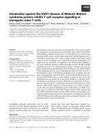
[7,8]. Overall, for the 251 sera that were compared, there
was a highly significant correlation (R = 0.777, P < 0.001)
between levels of antibodies against human CII and anti-
bodies against bovine CII (Fig. 1). The correlation was
higher for patients with RA (R = 0.875) than for patients
with PsA or OA (R = 0.556).
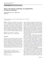
Purified CB peptides of bovine CII
After digestion of bovine CII with CNBr, the CB peptides
were separated on a carboxymethylcellulose column, using
an increasing salt gradient (Fig. 2a). The purity of the pep-
tides after chromatography was then checked by SDS-
PAGE (Fig. 2b). After purification and renaturation of the
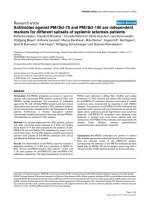
CB peptides by stepwise cooling, circular dichroism was
used to confirm that, upon renaturation, the CB peptides
had adopted a correctly folded triple-helical conformation.
Circular dichroism analysis of CB10 showed a large posi-
tive ellipticity peak at 220–225 nm, as reported for the
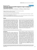
Figure 1
Correlation of antibodies against bovine type II collagen (CII) and human CIICorrelation of antibodies against bovine type II collagen (CII) and
human CII. Scatter plot of the levels of IgG antibodies against human
CII and bovine CII in sera of patients with rheumatoid arthritis (RA; n =
94), psoriatic arthritis (PsA; n = 33), osteoarthritis (OA; n = 31), and
normal human serum (NHS; n = 93). There is a highly significant corre-
lation between the levels of antibodies against human CII and bovine
CII (R = 0.777, P < 0.001). The points represent for individual sera the
number of standard deviations above the mean of normal sera for which
the mean is 0. A serum is regarded as positive if it is more than three
standard deviations above the mean (dashed lines).
0 5 10 15 20 25 30 35 40
0
5
10
15
20
25
RA
PsA
OA
NHS
Abs to human CII
Abs to bovine CII
Arthritis Research & Therapy Vol 6 No 5 Cook et al.
R480
native CII triple helix, that was lost on denaturation by heat
(Fig. 3a) [25]. Unfolding of CB10 started at about 20°C, as
shown by a decrease in the positive peak at 222 nm with
increasing temperature (Fig. 3b). These data confirm that,
after renaturation, CB10 assumes a triple-helical, collagen-
like conformation. Similar results were obtained for CB11
(data not shown). By ELISA, mAb M2139, which recog-
nizes native CB10 of CII, reacted strongly with the rena-
tured CB10 but not renatured CB11 (Fig. 4a), whereas
mAb CII-C1, which recognizes native CB11 of CII, reacted
strongly with CB11 but not CB10 (Fig. 4b). On denatura-
tion of CB10 and CB11, there was a loss of reactivity of
mAbs M2139 and CII-CI (Fig. 4).
Antibodies against the purified CB10 and CB11 of CII as
measured by ELISA
IgG antibodies against bovine CB10 were present in 84
(88%) of 96 patients with RA, 4 (12%) of 33 patients with
PsA, 2 (6%) of 34 patients with OA, and 3 (3%) of 93 con-
trol sera (Fig. 5a). Moreover, levels of antibodies against
CB10 were significantly higher in RA sera than in PsA, OA,
and control sera (P < 0.0001). In fact, the four anti-CB10-
positive PsA patients had levels between 3 and 4 SD
above the mean. The frequency of IgG antibodies against
bovine CB11 was lower, but still substantial, at 48 (50%)
of 96 patients with RA (Fig. 5b), but only 2 (6%) of 33
patients with PsA, 7 (21%) of 34 patients with OA, and 2
(2%) of 93 control sera (Fig. 5b). The concentrations of
antibodies against CB11 were significantly higher in RA
than in other diseases (P < 0.0001).
The sensitivity of the test for antibodies in RA was
increased from 24% to 88% when the intact CII molecule
was replaced by CB10 and, notably, this was not at the
expense of specificity, which was 94% when CB10 was
used.
Discussion
The most significant finding from our study was that in RA
the use in an ELISA of fragments of the intact CII molecule,
particularly CB10, as antigen markedly increased the sen-
sitivity of detection of antibodies in serum against NCII from
Figure 2
Purification of CB peptides of type II collagen (CII)Purification of CB peptides of type II collagen (CII). (a) Carboxymethylcellulose chromatogram and corresponding Coomassie blue protein stain
illustrating the separation of the CB peptides of bovine CII with a linear salt gradient (dotted line). (b) Coomassie blue staining of purified CB pep-
tides of bovine CII separated on a 15% Tricine gel.
Available online />R481
24% to 88%. Moreover, this increase in sensitivity was not
at the expense of the specificity, which remained high at
94%. The current study used CB peptides purified from
bovine CII. Although obtaining sufficient human CII pre-
cluded studies on the relative efficacy of human versus
bovine CB10, our data on the use of intact CII suggest that
human CB10 might be an even more precise reactant.
There have been several studies reporting that the fre-
quency of anti-NCII might be as high as 60–75% in
patients with RA when tested very early in the disease [14-
16], and thereafter frequencies tend to fall to those ascer-
tained in the earlier cross-sectional studies [17]. If the same
applies to anti-CB10, it is likely that collagen–related
autoimmunity occurs during early initiating events in the
development of RA, rather than later as a result of new
epitopes being exposed as a result of articular damage.
Our observations should therefore be applicable to routine
diagnostic testing for anti-CII in articular diseases and, in
particular, to those cases of early undifferentiated arthritis
that do not fulfil criteria for RA but might later evolve to typ-
ical disease. Moreover, tests for anti-CB10 in conjunction
with other assays including rheumatoid factor and anti-
cyclic citrullinated peptide (anti-CCP) could add further to
diagnostic precision, although this remains to be tested.
During analysis, epitopes of CII are detected usually by
Western blotting with CNBr digests of collagen. In RA,
studies using CB peptides of CII showed reactivity to mul-
tiple components [6,16,26,27], and there was no apparent
difference in the patterns of recognition of different CB
peptides with sera from, for example, patients with RA or
systemic lupus erythematosus [27], although anti-CII sera
from patients with relapsing polychondritis react uniquely
with CB9,7 [6]. Analytical methods alternative to immuno-
blotting on CB peptides have included epitope scanning of
CB11 by using overlapping sequential synthetic peptides.
This revealed as many as 21 different antibody-binding
sites on CB11 with the use of individual sera from rats
immunized with CII [28]. More recently, the use of recom-
binant chimeric preparations of CII has allowed the detec-
Figure 3
Circular dichroism (CD) spectroscopy of CB10Circular dichroism (CD) spectroscopy of CB10. (a) CD analysis of
CB10 (bold) showing positive ellipticity peak at 220–225 nm that was
lost after heat denaturation (non-bold). (b) Thermal melting profile of
CB10 followed at 222 nm. At about 20°C, unfolding of CB10 started.
(a)
(b)
-80
-60
-40
-20
0
20
40
190 200 210 220 230 240 250
Wa velength (nm)
CD( mdeg)
-20
-10
0
10
20
30
40
50
01020304050
Temperature
CD(mdeg)
Figure 4
Reactivity of monoclonal antibodies (mAbs) to CB10 and CB11Reactivity of monoclonal antibodies (mAbs) to CB10 and CB11. (a)
mAb M2139, reactive with native CB10; (b) mAb CII-C1, reactive with
native CB11. mAbs were tested by enzyme-linked immunosorbent
assay for reactivity on purified renatured CB10 (CB10), purified rena-
tured CB11 (CB11), denatured CB10 (dCB10), and denatured CB11
(dCB11).
(a)
M2139
0
0.2
0.4
0.6
0.8
1
1.2
1.4
200
4
0
0
8
0
0
1600
32
0
0
6400
1
280
0
2
560
0
5
120
0
1
0
2
4
0
0
2048
0
0
M2139 mAb dilution
Absorbance (O.D.)
CB10
CB11
dCB10
dCB11
(b)
CII-C1
0
0.5
1
1.5
2
2.5
20
0
40
0
8
0
0
16
0
0
32
00
6
4
0
0
12800
2
56
0
0
5
120
0
1
024
00
204
8
0
0
CII-CI mAb dilution
Abso rbance (O.D.)
CB10
CB11
dCB10
dCB11
Arthritis Research & Therapy Vol 6 No 5 Cook et al.
R482
tion of conformation-dependent epitopes; it is of interest
that antibodies against intact CII from patients with RA rec-
ognized several epitopes known to be arthritogenic for
murine CIA, whereas anti-CII from patients with diverse
diseases, OA, systemic lupus erythematosus, and relaps-
ing polychondritis, reacted with other sites that lacked
arthritogenic epitopes [29].
The above observations prompt inferences on the role of
antibodies against CII in the pathogenesis of RA. Consid-
ering the CIA model, anti-CII are directly pathogenic, well
exemplified by transfer experiments in mice [30-32]. In
addition, CII-specific B cells in the diseased synovium, and
in synovial fluids of patients with RA, produce IgG autoan-
tibodies that react with native CII, suggesting an intra-artic-
ular antigen-driven immune process [3,4,33]. Presumably
IgG antibodies form immune complexes with CII in the car-
tilage matrix and initiate the complement cascade to pro-
voke an inflammatory reaction within the joint that causes
degradation of the cartilage matrix, noting that purified anti-
CII from RA serum activates complement C5 to C5a when
bound to human cartilage in vitro [2]. Anti-CII in RA are pre-
dominantly of the complement-fixing IgG subclasses IgG1
and IgG3 [7,34], as found in CIA-susceptible mouse
strains [35], whereas in the other rheumatic diseases, in
which anti-NCII are present, the non-complement fixing
subclasses IgG2 and IgG4 are predominant. Furthermore,
anti-CII, when bound to cartilage in vitro, induces degrada-
tion and phagocytosis by leukocytes of the collagen/prote-
oglycan matrix [36]. These observations, including the C5
activation analyses, provide persuasive evidence that
cartilage-bound anti-CII can participate in the initiation or
perpetuation of RA. In view of the particular attention given
to the role of T lymphocytes among effector mechanisms in
RA, these observations on antibody-dependent damage
are germane; further, they are not limited to anti-CII,
because antibody transfer with sera induces a profound
inflammatory arthritis in naive recipients in the murine K/
BxN model of RA in which an entirely different autoanti-
genic molecule is implicated [37].
Conclusion
Current immunoassays to detect antibodies against intact
CII have insufficient specificity and/or sensitivity to be use-
ful for the diagnosis of RA. However, by using as substrate
the specific cleavage product CB10 of the CII molecule in
an ELISA, antibodies were detectable in as many as 88%
of patients with RA, without any loss of specificity. CB10 is
therefore a more sensitive substrate than the intact colla-
gen molecule and, combined with other assays
(rheumatoid factor, anti-cyclic citrullinated peptide) could
well comprise a panel with a highly reliable predictive value.
Moreover, it might also aid our understanding of the role of
collagen autoimmunity in the pathogenesis of RA.
Competing interests
MR has been paid by ThermElectron Corporation to per-
form further work directed to kit development over the past
2 years. Results reported in this manuscript are the subject
of Australian Patent No. 762471 Montech Medical
Developments Pty Ltd and Rondole Pty Ltd: 'Method for
the diagnosis of rheumatoid arthritis'; inventors: AC, MR
and IM.
Acknowledgements
We thank Ms Veronica Glattauer (CSIRO) for her assistance in the puri-
fication of the collagen fragments, and the rheumatologists at the Royal
Melbourne Hospital and Monash Medical Centre for providing the sera
used in this study. Financial support was from Rondole Pty Ltd, under
the Australian Federal Government Syndicated Research and Develop-
ment Scheme.
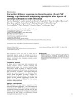
Figure 5
Levels of antibodies against CB10 (a) and CB11 (b) of bovine type II collagen in patients with rheumatoid arthritis (RA; n = 96), psoriatic arthritis (PsA; n = 33), osteoarthritis (OA; n = 34) and in normal human serum (NHS; n = 93)Levels of antibodies against CB10 (a) and CB11 (b) of bovine type II
collagen in patients with rheumatoid arthritis (RA; n = 96), psoriatic
arthritis (PsA; n = 33), osteoarthritis (OA; n = 34) and in normal human
serum (NHS; n = 93). The points represent, for individual sera, the
number of standard deviations (SD) above the mean of normal sera, for
which the mean is 0. A serum is regarded as positive if it is 3SD or
more above the mean (dashed line).
(a)
RA PsA OA NHS
0
10
20
30
40
No. of S.D.
(b)
RA PsA OA NHS
0
10
20
30
40
50
60
No. of S.D.
Available online />R483
References
1. Holmdahl R, Andersson M, Goldschmidt TJ, Gustafsson K, Jans-
son L, Mo JA: Type II collagen autoimmunity in animals and
provocations leading to arthritis. Immunol Rev 1990,
118:193-232.
2. Watson WC, Brown PS, Pitcock JA, Townes AS: Passive trans-
fer studies with type II collagen antibody in B10.D2/old and
new line and C57Bl/6 normal and beige (Chediak–Higashi)
strains: evidence of important roles for C5 and multiple inflam-
matory cell types in the development of erosive arthritis. Arthri-
tis Rheum 1987, 30:460-465.
3. Tarkowski A, Klareskog L, Carlsten H, Herberts P, Koopman WJ:
Secretion of antibodies to types I and II collagen by synovial
tissue cells in patients with rheumatoid arthritis. Arthritis
Rheum 1989, 32:1087-1092.
4. Rudolphi U, Rzepka R, Batsford S, Kaufmann SH, von der Mark K,
Peter HH, Melchers I: The B cell repertoire of patients with
rheumatoid arthritis. II. Increased frequencies of IgG+ and
IgA+ B cells specific for mycobacterial heat-shock protein 60
or human type II collagen in synovial fluid and tissue. Arthritis
Rheum 1997, 40:1409-1419.
5. Morgan K, Clague RB, Collins I, Ayad S, Phinn SD, Holt PJ: Inci-
dence of antibodies to native and denatured cartilage colla-
gens (types II, IX, and XI) and to type I collagen in rheumatoid
arthritis. Ann Rheum Dis 1987, 46:902-907.
6. Terato K, Shimozuru Y, Katayama K, Takemitsu Y, Yamashita I, Miy-
atsu M, Fujii K, Sagara M, Kobayashi S, Goto M, et al.: Specificity
of antibodies to type II collagen in rheumatoid arthritis. Arthritis
Rheum 1990, 33:1493-1500.
7. Cook AD, Mackay IR, Cicuttini FM, Rowley MJ: IgG subclasses of
antibodies to type II collagen in rheumatoid arthritis differ
from those in systemic lupus erythematosus and other con-
nective tissue diseases. J Rheumatol 1997, 24:2090-2096.
8. Cook AD, Stockman A, Brand CA, Tait BD, Mackay IR, Muirden
KD, Bernard CC, Rowley MJ: Antibodies to type II collagen and
HLA disease susceptibility markers in rheumatoid arthritis.
Arthritis Rheum 1999, 42:2569-2576.
9. Trentham DE, Kammer GM, McCune WJ, David JR: Autoimmunity
to collagen: a shared feature of psoriatic and rheumatoid
arthritis. Arthritis Rheum 1981, 24:1363-1369.
10. Choi EK, Gatenby PA, McGill NW, Bateman JF, Cole WG, York
JR: Autoantibodies to type II collagen: occurrence in rheuma-
toid arthritis, other arthritides, autoimmune connective tissue
diseases, and chronic inflammatory syndromes. Ann Rheum
Dis 1988, 47:313-322.
11. Charriere G, Hartmann DJ, Vignon E, Ronziere MC, Herbage D,
Ville G: Antibodies to types I, II, IX, and XI collagen in the serum
of patients with rheumatic diseases. Arthritis Rheum 1988,
31:325-332.
12. Rowley MJ, Gershwin ME, Mackay IR: Collagen antibodies in
juvenile arthritis and adult rheumatoid arthritis: differences in
levels and type-specificity. J Rheumatol 1988, 15:289-294.
13. Choi EK, Gatenby PA, Bateman JF, Cole WG: Antibodies to type
II collagen in SLE: a role in the pathogenesis of deforming
arthritis? Immunol Cell Biol 1990, 68:27-31.
14. Pereira RS, Black CM, Duance VC, Jones VE, Jacoby RK, Welsh
KI: Disappearing collagen antibodies in rheumatoid arthritis.
Lancet 1985, 2:501-502.
15. Fujii K, Tsuji M, Kitamura A, Murota K: The diagnostic signifi-
cance of anti-type II collagen antibody assay in rheumatoid
arthritis. Int Orthop 1992, 16:272-276.
16. Cook AD, Rowley MJ, Stockman A, Muirden KD, Mackay IR: Spe-
cificity of antibodies to type II collagen in early rheumatoid
arthritis. J Rheumatol 1994, 21:1186-1191.
17. Cook AD, Rowley MJ, Mackay IR, Gough A, Emery P: Antibodies
to type II collagen in early rheumatoid arthritis. Correlation
with disease progression. Arthritis Rheum 1996,
39:1720-1727.
18. Arnett FC, Edworthy SM, Bloch DA, McShane DJ, Fries JF, Cooper
NS, Healey LA, Kaplan SR, Liang MH, Luthra HS, et al.: The Amer-
ican Rheumatism Association 1987 revised criteria for the
classification of rheumatoid arthritis. Arthritis Rheum 1988,
31:315-324.
19. Sherritt MA, Tait B, Varney M, Kanaan C, Stockman A, Mackay IR,
Muirden K, Bernard CC, Rowley MJ: Immunosusceptibility
genes in rheumatoid arthritis. Hum Immunol 1996, 51:32-40.
20. Rowley MJ, Stockman A, Brand CA, Tait BD, Rowley GL, Sherritt
MA, Mackay IR, Muirden KD, Bernard CC: The effect of HLA-
DRB1 disease susceptibility markers on the expression of RA.
Scand J Rheumatol 1997, 26:448-455.
21. Scott PG, Veis A: The cyanogen bromide peptides of bovine
soluble and insoluble collagens. I. Characterization of pep-
tides from soluble type I collagen by sodium dodecylsulphate
polyacrylamide gel electrophoresis. Connect Tissue Res 1976,
4:107-116.
22. Miller EJ, Lunde LG: Isolation and characterization of the cyano-
gen bromide peptides from the alpha 1(II) chain of bovine and
human cartilage collagen. Biochemistry 1973, 12:3153-3159.
23. Terato K, Cremer MA, Hasty KA, Kang AH, Hasty DL, Townes AS:
Physicochemical and immunological studies of the renatured
alpha 1(II) chains and isolated cyanogen bromide peptides of
type II collagen. Collagen Relat Res 1985, 5:469-480.
24. Holmdahl R, Rubin K, Klareskog L, Larsson E, Wigzell H: Charac-
terization of the antibody response in mice with type II colla-
gen-induced arthritis, using monoclonal anti-type II collagen
antibodies. Arthritis Rheum 1986, 29:400-410.
25. Goodman M, Bhumralkar , Jefferson EA, Kwak J, Locardi E: Colla-
gen mimetics. Biopolymers 1998, 47:127-142.
26. Buckee C, Morgan K, Ayad S, Collins I, Clague RB, Holt PJ: Diver-
sity of antibodies to type II collagen in patients with rheuma-
toid arthritis: detection by binding to alpha-chains and to
cyanogen bromide peptides. Br J Rheumatol 1990, 29:254-258.
27. Rowley MJ, Mackay IR, Brand CA, Bateman JF, Chan D: Epitope
specificity of antibodies to type II collagen in rheumatoid
arthritis and systemic lupus erythematosus. Rheumatol Int
1992, 12:65-69.
28. Worthington J, Brass A, Morgan K: Identification of antibody
epitopes within the CB-11 peptide of type II collagen. I. Detec-
tion of antibody binding sites by epitope scanning. Autoimmu-
nity 1991, 10:201-207.
29. Burkhardt H, Koller T, Engstrom A, Nandakumar KS, Turnay J, Kra-
etsch HG, Kalden JR, Holmdahl R: Epitope-specific recognition
of type II collagen by rheumatoid arthritis antibodies is shared
with recognition by antibodies that are arthritogenic in colla-
gen-induced arthritis in the mouse. Arthritis Rheum 2002,
46:2339-2348.
30. Kerwar SS, Englert ME, McReynolds RA, Landes MJ, Lloyd JM,
Oronsky AL, Wilson FJ: Type II collagen-induced arthritis. Stud-
ies with purified anticollagen immunoglobulin. Arthritis Rheum
1983, 26:1120-1131.
31. Loutis N, Bruckner P, Pataki A: Induction of erosive arthritis in
mice after passive transfer of anti-type II collagen antibodies.
Agents Actions 1988, 25:352-359.
32. Holmdahl R, Jansson L, Larsson A, Jonsson R: Arthritis in DBA/1
mice induced with passively transferred type II collagen
immune serum. Immunohistopathology and serum levels of
anti-type II collagen auto-antibodies. Scand J Immunol 1990,
31:147-157.
33. Ronnelid J, Lysholm J, Engstrom-Laurent A, Klareskog L, Heyman
B: Local anti-type II collagen antibody production in rheuma-
toid arthritis synovial fluid. Evidence for an HLA-DR4-restricted
IgG response. Arthritis Rheum 1994, 37:1023-1029.
34. Collins I, Morgan K, Clague RB, Brenchley PE, Holt PJ: IgG sub-
class distribution of antinative type II collagen and antidena-
tured type II collagen antibodies in patients with rheumatoid
arthritis. J Rheumatol 1988, 15:770-774.
35. Holmdahl R, Jansson L, Larsson E, Rubin K, Klareskog L: Homol-
ogous type II collagen induces chronic and progressive arthri-
tis in mice. Arthritis Rheum 1986, 29:106-113.
36. Jasin HE, Taurog JD: Mechanisms of disruption of the articular
cartilage surface in inflammation. Neutrophil elastase
increases availability of collagen type II epitopes for binding
with antibody on the surface of articular cartilage. J Clin Invest
1991, 87:1531-1536.
37. Kyburz D, Corr M: The KRN mouse model of inflammatory
arthritis. Springer Semin Immunopathol 2003, 25:79-90.