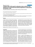
Báo cáo y học: "Collagen type II (CII)-specific antibodies induce arthritis in the absence of T or B cells but the arthritis progression is enhanced by CII-reactive T cells" ppsx
Bạn đang xem bản rút gọn của tài liệu. Xem và tải ngay bản đầy đủ của tài liệu tại đây (244.77 KB, 7 trang )
Open Access
Available online />R544
Vol 6 No 6
Research article
Collagen type II (CII)-specific antibodies induce arthritis in the
absence of T or B cells but the arthritis progression is enhanced by
CII-reactive T cells
Kutty Selva Nandakumar, Johan Bäcklund, Mikael Vestberg and Rikard Holmdahl
Section for Medical Inflammation Research, Lund University, Sweden
Corresponding author: Kutty Selva Nandakumar,
Received: 11 May 2004 Revisions requested: 26 May 2004 Revisions received: 16 Jun 2004 Accepted: 30 Jun 2004 Published: 23 Sep 2004
Arthritis Res Ther 2004, 6:R544-R550 (DOI 10.1186/ar1217)
http://arthr itis-research.com/conte nt/6/6/R544
© 2004 Nandakumar et al.; licensee BioMed Central Ltd.
This is an Open Access article distributed under the terms of the Creative Commons Attribution License ( />2.0), which permits unrestricted use, distribution, and reproduction in any medium, provided the original work is cited.
Abstract
Antibodies against type II collagen (anti-CII) are arthritogenic
and have a crucial role in the initiation of collagen-induced
arthritis. Here, we have determined the dependence of T and B
cells in collagen-antibody-induced arthritis (CAIA) during
different phases of arthritis. Mice deficient for B and/or T cells
were susceptible to the CAIA, showing that the antibodies
induce arthritis even in the absence of an adaptive immune
system. To determine whether CII-reactive T cells could have a
role in enhancing arthritis development at the effector level of
arthritis pathogenesis, we established a T cell line reactive with
CII. This T cell line was oligoclonal and responded to different
post-translational forms of the major CII epitope at position
260–270 bound to the A
q
class II molecule. Importantly, it cross-
reacted with the mouse peptide although it is bound with lower
affinity to the A
q
molecule than the corresponding rat peptide.
The T cell line could not induce clinical arthritis per se in A
q
-
expressing mice even if these mice expressed the major
heterologous CII epitope in cartilage, as in the transgenic MMC
(mutated mouse collagen) mouse. However, a combined
treatment with anti-CII monoclonal antibodies and CII-reactive T
cells enhanced the progression of severe arthritis.
Keywords: arthritis, B cells, collagen type II, monoclonal antibodies, T cells
Introduction
Collagen-induced arthritis (CIA) is a widely used animal
model for rheumatoid arthritis (RA). Immunization with
native collagen type II (CII) in adjuvant induces autoimmune
polyarthritis in susceptible rodents and primates [1]. The
separate roles of T cells and B cells in both the initial and
the progression phases of arthritis in this model are still
undefined. Clearly, immunization with heterologous CII acti-
vates both CII-reactive T cells and B cells. The T cell
response is dominated by reactivity to CII used for immuni-
zation, and T cells do not readily cross-react with mouse CII
[2]. In contrast, B cells produce high levels of autoreactive
and arthritogenic IgG antibodies reactive with both heterol-
ogous and homologous CII. The most likely scenario is that
the heteroreactive T cells give help to autoreactive B cells
that cross-react with mouse CII. Molecular identification of
the relevant epitopes supports this interpretation because
there is a critical difference in the T cell epitope but not in
the major B cell epitopes between mouse CII and heterol-
ogous CII. Furthermore, depletion of T cells with anti-CD4
or anti-T-cell receptor (anti-TCR) antibodies is more effec-
tive if given before immunization than if given afterwards
[3,4]. Finally, severe arthritis is readily induced with anti-CII
antibodies [5], whereas transfer with T cells induces only
synovitis and not clinical arthritis [6].
However, it is unlikely that CIA pathogenesis can be
reduced to mediation by anti-CII antibodies alone. The
question is whether autoreactive T cells might have an
additional role in CIA, in particular whether they have a role
in the further progression of arthritis and during the chronic
relapsing disease course that follows the initial arthritis in
some mouse strains. This possibility has also been high-
lighted by the finding that many heteroreactive T cells are
most probably potentially autoreactive to CII in vivo,
because a major difference is the binding of the peptide to
CAIA = collagen-antibody-induced arthritis; CIA = collagen-induced arthritis; CII = type II collagen; IFN-γ = interferon-γ ; IL = interleukin; MMC =
mutated mouse collagen; RA = rheumatoid arthritis; TCR = T cell receptor.
Arthritis Research & Therapy Vol 6 No 6 Nandakumar et al.
R545
the MHC rather than interaction with TCR [2,7]. The differ-
ence between the mouse and the heterologous immunodo-
minant peptide is dependent on differences in binding to
the MHC class II molecule A
q
. Thus, they recognize the
same peptide but different densities of the peptide are pre-
sented depending on whether the CII is of mouse or of het-
erologous origin. Interestingly, immunization with mouse CII
induces arthritis in a smaller number of mice but gives a
more chronic disease course than immunization with heter-
ologous CII [8,9]. Furthermore, in the mutated mouse colla-
gen (MMC) mouse, which expresses a mutated CII with the
heterologous CII – namely mutated at position 266, chang-
ing Asp to Glu – the heterologous CII is expressed in the
joints. In this mouse T cells are partly tolerized and the
development of arthritis is differently genetically controlled
[10,11].
The development of arthritis after injection of collagen anti-
bodies (collagen-antibody-induced arthritis; CAIA) is thus
likely to be different from the development of arthritis in CIA,
although the resulting clinical arthritis shares many com-
mon characteristics [5]. CAIA is known to develop inde-
pendently of MHC alleles, whereas CIA is crucially
dependent on MHC alleles, with the A
q
molecule as one of
the most susceptible alleles. This suggests that CAIA
develops independently of MHC-restricted T cells, and
thereby also independently of T cell-dependent B cells. To
confirm this assumption directly we investigated mice defi-
cient in B cells and T cells on backgrounds susceptible to
CIA. Interestingly, such mice not only developed CAIA but
had a more severe disease, suggesting that these cells
have a modifying role in this model. We also readdressed
the role of transferred T cells by using a T cell line reactive
with the major CII epitope 260–270 but with oligoclonal
reactivity to the various post-translational modifications. As
expected, these T cells could not induce clinical arthritis in
either wild-type or MMC mice. However, the transferred T
cells enhanced the CII-antibody-induced arthritis into a
more prolonged disease course.
Materials and methods
Animals
Male B10.Q and QD ([B10.Q × DBA/1]F
1
) mice at 4–6
months of age were used in the present study. The B10.Q
strain was obtained from Professor Jan Klein (Tübingen,
Germany), and DBA/1 mice were from Jackson Laborato-
ries (Ban Harbor, ME, USA). B cell-deficient mice (µMT
mice kindly provided by Dr Werner Muller [Cologne, Ger-
many]) on the (C57Bl/6 × 129)F
1
background were back-
crossed to B10.Q background (12n) and T cell-deficient
mice (lacking αβ T cells as a result of targeted germline
mutation in their TCRβ gene, obtained from Jackson Labo-
ratories) on the (C57Bl/6 × 129)F
1
background were back-
crossed to B10.Q background (6n). To obtain mice
deficient in both B and T cells, heterozygous female mice
deficient in B cells and T cells were crossed with doubly
deficient males, and offspring were investigated for the
absence of B cells and T cells by cytofluorimetric analysis.
Blood cells were stained with anti-CD45Ra (B220 coupled
to fluorescein isothiocyanate) and anti-TcR (145-2C11
coupled to phycoerythrin) before analysis. MMC transgenic
mice (previously named MMC-1), which originated on the
C3H.Q background as described previously [10], were
backcrossed for eight generations onto the B10.Q back-
ground. The transgene MMC is a mutated mouse CII gene
in which position 266 has a been changed from aspartic
acid (D) to glutamic acid (E), thereby expressing the rat
CII260–270 epitope in a CII-restricted fashion. All mice
were kept in a conventional but barrier animal facility (as
defined in
) with a climate-controlled
environment having 12 hours light/12 hours dark cycles in
polystyrene cages containing wood shavings; the mice
were fed with standard rodent chow and water ad libitum.
All animal experiments had been approved by the local ani-
mal welfare authorities.
CII-specific monoclonal antibodies
The CII-specific hybridomas were generated and charac-
terized as described in detail elsewhere [12-14]. From the
panel of monoclonal antibodies generated, a combination
of an IgG2b antibody of the clone M2139 binding to the J1
epitope (amino acids 551–564) and an IgG2a antibody of
the clone CIIC1 binding to the C1
I
epitope (amino acids
358–363) was found to be more arthritogenic [5], whereas
CIIF4 monoclonal antibody binding to F4 epitope (amino
acids 926–936) was found to be inhibitory [15]. Recent
studies in vitro also emphasize that these arthritogenic
monoclonal antibodies M2139 and CIIC1 suppressed the
self-assembly of CII into fibrils, whereas CIIF4 was found to
be inert [16]. Figure 1 illustrates the B cell and T cell
epitopes present in the CII α-chain recognized by the mon-
oclonal antibodies and the T cell line used in this study.
Monoclonal antibodies were generated as culture superna-
tants and purified by affinity chromatography with γ-bind
plus affinity gel matrix (Pharmacia, Uppsala, Sweden). The
IgG content was determined by freeze-drying. The antibody
solutions were filter-sterilized using syringe filters with a
pore size of 0.2 µm (Dynagard; Spectrum Laboratories,
CA, USA), aliquoted and stored at – 70°C until use. The
amount of endotoxin in the antibody solutions prepared
was found to be in the range 0.02–0.08 EU/mg of protein
as analysed with the Limulus amebocyte lysate (Pyro-
chrome) method (Cape Cod Inc., Falmouth, MA, USA).
Passive transfer of antibodies
The cocktail of M2139 and C1 monoclonal antibodies was
prepared by mixing equal concentrations of each of the
sterile filtered antibody solutions to get a final amount of 9
mg. Mice were injected intravenously twice with 0.25–0.4
ml of antibody solution, with a minimum interval of 3 hours.
Available online />R546
As a control, groups of mice received equal volumes of
PBS. On day 5, lipopolysaccharide (25 µg per mouse) was
injected intraperitoneally in all mice. A pair of irrelevant anti-
bodies of the same subclass (mouse anti-human HLA-DRα,
IgG2a [L243] and mouse anti-human parathyroid epithelial
cells, IgG2b [G11]) did not induce arthritis in the most sus-
ceptible strain, BALB/c mice [5].
Characteristics of CII-specific T cell line
A T cell line specific for rat CII was established as
described previously [17]. In brief, draining lymph nodes
from rat CII-immunized QD mice (on day 8) were stimulated
in vitro with rat CII for 4 days. These cells were allowed to
rest for a week in the presence of interleukin-2 (IL-2) with-
out antigen-presenting cells. T cells were subsequently re-
stimulated with irradiated (3000 rad) syngenic splenocytes
and rat CII for 3 days (5 × 10
5
T cells/ml, 5 × 10
6
antigen-
presenting cells/ml, 10 µg/ml rat CII) followed by 2 weeks
of resting in medium containing IL-2. At the time of re-stim-
ulation, an aliquot of the cell line was tested for antigen spe-
cificity. Lathyritic CII was used for the first in vitro re-
stimulation, to avoid contamination of pepsin-reactive T
cells. The cell line responded towards denatured CII, the
non-modified CII 256–270 peptide and the glycosylated
CII 256–270 peptide with proliferation and interferon-γ
(IFN-γ) production (Fig. 2), but no response towards pepsin
was observed. IFN-γ was measured by enzyme-linked
immunosorbent assay as described previously [18].
Arthritis development
Development of clinical arthritis was followed by means of
visual scoring of the mice. Mice were examined daily or on
alternate days for arthritis development until the end of the
experiment. Arthritis was scored with an extended scoring
protocol ranging from 1 to 15 for each paw, with a maxi-
mum score of 60 per mouse, based on the number of
inflamed joints in each paw, inflammation being defined by
Figure 1
Type II collagen (CII)-specific B and T cell epitopesType II collagen (CII)-specific B and T cell epitopes. Illustration of T
(residues 260–267) and B (C1
I
, residues 358–363; J1, residues 551–
564) epitopes present in the triple-helical form of the collagen type II
recognized by the monoclonal antibodies and the T cells used in this
study. As indicated, mouse CII differs from rat CII in position 266.
Aspartic acid (D) in mCII is replaced by glutamic acid (E) in rCII. Major
post-translational modifications in the CII peptide 260–267 occur in
lysine at position 264.
T (260-267)
C1
I
(358-363) J1 (551-564)
MPGERGAAGIAGPKGARGLT
CII triple helical structure
Figure 2
Characteristics of the type II collagen (CII)-specific T cell lineCharacteristics of the type II collagen (CII)-specific T cell line. CII-spe-
cific T cell line QDHT during passage 5 was used in this study. An aliq-
uot of the cell line used for transfer was tested for proliferation (a) and
interferon-γ secretion (b) after stimulation with different peptides.
Mouse and rat CII (mK and rK, respectively), hydroxylated rat CII (rHyK),
denatured rat CII (dCII) and mouse and rat galactosylated CII (mGal-
HyK and rGalHyK, respectively) peptides were used. Rat CII was heat
denatured at 50°C for 20 min. Results are representative of several
experiments performed with this cell line.
0
10000
20000
30000
40000
50000
CPM
10
–6
10
–5
10
–4
10
–3
10
–2
10
–1
10
–0
10
1
conc. [µg/ml]
mK
mGalHyK
rGalHyK
rHyK
rK
dCII
0
0.5
1
1.5
2
2.5
3
A405nm
conc. [µg/ml]
mK
mGalHyK
rGalHyK
rHyK
rK
dCII
(a)
(b)
10
–6
10
–5
10
–4
10
–3
10
–2
10
–1
10
–0
10
1
Arthritis Research & Therapy Vol 6 No 6 Nandakumar et al.
R547
swelling and redness. Each arthritic toe and knuckle was
scored as 1, with a maximum of 10 per paw, and an arthritic
ankle or mid-paw was given a score of 5.
Statistics
Arthritis incidence and severity were analysed by χ
2
analy-
sis and the Mann–Whitney U-test respectively. P ≤ 0.05
was considered as significant.
Results
Antibody-mediated arthritis in mice deficient in B and/or
T cells
To understand the role of B and T cells in antibody-medi-
ated inflammation, mice deficient in either B cells or T cells
or both were injected with a combination of two different
monoclonal antibodies against CII. The antibodies have
been shown to bind to cartilage surfaces shortly after intra-
venous injection [19] and the epitopes recognized are
depicted in Fig. 1. As shown in Fig. 3a, most of the B cell-
deficient (71%) and T cell-deficient (87%) mice developed
severe arthritis; 50% of the mice deficient in both B and T
cells also developed arthritis, whereas only 25% of litter-
mate controls developed the disease (B cell-deficient ver-
sus controls, cumulative incidence P ≤ 0.0354; T cell-
deficient versus controls, cumulative incidence P ≤
0.0117). Mice deficient in either B or T cells developed
more severe arthritis than mice deficient in both popula-
tions or than the control littermates (Fig. 3b); however, the
difference in arthritis severity between the groups on differ-
ent days was not significant. These data show that neither
T cells nor B cells are necessary for CAIA development.
Furthermore, the observed enhancement in the frequency
of arthritis in the T cell-deficient mice and the B cell-defi-
cient mice suggest that these cells might play regulatory
roles in the initiation of disease.
Effect of T cells transfer on CAIA
To ascertain whether a transfer of CII-specific T cells after
antibody injection induced more susceptibility or prolonged
the disease period, we established a rat CII-specific T cell
line. The line was established from rat CII-immunized QD
mice and re-stimulated in vitro four or five times with rat CII
before transfer. The established T cell line was A
q
-
restricted and oligoclonal because it responded to both the
non-modified and hydroxylated, as well as the glycosylated,
versions of the major CII peptide 260–270 containing var-
ious post-translational modifications at the major T cell rec-
ognition site on lysine 264 (Figs 1 and 2). The strongest
reactivity was seen to the galactosylated peptide, but the
hydroxylated peptide also mounted a strong response.
Interestingly, the T cell line cross-reacted to the glyco-
sylated mouse peptide, and the lower reactivity to the
mouse glycopeptide than to the rat glycopeptide is most
probably dependent on both the lower affinity of the mouse
peptide for A
q
and also a different reactivity of the clonally
distinct glycopeptide-reactive T cells [7].
To investigate the role of T cells in the acute effector stage
of clinical arthritis, newly activated T cells were injected into
QD mice intravenously 1 day after the antibody transfer. As
expected, injection of the antibodies alone was sufficient to
induce arthritis, but co-transfer of T cells did not enhance
the initiation of arthritis (Fig. 4). However, transfer of both
antibodies and T cells did result in persistent disease
Figure 3
Collagen-antibody-induced arthritis in mice deficient in B cells and T cellsCollagen-antibody-induced arthritis in mice deficient in B cells and T
cells. Frequency (a) and severity (b) of arthritis in groups (n = 6–14) of
B10.Q mice deficient in B cells (B KO), T cells (T KO), in both B and T
cells (B KO+T KO) and wild-type littermate controls (WT). Mice were
injected intravenously with 9 mg of monoclonal antibody cocktail on day
0. Another set of animals (n = 4–8) from each group were injected with
PBS on day 0 as control. All the mice received lipopolysaccharide (25
µg per mouse) intraperitoneally on day 5. All the animals were scored
for arthritis up to 1 month and were included in the calculations. None
of the control mice developed arthritis. Mice deficient in B cells or T
cells were compared with WT mice for statistical calculations. There
was no significant difference in arthritis frequency and severity between
B cell-deficient and T cell-deficient mice. The error bars in (b) indicate
SEM.
0
1
2
3
4
5
6
7
8
9
0 2 4 6 8 10121517212428
Days
Mean arthritis score
BKO TKO BKO+TKO WT
0
10
20
30
40
50
60
70
80
90
100
0
2
4
6
8
10
12
15
17
21
24
28
Days
% of incidence
BKO TKO BKO+TKO WT
(a)
(b)
Available online />R548
activity. As the T cell line was mainly considered as hetero-
reactive when transferred into wild-type mice (Fig. 2), co-
transfer of T cells and antibodies was also performed in
MMC mice (which express the rat CII epitope in cartilage)
to see whether the presence of truly autoreactive T cells
could have a different effect on the acute phase of arthritis.
However, T cells again did not affect the initiation phase of
the disease; instead, the effect was noted in the more
chronic phase of the disease. Still, co-transfer of T cells into
MMC mice resulted in an even more pronounced and sig-
nificant progression of arthritis than in mice that had anti-
bodies transferred. In contrast, wild-type (MMC-negative)
mice that had both T cells and antibodies transferred did
not show a significant difference from antibody-transferred
mice (Fig. 4). Furthermore, an ovalbumin-specific T cell line
failed to enhance and perpetuate the arthritis induced by
anti-CII antibodies (data not shown).
Discussion
As we show in the present study, anti-CII monoclonal anti-
bodies are capable of initiating disease independently of B
and T cells during the effector phase of arthritis. This is not
an unexpected finding because CAIA is induced in naive
mice by preformed anti-CII antibodies. Interestingly, how-
ever, immune cells might have a regulatory role in CAIA
because mice deficient in both T cells and B cells are more
susceptible to arthritis than their control littermates. There
are several possible explanations for this observation.
Clearly, B cells could be regulatory owing to the secretion
of a cytokine such as IL-10 [20], the expression of inhibitory
receptors such as FcgRIIb [21] or the secretion of regula-
tory antibodies such as anti-CII antibodies [15,22]. Simi-
larly, there are several ways in which T cells might be
regulatory in an effector state like this: for example, they
might control bone destruction through interaction with the
osteoprotegrin system [23] or through the regulation of
cytokines such as IFN-β, tumour necrosis factor-α or IL-4
[24-26]. However, a surprising finding was that mice defi-
cient in both cell types were not as susceptible as the
respective single-cell deficient mice. In the doubly deficient
mice a complete absence of the adaptive immune response
could have led to a more predominant role for the innate
immune system in the regulation of the antibody-mediated
inflammatory response. In addition, we have shown here
that already activated CII-reactive T cells reactive to glyco-
sylated CII could prolong the disease initiated by antibod-
ies, a finding that is highly relevant for comparison with the
CIA model.
As in RA, susceptibility to CIA is linked to the expression of
certain class II MHC alleles, explaining the crucial role
depicted to T cells. The predominant role of T cells in CIA
development was demonstrated by using anti-CD4 or anti-
TCRαβ monoclonal antibodies and T cell-deficient mice
[3,4,27]. Mice deficient in the co-stimulatory molecule
CD28 were found to be resistant to CIA [28]. Similarly,
administration of CTLA4Ig at the time of immunization pre-
vented the development of CIA [29]. These studies demon-
strate the importance of T cell activation in CIA
pathogenesis. Depletion of CD4
+
T cells has a major influ-
ence during the priming phase of arthritis [3] and sup-
pressed the adoptive transfer of disease to severe
combined immunodeficient mice using spleen cells from
CII-immunized mice [30]. Partial protection of CD4-defi-
cient B10.Q mice and significantly reduced incidence in
CD8-deficient mice from CIA suggested an initiating role
for the T cells during the priming phase of CIA [31]. How-
ever, T cell reactivity alone could not explain the disease
Figure 4
Role of co-transferred type II collagen (CII)-specific T cells in collagen-antibody-induced arthritisRole of co-transferred type II collagen (CII)-specific T cells in collagen-
antibody-induced arthritis. Incidence (a) and severity (b) of arthritis in
different groups of age-matched male mutated mouse collagen (MMC)-
positive and MMC-negative littermates (n = 7–11) that were injected
with either monoclonal antibodies (day 0), T cells (10
7
, day 1) or anti-
bodies and T cells. All mice were monitored for the development of
arthritis for 38 days and were included in the calculations. MMC-posi-
tive mice injected with antibodies were compared with mice co-trans-
ferred with antibodies and T cells. *P ≤ 0.05; **P ≤ 0.01; ***P ≤ 0.005.
The error bars in (b) indicate SEM.
0
5
10
15
20
25
0 2 4 6 8 101316192226303338
Days
Mean arthritis score
Ab in MMC+ Ab in MMC-
Ab+TCL in MMC+ Ab+TCL in MMC-
*
*
*
**
**
**
**
0
10
20
30
40
50
60
0 2 4 6 8 10 13 16 19 22 26 30 33 38
Days
%ofincidence
Ab in MMC+ Ab in MMC- Ab+TCL in MMC+
Ab+TCL in MMC- TCL in MMC+ TCL in MMC-
****
***
**
(a)
(b)
Arthritis Research & Therapy Vol 6 No 6 Nandakumar et al.
R549
pathology in CIA. Transfer of synovitis but not clinical arthri-
tis using CII-specific T cells has previously been shown. In
contrast, the high incidence of arthritis induced by native
but not denatured collagen indicated the importance of B
cells in CIA. It has been shown that mice pre-sensitized
with heat-denatured collagen developed progressive arthri-
tis after the transfer of anti-CII antibodies. In addition, adop-
tive transfer of lymphoid cells together with antibody in the
T cell-depleted mice was shown to induce arthritis, and the
effector cells were identified as Thy-1
+
and L3T4
+
Lyt-2
-
[32].
The recognition of CII by T cells is critical to the establish-
ment of autoimmune arthritis in CIA. However, it is debata-
ble whether T cells are capable of recognizing tissue
antigens such as insoluble CII in the cartilage tissue. It
therefore becomes all the more important to understand
the role of antigen-specific T cells in arthritis pathogenesis.
Antigen-specific T cells might have important roles either
during the initiation phase of arthritis or in the perpetuation
and exacerbation of the disease after the onset, or they
might simply maintain immunity to CII and perpetuate anti-
body production. Results from the present study demon-
strate that CII-specific T cells could have a role in the
perpetuation and exacerbation of already established dis-
ease rather than having any direct influence on the initiation
phase of arthritis.
It is possible that the pro-inflammatory cytokines induced
and/or secreted by the co-transferred CII-specific cells
could provide a constant cytokine milieu in or near the joints
for exacerbating the events induced by the formation of col-
lagen–IgG immune complexes. It should also be noted that
the ovalbumin-specific T cell line failed to enhance and per-
petuate the arthritis induced by anti-CII antibodies. With
the use of CII-specific monoclonal antibodies, it has been
shown that IL-1 and tumour necrosis factor-α are the impor-
tant cytokines for disease development [33], similar to anti-
glucose-6-phosphate isomerase antibody-induced disease
[34]. The observed enhancement of arthritis in the T cell
and B cell singly deficient mice also suggests that these
cells might have regulatory roles in the initiation of disease
by modulating the cytokine environment. Despite a prolon-
gation of arthritis, co-transfer of the CII-specific T cell line
with the monoclonal antibodies did not alter the acute
phase of antibody-mediated disease into a chronic disease
course, suggesting the importance of other cellular media-
tors in the pathogenesis of arthritis. However, experiments
to understand the factor(s) and cells involved during the
progression of arthritis from the initiation stage will provide
tools for effective intervention in arthritis progression in
patients with RA.
Conclusions
We demonstrated that anti-CII monoclonal antibodies are
capable of initiating arthritis independently of B and T cells
during the effector phase of arthritis. Already activated CII-
reactive T cells, especially reactive to glycosylated CII,
could prolong the disease initiated by antibodies, a finding
that is highly relevant for comparison with the CIA model.
Experiments to understand the factor(s) and cells involved
during the progression of arthritis from the initiation stage
could therefore provide tools for effective intervention in
arthritis progression in patients with RA.
Competing interests
None declared.
Acknowledgements
We are grateful to Carlos Palestro for taking excellent care of the mice.
We also thank the Crafoord, the Gustav V, the Kock and Österlund
Foundations, the Swedish Association against Rheumatism, the Sci-
ence Research Council and the Swedish Foundation for Strategic
Research.
References
1. Trentham DE, Townes AS, Kang AH: Autoimmunity to type II col-
lagen an experimental model of arthritis. J Exp Med 1977,
146:857-868.
2. Michaelsson E, Andersson M, Engstrom A, Holmdahl R: Identifi-
cation of an immunodominant type-II collagen peptide recog-
nized by T cells in H-2q mice: self tolerance at the level of
determinant selection. Eur J Immunol 1992, 22:1819-1825.
3. Ranges GE, Sriram S, Cooper SM: Prevention of type II colla-
gen-induced arthritis by in vivo treatment with anti-L3T4. J Exp
Med 1985, 162:1105-1110.
4. Moder KG, Luthra HS, Kubo R, Griffiths M, David CS: Prevention
of collagen induced arthritis in mice by treatment with an anti-
body directed against the T cell receptor alpha beta
framework. Autoimmunity 1992, 11:219-224.
5. Nandakumar KS, Svensson L, Holmdahl R: Collagen type II-spe-
cific monoclonal antibody-induced arthritis in mice: descrip-
tion of the disease and the influence of age, sex, and genes.
Am J Pathol 2003, 163:1827-1837.
6. Holmdahl R, Klareskog L, Rubin K, Larsson E, Wigzell H: T lym-
phocytes in collagen II-induced arthritis in mice. Characteriza-
tion of arthritogenic collagen II-specific T-cell lines and clones.
Scand J Immunol 1985, 22:295-306.
7. Huang JC, Vestberg M, Minguela A, Holmdahl R, Ward ES: Anal-
ysis of autoreactive T cells associated with murine collagen-
induced arthritis using peptide-MHC multimers. Int Immunol
2004, 16:283-293.
8. Holmdahl R, Jansson L, Larsson E, Rubin K, Klareskog L: Homol-
ogous type II collagen induces chronic and progressive arthri-
tis in mice. Arthritis Rheum 1986, 29:106-113.
9. Malfait AM, Williams RO, Malik AS, Maini RN, Feldmann M:
Chronic relapsing homologous collagen-induced arthritis in
DBA/1 mice as a model for testing disease-modifying and
remission-inducing therapies. Arthritis Rheum 2001,
44:1215-1224.
10. Malmstrom V, Michaelsson E, Burkhardt H, Mattsson R, Vuorio E,
Holmdahl R: Systemic versus cartilage-specific expression of
a type II collagen-specific T-cell epitope determines the level
of tolerance and susceptibility to arthritis. Proc Natl Acad Sci
USA 1996, 93:4480-4485.
11. Backlund J, Nandakumar KS, Bockermann R, Mori L, Holmdahl R:
Genetic control of tolerance to type II collagen and develop-
ment of arthritis in an autologous collagen-induced arthritis
model. J Immunol 2003, 171:3493-3499.
12. Holmdahl R, Rubin K, Klareskog L, Larsson E, Wigzell H: Charac-
terization of the antibody response in mice with type II colla-
Available online />R550
gen-induced arthritis, using monoclonal anti-type II collagen
antibodies. Arthritis Rheum 1986, 29:400-410.
13. Karlsson R, Mo JA, Holmdahl R: Binding of autoreactive mouse
anti-type II collagen antibodies derived from the primary and
the secondary immune response investigated with the biosen-
sor technique. J Immunol Methods 1995, 188:63-71.
14. Schulte S, Unger C, Mo JA, Wendler O, Bauer E, Frischholz S, von
der Mark K, Kalden JR, Holmdahl R, Burkhardt H: Arthritis-related
B cell epitopes in collagen II are conformation-dependent and
sterically privileged in accessible sites of cartilage collagen
fibrils. J Biol Chem 1998, 273:1551-1561.
15. Burkhardt H, Koller T, Engstrom A, Nandakumar KS, Turnay J, Kra-
etsch HG, Kalden JR, Holmdahl R: Epitope-specific recognition
of type II collagen by rheumatoid arthritis antibodies is shared
with recognition by antibodies that are arthritogenic in colla-
gen-induced arthritis in the mouse. Arthritis Rheum 2002,
46:2339-2348.
16. Gray RE, Seng N, Mackay IR, Rowley MJ: Measurement of anti-
bodies to collagen II by inhibition of collagen fibril formation in
vitro. J Immunol Methods 2004, 285:55-61.
17. Corthay A, Backlund J, Broddefalk J, Michaelsson E, Goldschmidt
TJ, Kihlberg J, Holmdahl R: Epitope glycosylation plays a critical
role for T cell recognition of type II collagen in collagen-
induced arthritis. Eur J Immunol 1998, 28:2580-2590.
18. Svensson L, Jirholt J, Holmdahl R, Jansson L: B cell-deficient
mice do not develop type II collagen-induced arthritis (CIA).
Clin Exp Immunol 1998, 111:521-526.
19. Holmdahl R, Mo JA, Jonsson R, Karlstrom K, Scheynius A: Multiple
epitopes on cartilage type II collagen are accessible for anti-
body binding in vivo. Autoimmunity 1991, 10:27-34.
20. Mauri C, Gray D, Mushtaq N, Londei M: Prevention of arthritis by
interleukin 10-producing B cells. J Exp Med 2003,
197:489-501.
21. Nandakumar KS, Andren M, Martinsson P, Bajtner E, Hellstrom S,
Holmdahl R, Kleinau S: Induction of arthritis by single mono-
clonal IgG anti-collagen type II antibodies and enhancement
of arthritis in mice lacking inhibitory FcγRIIB. Eur J Immunol
2003, 33:2269-2277.
22. Staines NA, Hardingham T, Smith M, Henderson B: Collagen-
induced arthritis in the rat: modification of immune and
arthritic responses by free collagen and immune anti-collagen
antiserum. Immunology 1981, 44:737-744.
23. Kong YY, Yoshida H, Sarosi I, Tan HL, Timms E, Capparelli C,
Morony S, Oliveira dos Santos AJ, Van G, Itie A, et al.: OPGL is a
key regulator of osteoclastogenesis, lymphocyte development
and lymph-node organogenesis. Nature 1999, 397:315-323.
24. Takayanagi H, Kim S, Matsuo K, Suzuki H, Suzuki T, Sato K, Yoko-
chi T, Oda H, Nakamura K, Ida N, et al.: RANKL maintains bone
homeostasis through c-Fos-dependent induction of
interferon-β. Nature 2002, 416:744-749.
25. Redlich K, Hayer S, Ricci R, David JP, Tohidast-Akrad M, Kollias G,
Steiner G, Smolen JS, Wagner EF, Schett G: Osteoclasts are
essential for TNF-alpha-mediated joint destruction. J Clin
Invest 2002, 110:1419-1427.
26. Svensson L, Nandakumar KS, Johansson A, Jansson L, Holmdahl
R: IL-4-deficient mice develop less acute but more chronic
relapsing collagen-induced arthritis. Eur J Immunol 2002,
32:2944-2953.
27. Corthay A, Johansson A, Vestberg M, Holmdahl R: Collagen-
induced arthritis development requires αβ T cells but not γδ T
cells: studies with T cell-deficient (TCR mutant) mice. Int
Immunol 1999, 11:1065-1073.
28. Tada Y, Nagasawa K, Ho A, Morito F, Ushiyama O, Suzuki N, Ohta
H, Mak TW: CD28-deficient mice are highly resistant to colla-
gen-induced arthritis. J Immunol 1999, 162:203-208.
29. Webb LM, Walmsley MJ, Feldmann M: Prevention and ameliora-
tion of collagen-induced arthritis by blockade of the CD28 co-
stimulatory pathway: requirement for both B7-1 and B7-2. Eur
J Immunol 1996, 26:2320-2328.
30. Kadowaki KM, Matsuno H, Tsuji H, Tunru I: CD4+ T cells from col-
lagen-induced arthritic mice are essential to transfer arthritis
into severe combined immunodeficient mice. Clin Exp Immunol
1994, 97:212-218.
31. Ehinger M, Vestberg M, Johansson AC, Johannesson M, Svensson
A, Holmdahl R: Influence of CD4 or CD8 deficiency on collagen-
induced arthritis. Immunology 2001, 103:291-300.
32. Seki N, Sudo Y, Yoshioka T, Sugihara S, Fujitsu T, Sakuma S,
Ogawa T, Hamaoka T, Senoh H, Fujiwara H: Type II collagen-
induced murine arthritis. I. Induction and perpetuation of
arthritis require synergy between humoral and cell-mediated
immunity. J Immunol 1988, 140:1477-1484.
33. Kagari T, Doi H, Shimozato T: The importance of IL-1 beta and
TNF-alpha, and the noninvolvement of IL-6, in the develop-
ment of monoclonal antibody-induced arthritis. J Immunol
2002, 169:1459-1466.
34. Ji H, Pettit A, Ohmura K, Ortiz-Lopez A, Duchatelle V, Degott C,
Gravallese E, Mathis D, Benoist C: Critical roles for interleukin 1
and tumor necrosis factor alpha in antibody-induced arthritis.
J Exp Med 2002, 196:77-85.