báo cáo khoa học: "Primary malignant mixed müllerian tumor of the peritoneum a case report with review of the literature" doc
Bạn đang xem bản rút gọn của tài liệu. Xem và tải ngay bản đầy đủ của tài liệu tại đây (450.87 KB, 4 trang )
CAS E REP O R T Open Access
Primary malignant mixed müllerian tumor of the
peritoneum a case report with review of the
literature
Fisnik Kurshumliu
1
, Helle Rung-Hansen
2
, Vibeke Ravn Skovlund
3
, Lumturije Gashi-Luci
1
, Thomas Horn
3*
Abstract
Malignant mixed Müllerian tumor is a rare malignancy of the genital tract and extremely uncommon in
extragenital sites. This report describes a case of malignant mixed Müllerian tumor arising in the lower peritoneum
of a 72-year-old female patient. The patient presented with ascites, lower abdominal mass and pleural effusion. The
serum level of CA125 was elevated. At operation a diffuse carcinosis associated with tumor mass measuring 20 ×
15 × 10 cm in the vesicouterine and Duglas’ pouch were found. The uterus and the adnexa were unremarkable.
Histopathology revealed a typical malignant mixed Müllerian tumor, heterologous type. The epithelial component
was positive for cytokeratin 7 and vimentin whereas the mesenchymal component was positive for Vimentin, S100
and focally for CK7. The histogenesis of this tumor arising from the peritoneum is still speculative. Based on the
previous reports and the immunohistochemical analysis of our case, we believe that this is a monoclonal tumor
with carcinoma being the “precursor” element. Nevertheless, further molecular and genetic evidence is needed to
support such a conclusion.
Background
Malignant mixed Müllerian tumor (MMMT) is a rare
entity that arises from structures that are embryologi-
cally related to the Müllerian system [1,2]. The usual
location of MMMT i s the female genital tract. Extrage-
nital origin is extremely rare [3-5]. Histologically and by
immunohistochemistry, the tumor exhibits both epithe-
lial and mesenchymal components.
Sincethefirstreportin1955byOberandBlack[5],
to our knowledge there have been only 30 well docu-
mented reports of extragenita l malignant mixed Müller-
ian tumors [6-15]. This prompted us to repo rt on a
tumor with primary perit oneal location and to review
the relevant existing literature.
Case Report
A 72-year-old woman, with unremarkable gynaecological
history presented with chest pain and dyspnoea, increas-
ing in intensity over the last three weeks. A chest X-ray
showed pleural effusion and the subsequent fine needle
aspiration cytology revealed malignant epithelial cells.
The serum level of CA125 was 712 U/ml.
CT scan of abdomen and chest revealed ascites and
pleural effusion but no tumor mass. Pelvic ultrasonogra-
phy, however, revealed excrescences adjacent to the
interior surface of the abdominal wall and tumor load in
the lower part of abdomen. The uterus and the right
ovary were described as normal; the left ovary was not
visualised.
An ultrasound-guided biopsy from the tumor reported
a carcinosarcoma (see later).
The patient underw ent exploratory laparotomy. A
widely spread peritoneal carcinosis and a tumor measur-
ing20×15×10cminthevesicouterineandDuglas’
pouch were found.
Biopsy samples were taken from the tumor as well as
from the serosa of the urinary bladder. Also, complete
hysterectomy with partial omentectomy was performed.
There was no suspicion of intrahepatic metastasis. Gall-
bladder, stomach, pancreas and appendix were
unremarkable.
Histopathology was consistent with the diagnosis of a
primary peritoneal malignant mixed M üllerian tumor
given that the uterus and the adnexa were unremarkable.
* Correspondence:
3
Institute of Anatomic Pathology, Herlev University Hospital, Copenhagen,
Denmark
Full list of author information is available at the end of the article
Kurshumliu et al. World Journal of Surgical Oncology 2011, 9:17
/>WORLD JOURNAL OF
SURGICAL ONCOLOGY
© 2011 Kurshumliu et al; licensee BioMed Central Ltd. This is an Open Access article distributed under the terms of the Creative
Commons Attribution License ( which permits unrestricted use, distribution, and
reproduction in any medi um, provided the original wor k is properly cited.
Postoperatively, the patie nt underwent chemotherapy
(Carboplatin in doses of 468 mg/360 mg every third
week, as it was not felt that the patient was fit for more
aggressive treatment). The disease progressed despite
treatment and subsequent introduction of Treosulfan.
The patient passed away 12 months after diagnosis.
No autopsy was performed.
Material and methods
Gross pathology
The tumor tissue submitted for pathology were frag-
mented, irregular tissue masses measuring altogether 16
× 12 × 4.5cm. All the fragments were tan-white, irregu-
lar fleshy masses with areas of necrosis and hemorrhage.
The uterus and the Fallopian tubes were unremark-
able. The right and the left ovary were of normal dimen-
sions. Numerous sections were taken from the tumor as
well as from the uterus and adnexa.
Histology and Histochemistry
The tissue was fixed in 10% neutral buffered formalin
(pH 7,0), routinely processed, and embedded in paraffin
using standard methods. Four-micrometer sections were
stai ned with hematoxylin and eosin, Periodic acid-Schiff
(PAS) with diastase predigestion, and Alcian/PAS.
Immunohistochemistry
Formalin-fixed, paraffin-embedded tissue was stained
with the peroxidase method using the EnVision visuali-
sation system (Additional file 1).
Results
Histology and histochemistry
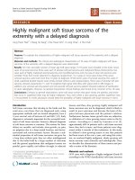
Most of the tumor tissue had the characteristics of poorly
differentiated carcinoma (Figure 1). There was no
squamous cell or glandular differentiation. The mesench-
ymal component was composed of sheets of un differen-
tiated spindle cells and areas of cartilage (Figure 2).
Numerous sections from theuterusandtheadnexae
showed no evidence of tumor.
Immunohistochemistry
Immunohistochemical staining for cytokeratin 7 deco-
rated cells of the epithelial component and scattered
cells within the mesenchymal component (Figure 3 and
Figure 4). Vimentin was strongly positive in mesenchy-
mal component and sporadicaly in the epithelial areas
(Figure 5). Cytokeratin 20 and calretinin were negative
in both epithelial and mesenchymal elements.
Discussion and the review of Literature
Malignant mixed Müllerian tumor (MMMT) is a
highly aggressive tumor consisted of malignant
Figure 1 Histological picture showing the admixed carcinoma
and spindle cell sarcomatous elements (HE × 200).
Figure 2 Histological picture showing malignant cartilage
admixed with sheets of undifferentiated spindle cells (HE ×
400).
Figure 3 Immunohistochemical stain with Cytokeratin 7 shows
strong immunoreactivity of the undifferentiated epithelial cells
(× 400).
Kurshumliu et al. World Journal of Surgical Oncology 2011, 9:17
/>Page 2 of 4
epithelial and stromal cells. MMMTs were traditionally
regarded as a subtype of uterine sarcomas or a mixture
of true carcinoma and sarcoma, however several
reports suggested a monoclonal origin of t hese tumors
[1-3] Interestingly, molecular data published by Wada
and co-workers [ 4] suggested that although most carci-
nosarcomas are combination tumors, some develop as
collision tumors.
The morphology of the present tumor is consistent
with malignant mixed Müllerian tumor. The epithel ial
component exhibited positivity for cytokeratin 7 and
Vimentin and was negative for Cytokeratin 20. More-
over, the mesenchymal component was diffusely positive
for Vimentin, focally for CK 7 and exhibited areas of
heterologous malignant cartilage.
The primary peritoneal location and origin was con-
firmed after thorough gross and microscopic
examination of the uterus and adnexae. Furthermore,
absence of calretinin expression suggested that the
tumor was of Müllerian rather than pure mesothelial
origin [6]. A search of the literature revealed 30 pre-
viously reported cases of extragenital MMMT (Addi-
tional file 2) with the majority of the patients being in
the postmenopausal age [5-19]. Twenty-two of the
reported cases were of primary peritoneal origin and
most of them arose i n the pelvis [19]. Thirteen were
heterologous type [19].
In the homologous type MMMT, mesothelioma would
rightfully be considered as a possibility [6]. However, in
this case a positive reaction of the cells to Calretinin
would b e expected [6]. Immunoreactivity of the epithe-
lial cells to Cytokeratin 7 and negative reaction for cyto-
keratin 20 also points toward Müllerian origin. A
theoretical possibility is of course the origin in endome-
triosis or endosalpingiosis. However, although impossi-
ble t o rule out, the lack of demonstrable endometriosis
associated with the patient’s current tumor makes this
hypothesis unlikely [16].
Several theories have emerged in the attempt to
explain the biphasic appearance of the tumor, the most
important of which are the “collision”, “ conversion ” and
the “combination” theory [1]. Sternberg et al. [20] was
the first to suggest the conversion, stating that sarcoma-
tous el ements may devel op from carcinoma. They
described a case of metastatic heterologous type carci-
nosarcoma of the omentum with primary endometrial
origin. There was no evidence of sarcoma component in
the primary tumor. From that time on ward, a number
of cases with metachronous or synchronous gynaecolo-
gic carcinoma have b een reported [11,14,16]. Masuda et
al. [21] further supported the conversion theory with
their study in which cell lines established from malig-
nant mixed Müllerian tumors showed the ability of t he
epithelial tumor cells to t ransform into epithelial,
mesenchymal or both types of differentiation in vitro,
while the mesenchymal cells did not show similar
capabilities.
MMMT has a poor prognosis with most of the patients
following a rapidly fatal course regardless of the initial
tumor stage [9]. A review done by Garamvoelgyi et al. [14]
showed that most patients passed away within one year
with median postoperative survival time being 14 months.
Due to the rarity of the disease, limited data regarding the
management of peritoneal MMMT exist. Treatment mod-
alities include surgery, chemotherapy and irradiation with
various survival outcomes. Ko et al. [19] reported on a
patient that was treated with optimal tumor debulking and
a combination of chemotherapy with Ifosfamide and Cis-
platin, followed by pelvic irradiation. There were no signs
of recurrence for 48 months and was the case with the
longest disease-free survival in the reported literature.
Figure 5 Immunohistochemical stain with Vimentin shows
areas with strong immunoreactivity of the epithelial and
mesenchymal cells (× 400).
Figure 4 Immunohistochemical stain with Cytokeratin 7 shows
scattered Cytokeratin 7-positive cells in the mesenchymal
component (× 400).
Kurshumliu et al. World Journal of Surgical Oncology 2011, 9:17
/>Page 3 of 4
Conclusion
Based on the ample evidence of the previous report s,
and the immunohistochemical analysis of our case, we
believe that this is a monoclonal tumor with the carci-
noma being the “ precursor” element. Nevertheless,
further molecular and genetic evidence is needed to
support such a conclusion.
Consent
Informed consent was obtained from the patient for
publication of this case r eport and accompanying
images.
Additional material
Additional file 1: Immunohistochemical stains.
Additional file 2: Primary peritoneal MMMT reported in the
literature.
Author details
1
Institute of Anatomic Pathology, Medical School, University Clinical Center,
Prishtina, Kosovo.
2
Department of Gynecology and Obstetrics, Herlev
University Hospital, Copenhagen, Denmark.
3
Institute of Anatomic Pathology,
Herlev University Hospital, Copenhagen, Denmark.
Authors’ contributions
FK prepared the manuscript and reviewed the literature. HRH provided the
clinical data. VRH reviewed the slides, LGL reviewed the manuscript, TH
reviewed the slides and supervised the preparation of the manuscript.
Competing interests
The authors declare that they have no competing interests.
Received: 2 May 2010 Accepted: 4 February 2011
Published: 4 February 2011
References
1. McCluggage WG: Malignant biphasic uterine tumours: carcinosarcomas
or metaplastic carcinomas? J Clin Pathol 2002, 55:321-325.
2. Mayall F, Rutty K, Campbell F, Goddard H: p53 immunostaining suggests
that uterine carcinosarcomas are monoclonal. Histopathology 1994,
24(3):211-214.
3. Watanabe M, Shimizu K, Kato H, Imai H, Nakano H, Sugawa M, Shiraishi T:
Carcinosarcoma of the uterus: immunohistochemical and genetic
analysis of clonality of one case. Gynecol Oncol 2001, 82(3):563-567.
4. Wada H, Enomoto T, Fujita M, Yoshino K, Nakashima R, Kurachi H, Haba T,
Wakasa K, Shroyer KR, Tsujimoto M, Hongyo T, Nomura T, Murata Y:
Molecular evidence that most but not all carcinosarcomas of the uterus
are combination tumors Cancer Res. 1997, 57:5379-5385.
5. Ober WB, Black MB: Neoplasms of the subcoelomic mesenchyme: report
of two cases. Arch Pathol 1955, 59:698-705.
6. Sumathi VP, Murnaghan M, Dobbs SP, McCluggage WG: Extragenital
Müllerian carcinosarcoma arising from the peritoneum: report of two
cases. Int J Gynecol Cancer 2002, 12:764-767.
7. Lauchlan SC: The secondary Müllerian system. Obstet Gynecol Surg 1972,
27:133-146.
8. Zelmanowicz A, Hildesheim A, Sherman ME, Sturgeon SR, Kurman RJ,
Barrett RJ, Berman ML, Mortel R, Twiggs LB, Wilbanks GD, Brinton LA:
Evidence for a common etiology for endometrial carcinomas and
malignant mixed Müllerian tumors. Gynecol Oncol 1998, 69:253-257.
9. Friedrich M, Villena-Heinsen C, Mink D, Bonkhoff H, Schmidt W:
Carcinosarcoma, endometrial intraepithelial carcinoma and
endometriosis after tamoxifen therapy in breast cancer. Eur J Obstet
Gynecol 1999, 82:85-87.
10. Nascimento MC, Choo PS, Bligh J, Obermair A: Primary peritoneal
malignant mixed Müllerian tumor (MMMT): a case report. Cancer Therapy
2004, 2:571-574.
11. Shen DH, Khoo US, Xue WC, Ngan HYS, Wang JL, Liu VWS, Chan YK,
Cheung ANY: Primary peritoneal malignant mixed Müllerian tumors: a
clinicopathologic, immunohistochemical and genetic study. Cancer 2001,
91:1052-1060.
12. Shintaku M, Matsumoto T: Primary Müllerian carcinosarcoma of the
retroperitoneum: report of a case. International J Gynecol Pathol 2001,
20:191-195.
13. Ibanez-Manlapaz IG, McCoy D, Vincent V III, Ule UJ: Malignant mixed
Müllerian tumor of the extra-genital coelomic epithelium: report of two
cases. Pathology Oncology Research 1997, 3:130-134.
14. Garamvoelgyi E, Guillou I, Gebhard S, Salmeron M, Seematter RJ, Hadji MH:
Primary malignant mixed Müllerian tumor (metaplastic carcinoma) of
the female peritoneum. Cancer 1994, 74:854-863.
15. Choong SYM, Scurry JP, Planner RS, Grant PT: Extrauterine malignant
mixed Müllerian tumor of primary peritoneal origin. Pathology
1994,
26:497-498.
16. Garde JR, Jones MA, McAfee R, Tarraza HM: Extragenital malignant mixed
Müllerian tumor. Gynaecol Oncol 1991, 43:186-190.
17. Weisz-Carrington P, Bigelow B, Schinella RA: Extragenital mixed
heterologous tumor of Müllerian type arising in the cecal peritoneum:
report of a case. Dis Colon Rectum 1977, 20(4):329-333.
18. Hasiuk AS, Petersen RO, Hanjani P, Griffin TD: Extragenital malignant mixed
Müllerian tumor: case report and review of the literature. Am J Clin
Pathol 1984, 81:102-105.
19. Ko ML, Jeng CJ, Huang SH, Shen J, Tzeng CR, Chen SC: Primary peritoneal
carcinosarcoma (malignant mixed Müllerian tumor): Report of a case
with five-year disease free survival after surgery and chemoradiation
and a review of literature. Acta Oncologica 2005, 44(7):756-760.
20. Sternberg WH, Clark WH, Smith RC: Malignant mixed Müllerian tumor
(malignant mixed mesodermal tumor) of the uterus: a study of 21 cases.
Cancer 1954, 7:704-724.
21. Masuda A, Takeda A, Fukami H, Yamada C, Matsuyama M: Characteristics
of cell lines established from mixed mesodermal tumor of the human
ovary: carcinomatous cells are changeable to sarcomatous cells. Cancer
1987, 60:2969-2703.
doi:10.1186/1477-7819-9-17
Cite this article as: Kurshumliu et al.: Primary malignant mixed müllerian
tumor of the peritoneum a case report with review of the literature.
World Journal of Surgical Oncology 2011 9:17.
Submit your next manuscript to BioMed Central
and take full advantage of:
• Convenient online submission
• Thorough peer review
• No space constraints or color figure charges
• Immediate publication on acceptance
• Inclusion in PubMed, CAS, Scopus and Google Scholar
• Research which is freely available for redistribution
Submit your manuscript at
www.biomedcentral.com/submit
Kurshumliu et al. World Journal of Surgical Oncology 2011, 9:17
/>Page 4 of 4