báo cáo khoa học: "Ectopic thymoma presenting as a giant intrathoracic tumor: A case report" ppt
Bạn đang xem bản rút gọn của tài liệu. Xem và tải ngay bản đầy đủ của tài liệu tại đây (1.25 MB, 4 trang )
CAS E REP O R T Open Access
Ectopic thymoma presenting as a giant
intrathoracic tumor: A case report
Masahiro Kitada
1*
, Kazuhiro Sato
1
, Yoshinari Matsuda
1
, Satoshi Hayashi
1
, Yoshihiko Tokusashi
2
, Naoyuki Miyokawa
2
and Tadahiro Sasajima
1
Abstract
Ectopic thymoma rarely presents as an intrathoracic tumor. We report a case of ectopic thymoma presenting as a
giant right intrathoracic tumor that was treated with resection. The patient was a 50-year -old Japanese woman
who presented with the chief complaint of chest pain. Detailed examination revealed a solid tumor measuring 15
× 10 × 8 cm in diameter, with a clear border. The Imaging findings suggested a solitary fibrous tumor, and surgery
was performed. At surgery, the tumor was found to beadherent to the diaphragm, mediastinal pleura, and lower
lobe of the lung, although it could be dissected with relative ease and was removed. Pathological diagnosis
indicated a type B1 tumor with no capsular invasion according to the World Health Organization classification, and
a diagnosis of Masaoka stage I thymoma was made. No continuity with the normal thymus tissue was seen, and
the thymoma was considered to be derived from ectop ic thymic tissue in the pleura.
Background
Thymomas usually manifest in the anterior-superior
mediastinum, and ectopic thymomas account for only 4%
of all thymomas. Among ectopic thymoma, intrathoracic
tumors of pleural origin are rather rare. We report,
herein, a patient with a giant intrathoracic tumor that
was discovered during a clinical workup to determine the
cause of chest pain in the patient. The tumor was diag-
nosed as a thymoma t hat was difficult to differentiate
from solitary fibrous tumor (SFT) by diagnostic imaging.
Case
A 50-year-old Japanese woman with the chief complaint
of chest pain was examined at a loc al hospital. A chest
radiograph revealed a giant tumor in the right lower lung
field, and the patient was referred to our department.
The patient had no pertinent personal or family history,
and had never smoked. Respiratory sounds in the right
lower lung field were diminished, but no other abnormal-
ities were detected on physical examination. Respiratory
function tests revealed a vital capacity (VC) of 1,470 mL
and a percent predicted VC of 46.7%, indicative of
restrictive pulmonary disease. No abnormalities were
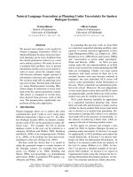
identified on blood biochemistry. A plaine chest x-ray
(Figure 1) showed a giant tumor shadow measuring 15 ×
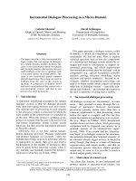
13 cm in the right lower l ung field, and chest computed
tomography (Figure 2) showed a solid tumor measuring
15 × 10 × 8 c m in the right thoracic c avity. The tumor
showed a clear borders and internal calcification, and was
found todisplace the diaphragm downward and th e heart
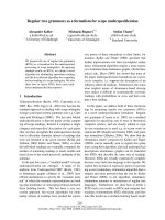
to the left. In addition, on chest magnetic resonance ima-
ging (MRI) (Figure 3), the tumor was visualized as an iso-
intensity relative to the skeletal muscle on T1-weighted
images, while on T2-weighted images, partial inclusion of
weak signals hypointensity of moderate signal strength.
Moreover, diffusion-weighted imaging revealed slightly
heterogeneous signal hyperintensity, but no findings sug-
gestive of degeneration or necrosis. A fibrous septum was
found within the tumor, which showed a trabecular
growth pa ttern, which led to t he diagnosis of SFT, and
surgery was performed. A small, the sixt h intercostal
video-assisted thoracotomy was performed, and the tumor
was found to be slightly adherent to the diaphragm, med-
iastinal pleura, and lower lobe of the right lung. However,
the tumor could be relatively easily dissected from these
organs and removed. No continuity with normal thymic
tissue was seen. Intraoperative rapid pathological diagnosis
indicated a thymoma or lymphoma, and the surgery was
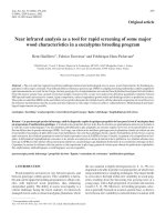
terminated. The excised specimen (Figure 4) showed a
tumor measuring 5 × 10 × 8 cm and weighing 430 g,
* Correspondence:
1
Department of Surgery, Asahikawa Medical University, Midorigaoka-Higashi
2-1-1-1, Asahikawa, Hokkaido 078-8510, Japan
Full list of author information is available at the end of the article
Kitada et al. World Journal of Surgical Oncology 2011, 9:66
/>WORLD JOURNAL OF
SURGICAL ONCOLOGY
© 2011 Kitada et al; licensee BioMed Central Ltd. This is an Open Access article distributed under the terms of the Creative Commons
Attribution License ( which permits unrestricted use, distribution, and reproduction in
any m edium, provided the original work is properly cited.
covered by a thin, fibrous membrane. The mass was elastic
and soft, and the cut surface was lobulated and pale brown
in color. Histopathological examination of sections stained
with hematoxylin-eosin (HE) (Figure 5) revealed abundant
lymphocytes and large, bright tumor cells. No invasion of
the capsule was evident, leading to the diagnosis of a
Masaoka stage I thymoma. Immunohistochemical staining
(Figure 6) showed mature lymphocytes mainly composed
of T cells, mixed in a complex pattern with cytokeratin-
positive epithelial cells. These findings led to the diagnosis
of lymphocyte-predominant thymoma (type B1 thymoma).
The postoperative course was good, and the patient has
shown no evidence of recurrence as at the time of writing.
Discussion
Thymomas are tumors developing mainly in the thymus,
are located in the anterior mediastinum, with 96% of the
tumors occurring in the anterior or anterosuperior med-
iastinum, and only 4% being ectopic tumors [1,2]. Ecto-
pic thymomas have been described in the neck [3],
middle mediastinum [4,5] posterior mediastinum, lung
[6], and pleura [7,8], few reports have described giant
intrathoracic tum ors. Thymomas are generally asympto-
matic, but symptoms such as chest pain and respiratory
discomfort can be caused by compression of the sur-
rounding organs due to growth of the tumor. Symptoms
such as superior vena cava syndrome can also be coused
by tu mor invasion of t he surrounding tissues, myasthe-
nia gra vis, pure red cell apalasia, hypogammaglobuline-
mia. These symptoms/complications can lead to the
discovery of the tumor. In addition, some patien ts show
Figure 1 Plain chest radiograph showing a mass lesion in the right lower lung.
Figure 2 Chest computed tomograph showing a solid tumor
(15 × 10 × 8 cm) with a clear borders and internal calcification
in the right thoracic cavity.
Kitada et al. World Journal of Surgical Oncology 2011, 9:66
/>Page 2 of 4
multiple lung metastases or pleural dissemination arising
from recurrence or metastasis. The intrathoracic tumor
in the present patient was discovered during the course
of a clinical workup for chest pain, caused by compres-
sion of the surrounding organs. The patient also showed
restricted impairment of pulmonary function due to the
pressure on normal lung tissue, and surgical removal of
the t hymoma as quickly as possible was therefo re con-
sidered necessary.
Definitive diagnosis is needed before surgical removal
of a thymoma is planned. The differential diagnosed for
giant intrathoracic tumors include SFTs, tumors o f
pleural origin, such as malignant pleural me sothel ioma
or sarcoma, chest wall tumors, and metastatic tumors.
In our patient, SFT was initially suspected on the basis
of the MRI findings, including the shape, signal status
under various weightings, and the presence of numerous
linear non-signals that were considered to indicate flow
voids within the lesion. It was considered that percuta-
neous needle biopsy would yield a definitive diagnosis,
but this procedure was not performed considering the
risk of tumor cell dissemination and bleeding from the
tumor. Hemorrhagic shock induced by spontaneous rup-
ture of a giant thymoma has been reported [9], and cau-
tion is warranted when considering biopsy.
Figure 3 Chest magnetic resonance imaging. The tumor appeared isointense relative to the skeletal muscle on T1-weighted image, while T2-
weighted images show partial inclusion of weak signal hypointensities of moderate signal strength.
Figure 4 Excised specimen of the tumor showi ng a smooth
margin and calcification within.
Figure 5 Hi stopathological exa mination (hematoxylin and
eosin, × 400) revealed abundant lymphocytes and large, bright
tumor cells. Cells with chromatin-poor nuclei are evident.
Kitada et al. World Journal of Surgical Oncology 2011, 9:66
/>Page 3 of 4
Consent statement
Informed consent was obtained from the patient for
publication of this case report and of the accompanying
images. A copy of the written consent is available for
review by the Editor-in-Chief of this journal.
Author details
1
Department of Surgery, Asahikawa Medical University, Midorigaoka-Higashi
2-1-1-1, Asahikawa, Hokkaido 078-8510, Japan.
2
Department of Clinical
Pathology, Asahikawa Medical University, Midorigaoka-Higashi 2-1-1-1,
Asahikawa, Hokkaido 078-8510, Japan.
Authors’ contributions
MK operated on this case and analyzed all the data. KO, KS, YM, and SH
assisted in the operation. YT and NM diagnosed the pathology of this case.
TS did a professor of the Department of Surgery and had the guide of this
paper. All authors read and approved the final manuscript.
Competing interests
The authors declare that they have no competing interests.
Received: 27 February 2011 Accepted: 28 June 2011
Published: 28 June 2011
References
1. Detterbeck FC, Parsons AM: Thymic tumors. Ann Thorac Surg 2004,
77:1860-69.
2. Fukayama M, Maeda Y, Funata N, Koike M, Saito K, Sakai T, Ikeda T:
Pulumonary and pleural thymoma. Diagnostic application of lymphocyte
markers to the thymoma of unusual site. Am J Clin Pathol 1998,
89:617-21.
3. Yan B, Lim D, Petersson F: Ectopic cervical thymoma: a report of two
cases of a rare entity frequently misdiagnosed on fine needle aspiration
cytology and frozen section. Head Neck Pathol 2010, 4:152-6.
4. Nakamura Hiroshige, Adachi Yoshi, Fukuoka Shinji, Miwa Ken,
Haruki Tomohiro, Taniguchi Yuji: Thoracoscopic Resection of middle
mediastinal noninvasive thymoma; report of a case. Surg Today 2007,
37:787-789.
5. Minniti Salvatore, Valentini Marvi, Pinali Lucia, Malago Roberto,
Lestani Maurizio, Procacci Carlo: Thymic mass of the middle mediastinum-
Report of 2 cases and review of the literature. J Thoracic Imaging 2004,
19:192-195.
6. Moran CA, Suster S, Fishback NF, et al: Primary intrapulmonary thymoma.
A clinicopathologic and immunohistochemical study of eight cases. Am
J surg Pathol 1995, 19:304-12.
7. Moran CA, Travis WD, Rosado-de Christenson M, Koss MN, Rosai J:
Thymomas presenting as pleural tumors: report of eight cases. Am J
Surg Pathol 1992, 16:138-44.
8. Yamazaki K, Yoshino I, Oba T, Yohena T, Kameyama T, Tagawa T, Kawano D,
Koso H, Maehara Y: Ectopic pleural thymoma presenting as a giant mass
in the thoracic cavity. Ann Thorac Surg 2007, 83:315-17.
9. Santoprete Stefano, Ragusa Mark, Urbani Moira, Puma Francesco: Shock
induced by spontaneous rupture of a giant thymoma. Ann Thorac Surg
2007, 83:1526-28.
doi:10.1186/1477-7819-9-66
Cite this article as: Kitada et al.: Ectopic thymoma presenting as a giant
intrathoracic tumor : A case report. World Journal of Surgical Oncology
2011 9:66.
Submit your next manuscript to BioMed Central
and take full advantage of:
• Convenient online submission
• Thorough peer review
• No space constraints or color figure charges
• Immediate publication on acceptance
• Inclusion in PubMed, CAS, Scopus and Google Scholar
• Research which is freely available for redistribution
Submit your manuscript at
www.biomedcentral.com/submit
Figure 6 Immunohistochemical staining (keratin staining ×
400) showing epithelial cells distributed in a mesh-like form,
mixed among lymphocytes at a ratio of nearly 1:1.
Kitada et al. World Journal of Surgical Oncology 2011, 9:66
/>Page 4 of 4