Báo cáo khoa học: "Recent advances in the surgical care of breast cancer patients" pps
Bạn đang xem bản rút gọn của tài liệu. Xem và tải ngay bản đầy đủ của tài liệu tại đây (579.53 KB, 17 trang )
REVIEW Open Access
Recent advances in the surgical care of breast
cancer patients
Alessandra Mascaro, Massimo Farina, Raffaella Gigli, Carlo E Vitelli, Lucio Fortunato
*
Abstract
A tremendous improvement in every aspect of breast cancer management has occurred in the last two decades.
Surgeons, once solely interested in the extipartion of the primary tumor, are now faced with the need to incorpo-
rate a great deal of information, and to manage increasingly complex tasks.
As a comprehensive assessment of all aspects of breast cancer care is beyond the scope of the present paper, the
current revi ew will point out some of these innovations, evidence some controversies, and stress the need for the
surgeon to specialize in the various aspects of treatment and to be integrated into the multisciplinary breast unit
team.
Introduction
No other solid cancer has witnessed such a tremendous
change and improvement in terms of diagnosis and
management as breast cancer in the last 2 decades. This
remains the most common cancer among women
worldwide [1].
Breast cancer management has become increasingly
complex, a nd requires a comprehensive assessment of
multiple tasks in addition to the simple extirpation of
the primary tumor, including breast imaging, advanced
pathology, nuclear medicine and a variety of adjuvant
therapies, both local a nd systemic. This has shifted
breast cancer treatment into a multidisciplinary science.
Only a few decades ago, women with breast cancer
were uniformly treated with radical mastectomy and
total axillary dissection to achieve good loco-regional
control and the possibility of full recovery. Conservative
and selective surgical approaches to the breast and to
the axilla, once viewed with scepticism, have now
become standard of therapy for most patients [2,3].
Earlier detection and more effective treatments have
resulted both in an increasing percentage of small breast
cancers found at the initial diagnosis and in a small
decline in mortality [2].
Howewer, as the current goal for breast cancer
patients is “conservation” instead “the more radical exci-
sion the better” , the impact of local recurrence on
surviv al remains a relevant issue, and is presently a sub-
ject of research and debate.
The aims of this review are to analyze t he most
important changes which have occurred in the last sev-
eral years in the surgical management of breast cancer
patients and to rev iew some relevant issues such as sen-
tinel lymph node biopsy, the impact of local therapy on
survival, and the aesthetic results.
Non Palpable Lesions and Localization Techniques
Breast c ancer screening has dramatically increased the
diagnosis of suspicious, non-pa lpable breast lesions, and
therefore also the need to localize them in order to plan
surgical treatment [4]. Furthermore, patients with a
breast cancer removed with clear margins at the first
excision seem to have a decreased risk of local recur-
rence compared with patien ts who need further re-exci-
sions to achieve negative margins [5].
This represents a “ hot” topic in breast surgery, since
appr oximat ely 50% of breast cancers in modern surgical
practices are non palpable, and this incidence is cer-
tainly destined to increase [6].
Today, pre-operative confirmation of malignancy is
almost always achieved by fine-needle or core-biopsy,
and therefore, we need to localize these small cancers to
allow a one-step precise and directed excision.
Compared with their palpable counterpart, non-palp-
able lesions are associated with both a lower stage of
disease and a substantially decreased incidence of lymph
node involvement [7].
* Correspondence:
Department of Surgery, Senology Unit, San Giovanni-Addolorata Hospital, Via
Amba Aradam, 9, 00187 Rome, Italy
Mascaro et al. World Journal of Surgical Oncology 2010, 8:5
/>WORLD JOURNAL OF
SURGICAL ONCOLOGY
© 2010 Mascaro et a l; licensee BioMed Central Ltd. This is an Open Access article distributed under the terms of the Creative Commons
Attribution License ( censes/by/2.0), which permits unrestricted use, distribution, and reproduction in
any mediu m, provided the original work is properly cited.
Wire localization (WL) is the most common technique
used to identify small nodules, microcalcifications or
parenchymal distorsions. Howewer, it has some disad-
vantages such as pain and discomfort in some patients,
and occasiona lly carries risks of complications including
dislodgement of the wire, intraoperative wire transec-
tion, retention o f wire fragments, thermal injury with
the use of cautery, hematoma and even syncope. WL is
performed in most institutions as an additional proce-
dure, outside the operating room, with further problems
related to organization and scheduling.
Successful localization with free margins of resection
is not always achieved with this technique and failures,
with consequent r e-excisions, are reported in up to 33%
of cases [6-10].
A precise localization of a br east tumor with the wire
is not always possible, and the angle of access and tra-
jectory depends, in part, on the radiologist’ sability.
Further more, the introduction of the wire directly above
the lesion may be technically problematic, especially
under stereotaxic guidance in locations such as the
inferior quadrants.
For this reasons several new techniques have been
introduced in order to achieve breast tumor localization.
Radioguided occult lesion localization (ROLL) is a use-
ful method to detect nonpalpable lesions through the
injection of a nuclear tracer (99 m TC-labelled colloidal
albumin) directly around the tumor under ultrasound or
stereotaxic guida nce. Then, the excision of the primary
tumor is guided by a gamma probe, and a sentinel node
biopsy can be performed at the same time if needed
[11-13].
Unlike the WL, the procedure is generally more
straightforward and well tolerated, and the success rate
is reported to be very high [14-23] (Table 1).
Although RO LL has been shown to be comparab le to
WL in at least 2 restrospective [24,25] and four prospec-
tive-randomized studies [19,20,22,26] with regards to the
ability to identify the lesion, four reports have demon-
strated a statistical difference in achieving tumor-free
margins in favor of the former technique [17,24,26,27].
Another technique for loca lization of non-palpable
breast tumors is represented b y intraoperative ultra-
sound (IOUS).
It satisfies most requirements for an ideal technique to
localize non-palpable breast tumors which are well
visualized by ultrasound, while directing planes of sur-
gery during the exc ision. This in turn is helpful in guar-
anteeing both negative margins and an adequate
contour of resection in order to minimize the volume of
excision.
Identification rate of non-palpable lesions and free
margins of resection obtained through this procedure
are extremely high [28-34] (Table 2).
Furthermore, microcalcifications, usually visible only
by mammography, are sometimes associated with sono-
graphic alterations that can be detected, and removal of
such lesions under ultrasound guidance can sometimes
be performed [35].
Implications of Local Therapy
As conser vative approaches have developed in the last
three decades and represent the standa rd of care for
breast cance r patients around the world, the incidence
of local recurrence (LR) has been widely studied. It
occurs in 5-10% of patients at 10 y ears, and it is more
pronounced in the first 3 or 4 years after primary sur-
gery [36,37].
Although several factors have been associated with the
risk of LR, at the multivariate analysis only age, status of
surgical margins and postoperative radiotherapy seem to
be independently correlated with it [38]. Patients with
multifocal tumors, once uniformely thought to be asso-
ciated with a higher risk of LR, and therefore treated
with mastectomy, are now often offered breast conserva-
tion, when technically feasible, as most studies seem to
indicate that the LR rate is not h igher in these cases
than previous reports for unifocal can cers [39]. Simi-
larly, infiltrating lobular carcinoma is probably not asso-
ciated with a higher i ncidence of LR compared to the
ductal counterpart if resected with negative margins
[40].
Table 1 Complete excision rate of non palpable lesions
by ROLL
Author Year N Complete excision rate (%)
Gennari [14] 2000 647 99
Tanis [15] 2001 45 87
Ronka [16] 2004 215 93
Thind [17] 2005 68 84
Van Rijk [18] 2007 293 89
Moreno [19] 2008 61 93
Medina-Franco [20] 2008 50 89
Lavouè [21] 2008 72 85
Van Esser [22] 2008 40 78
Sarlos [23] 2009 100 98
Table 2 Identification rate of small lesions by US
Author Year N Identification (%) Free Margins (%)
Harlow [28] 1999 65 100 97
Smith [29] 2000 81 100 96
Kaufman [30] 2003 100 100 90
Bennett [31] 2005 103 100 93
Ngo [32] 2007 70 96 94
Haid [33] 2007 299 100 100
Fortunato [34] 2008 77 100 97
Mascaro et al. World Journal of Surgical Oncology 2010, 8:5
/>Page 2 of 17
The influe nce of age on th e risk of LR is striking, and
many reports have shown that this is increased three-
fold for women less than 40 years of age [38,41-43].
Furthermore, younger patients show a statistically signif-
icant reduction of LR in several “ boost trials” ,again
demonstrating the importance of an appropriate l ocal
therapy particularly in this age group [44].
It is interesting t o note that despit e the wide spread
use of conservative approaches in breast cancer patients,
there is no general agreement even on the definition of
“ negative” margins, and many describe such as the
absence o f tumor at the microscopic or inked margin,
or with 1- 3 mm clearance. It is clear that a high percen-
tage of patients whose tumors are 2-5 mm from the
radial margins have residual disease at re-excision [45].
For this reason, and despite best efforts, as many as 20-
25% of patients in many institutions around the world
return to the operating room after initial surgery for re-
excision [46]. While many reports fail to describe a sta-
tistically significant impact of margins on LR, most
would agree that one of the primary goals of conserva-
tive surgery is the removal of the primary tumor with a
portion of normal breast tissue, so as to maintain a
good breast shape [47-54].
Although the results of six prospective randomized
trials in patients with invasive breast cancer have
demonstrated that lumpectomy/quadrantectomy plus
RT and mastectomy have equivalent survival results
[55-60], it is worthwhile to remember that the first
conservation trial, the Guy’ s wide excision study
initiated in the 60’s, has shown a decreased survival in
the group treated conservatively [61]. This suggests
that poor surgical removal of t he primary tumor, pos-
sibly with dubious margins and without inking of the
specimen, together with employment of suboptimal
post-operative radiothera py, may lead to a negative
impact not only on local control but also on survival
[57,61].
Although additional retrospective data has been accu-
mulated in the last few years suggesting that failure of
local control has an impact on survival [62], the most
strikin g evidence comes from the EBCTCG meta-ana ly-
sis [63].
This ha s shown that adj uvant RT after BCS not only
may improve local control, but it may also reduce 15-
year breast cancer mortality. The effect of radiation on
LC seems more pronounced in node positive patients,
while the effect on surv ival remains important both for
node-negative and node-positive patients [64,65].
This has lead man y to suggest that for every four
women for whom local failure is prevented, one life
can be saved. As this disease is prevalent, and LR after
quadrantectomy and radiation is far from being an
exceptional event, this seems quite an important i ssue.
Minimalistic approaches are no longer viable for
women with breast cancer, a nd the aim of the contem-
porary surgeon is to team up with all available specia-
lists, and t o coordinate efforts to reach the goal of local
control.
Skin Sparing Mastectomy
Although breast conservation surgery (BCS) has become
the gold standard for patients with early breast cancer,
mastectomy remains an option and it is necessary in at
least 20% of those women with multicentric tumors,
widespread DCIS, and large or recurrent tumors [66].
Sometimes the risk of an unpleasant cosmetic result
with conservative surgery to achieve tumor-free margins,
or personal desire to avoid radiation therapy plays a role
in the decision process.
New options are now available for these women and
they repre sent the forefront of the surgical therapy for
breast cancer patients.
Oncologic need to remove the skin envelope or the
nipple-areola complex has never been proved, and has
been lately challenged on solid evidence and back-
ground. Immediate breast reconstruct ion (IBR), a proce-
dure once discouraged for some years after primary
surgery because of fear of rel apse, is now performed
routinely for an increasing number of patients. This has
a profoundly positive psychological effect, and allows for
a more s olid recovery of these women so touched by
this disease [67].
Skin sparing mastectomy (SSM) has been increasingly
used in the last 15 years to improve cosmesis because
the skin envelope is preserved and the surgical access is
limited to a small elliptical incision around the areola
[68]. Our understanding that skin involvement is rare is
corroborated by pathologic studies, and when present, it
is usually over the primary tumor site, or is found in
cases with advanc ed disease, skin tethering, or lymphatic
emboli [69]. However, as maximal skin preservation is
desirable, special technical considerations are to be
addressed by the surge on because the risk of leaving
some glandular tissue behind can be as high 10% if skin
flaps are more than 5 mm thick [70].
Clinical experience has confirmed so far that SSM has
very acceptable results in terms of local control even in
those studies with longer follow-up and is comparable
to modified radical mastectomy bo th in terms of local
control and survival [71-86] (Table 3).
Complications after SSM and immediate breast recon-
struction are reported in about 15% of cases, and
include flap necrosis and implant loss [87-89].
However, this risk must be weighed with the advan-
tage in cosmetic result and in patient satisfaction (as
defined by perception of body image, social activity and
sexual aspects), because these outcomes are better in
SSM with IBR compared with radical mastectomy [84].
Mascaro et al. World Journal of Surgical Oncology 2010, 8:5
/>Page 3 of 17
We favor IBR in almost all c ases, and therefor e routi-
nely perform SSM to allow the plastic surgeon to inter-
vene more comfortably at the same time. Sometimes,
post-operative radiation therapy may be needed, and
although several studies and current clinical recommen-
dation report that the rate of complication is too high if
an implant is inserted in this setting [74,90-92], in
recent years a few studies have reassessed this issue
[93-96]. We believe that this is still an option in selected
cases, as it allows the patient to start more readily adju-
vant systemic therapies if nee ded, and when it fails, it
does not preclude or negatively influence possible auto-
logus conversion or final outcome.
Nipple-Sparing Mastectomy
“ Nipple sparing mastectomy” (NSM) is the ultimate
challenge of this process which aims for an interaction
between conservative techniques and radical surgery. In
this procedure, the skin flap covering the breast gland
and the nipple-areola complex (NAC) are preserved. In
some cases the major ducts are removed.
In the past, the nipple has be en routinely removed for
fear of occult tumor involvement, although this has
probably be en overestimated. Many clinical studies have
shown that this involvement varies from 6 to 23%
depending on the size of the primary tumor, its location,
multicentricity, lymph node positivity and the presence
of extensive intraductal component [97-102] (Table 4).
We believe that this occurrence is rare in modern
clinical practice and although the risk is real, patients
can probably be safely selected for this approach.
Nevertheless, exact indications and contraindications
to this procedure are not well defined, and the incidence
of nipple involvement is reported to be as high as 50%
for tumors measuring more than 4 cm or located closer
than 2 cm from the nipple [103]. Therefore, the best
candidates for NSM are patients with no large tumor
(T1-T2), with lesions at least 1 cm from the areola or 2
cm from the nipple, or small multicentric carcinomas
[101].
Further more, nipple involvement is rare if the retro ar-
eolar margin is free of disease [104].
A strategic issue is to avoid partial or total nipple or
areola necrosis because, although this can be easily trea-
ted postoperatively and under local anesthesia, it results
in psychological distress to the patient, and it must be
considered a failure of the procedure itself.
Therateofnipplenecrosisvariesfrom0to15%
[101,105-110] (Table 5).
Surgical technique is extre mely important. It is now
well understood that the use of periareolar incisions
should be abandoned, as it negatively affects the vascu-
lar supply of the nipple-areola complex, and that either
a radial or a lateral incision seem to be more effective in
this regard [109].
Although it is not clear how much tissue can or
should be left under the NAC, or if “ nipple coring”
(removal of the terminal ducts from the inside of the
nipple papilla) should be performed (and how aggres-
sively), results of NSM can been examined in a few ret-
rospective studies published so far, and the local
recurrence rate is shown to be quite low in the majority
of them [103,106,107,110-115] (Table 6).
The role of post-operative radiotherapy following
NSM is unknown at the present, although a three-fold
decrease in the rate of locoregional failure has been
reported in one series [116]. However, in this retrospec-
tive study only large tumors (> 3 cm) were included,
and the site of failure is not clearly described.
Proponents at the European Institute of Oncology
have recently updated their experience reporting on
1,001 patients treated with a single intra-operative radio-
therapy treatment (21 Gy) with electrons (ELIOT) to the
NAC after NSM in the assumpt ion that this single
radiation dose may sterilize occult cancer foci eventually
left in the glandular tissue behind the areola [115]. This
is the largest experience with NSM, to date, and the
incidence of local recurrence is reported at 1.4% with a
Table 3 Recurrence Rates after SSM
Author Year N LR (%) F/U (mo)
Slavin [71] 1998 51 2 45
Newman [72] 1998 372 6 25
Simmons [73] 1999 77 4 60
Kroll [74] 1999 114 7 72
Rivadeneira [75] 2000 71 6 49
Medina-Franco [76] 2002 176 4 73
Foster [77] 2002 67 4 49
Carlson [78] 2003 565 5 65
Greenway [79] 2005 225 2 49
Margulies [80] 2005 50 0 8
Yano [81] 2007 124 2 34
Patani [82] 2008 83 0 34
Scholz [83] 2008 72 0 42
Ueda [84] 2008 74 5 50
Garwood [85] 2009 64 1 13
Gerber [86] 2009 238 10 101
Table 4 Occult Histologic Nipple Involvement
AUTHOR YEAR PATIENTS (N) NIPPLE INVOLVEMENT (%)
Santini [97] 1989 1291 12
Laronga [98] 1999 286 6
Sikand [99] 2005 220 7
Vlajciz [100] 2005 108 23
Petit [101] 2006 106 10
KG [102] 2008 397 15
Mascaro et al. World Journal of Surgical Oncology 2010, 8:5
/>Page 4 of 17
median follow-up of 20 months. Although some con-
cerns have been raised regarding the possible negative
effects (even long-term) on the vascularity of the NAC
after a single large dose of radiotherapy, the usefulness
of this approach is appealing but currently unproven. Of
interest, in a subgroup of patients, treated with ELIOT,
with very close tumor margins under the areola, no
local recurrence was observed.
Oncoplasty
Onc oplasty has been developed in the last 15 years as a
new surgical approach and incorporates a variety of
relatively simple, common plastic techniques. This has
generated much enthusiasm around the world, among
both by breast and plastic surgeons, and in the UK for-
mal oncoplasty training has been developed [117].
Indeed, oncoplastic surgery represents a step forward
in breast conservation, allowing us to treat tumors in
problematic locations (for example in the lo wer quad-
rants), to avoid poor cosmetic results, asymmetry or
unpleasant scarring in the upper quadrants, and to
obtain wider excisions and tumor free margins [118].
Oncoplasty is safe, as no statistical differences in
terms of local relapse and disease-free survival are evi-
denced when comparing classic quadrantectomies and
oncoplastic approaches [118-120]. It should be consid-
ered for those patients where adequate local excision
cannot be achieved without a significant risk of local
deformity, as it frequently occurs in resection of more
than 20% of breas t volume, or for tumors located in the
central, medial or inferi or quadrants. Other indic ations
include women considering a breast reduction in addi-
tion to excision.
Several volume displacement techniques can be
employed, including glandular remodelling, inferior or
superior pedicle flaps, round block excision, and the
Grisotti flaps. Their description is beyond the scope of
this review.
Centrally located tumors account for 5 to 20% of
breast cancer cases and have long been thought to be
associated with a higher incidence of multicentricity and
multifocality [121,122]. However, other more recent
reports have failed to substantiate a specific correlation
between location of the tumor and multicentricity
[123,124]. For this reason, they represent an important
challenge for breast surgeons, as they have been classi-
cally treated with a mastectomy, and until few years ago
only 7% of central breast cancers were treated with con-
servative surgery [119].
Several studies on the l ocal recurrence rate after cen-
tral quadrantectomy, each with a small number of
patients, show very accept able results even long-term
[125-132] (Table 7).
A d irect comparison between central quadrantect omy
and mastectomy has seldom been studied, and no signif-
icant differences in terms of local failure and overall sur-
vival have been reported [119,127,133-135]. However,
these reports are limited by their retrospective nature
and may not be comparable b ecause mastectomy wa s
Table 5 Nipple Necrosis after NSM
AUTHOR YEAR N PARTIAL NECROSIS % TOTAL NECROSIS %
Crowe [105] 2004 48 6 0
Caruso [106] 2006 50 2 0
Sacchini [107] 2006 192 7 4
Petit [101] 2006 106 10 5
Komorowski [108] 2006 38 5 8
Stolier [109] 2008 82 0 0
Voltura [110] 2008 51 6 0
Table 6 Nipple Sparing Mastectomy: Local Recurrence
AUTHOR YEAR N LOCAL RECURRENCE % FOLLOW-UP (months)
Simmons [111] 2004 17 0 24
Caruso [106] 2006 50 2 66
Sacchini [107] 2006 123 2 25
Denewer [112] 2007 41 0 8
Crowe [113] 2008 149 3 41
Voltura [110] 2008 51 6 18
Sookhan [103] 2008 18 0 11
Gerber [114] 2009 61 12 101
Petit [115] 2009 1001 1.4 20
Mascaro et al. World Journal of Surgical Oncology 2010, 8:5
/>Page 5 of 17
usually performed for larger tumors. Only one prospec-
tive non-randomized study has been published so far,
and it has confirmed an equivalen t outcome in te rms of
local or systemic disease [132].
We believe that by adhering to the principles of
breast-conserving surgery, including comp lete resection
of the primary tumor with a negative margin, these cen-
trally located tumors can be treated adequately by nip-
ple-areolar resection. Adjuvant radiation therapy to the
remainder of the breast can treat subclinical microscopic
disease, if present, with accepTable local control and
adequate cosmesis.
Sentinel Lymph node Biopsy, and Management of Special
Circumstances
Lymph node involvement is the single most important
prognostic factor for survival i n breast cancer patients,
and consequen tly information about it provide both sta-
ging information and guidance regarding treatment
options [136].
SLN biopsy is now considered an adequate axillary
staging procedure for patients who have breast cancer
because it is easy and reproducible if carried out by
experienced clinicians, and carries less morbidity com-
pared to axillary node dissection [137].
Many concerns were raised in the past because SLN
biopsy can result in some false-negative cases. A recent
meta-analysis of 69 trials found the rate of false nega-
tives to be about 7% of the node-positive patients [138].
Much of what is known today regarding SLN biopsy
in breast cancer does not result from randomized trials.
The procedure has been accepted quickly by most dedi-
cated surgeons around the world on the basis of a grow-
ing body of evidence that SLN is effective. Often, patient
demand has overcome the ca ution that surgeons usually
demonstrate before abandoning a well-tested procedure,
such as axillary node dissection. In some cases, rando-
mized trials have been prematurely closed because of
problems in accrual, either because randomization was
not acceptable to patients, or because s urgeons, after
acquiring sufficient experience with SLN biopsy, w ere
unwilling to allow their patients to enter the trial.
Enhanced pathology of the SLN ha s generated much
confusion and even controversy, but it is a key point as
different results can be obtained by different groups
using different protocols. A survey of the European
Working Group for Breast Screening Pathology reported
that 240 pathologists replying to a questionnaire
described some 123 different pathology protocols [139].
The authors’ group recently has proposed a simple,
practical standardized protocol, with slicing at three
levels at 100-micron intervals and double staining with
both hematoxylin and eosin (H&E) and immunohisto-
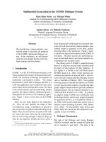
chemistry (MNF116) (Fig ure 1) [140]. This protocol has
allowed our pathologists to increase the diagnosis of
additional nodal disease by nearly two-thirds compared
with standard, single-section analysis of the lymp h
nodes stained with H&E, although the majority of this
gain is represented by minimal disease, micrometastases
or isolated tumor cells. (Figure 1)
Some important issues, such as the prognostic influ-
ence of SLN micrometastases, and the use of SLN
biopsy in special circumstances are still subject of open
debate among clinicians.
Theprognosticsignificanceofmicrometastasesin
SLN is controversial. Its diagnosis is rapidly increasing
(17% per annum since 1997) as reported by a recent
analysis of the SEER database of 175,000 patients treated
between 1990 and 2002 [141]. This probably results
from a combination of factors, including the diagno sis
of smaller tumors by mammographic screeni ng, and the
implementation of SLN biopsy with more frequent diag-
nosis of minimal node involvement by step sectioning.
In the most important retrospective study, conducted
by the International (Ludwig) Breast Cancer Study
Group, 9% of 921 patients who had negative axillary
lymph nodes on routine H&E single-section analysis
werefoundtobenodepositiveonserialsectioning
[142]. In some, but not in all, groups these women had
a significantly poorer 5-year disease-free and overall sur-
vival rate. Recent data seem to confirm the hypothesis
that micrometastases are indeed a marker of poorer
prognosis.
In a review of the published literature in 1997, Dow-
latshahi [143] analyzed all large and long-term studies
and confirmed a statistically significant decrease in sur-
vival associated with the presence of axillary node
micrometastases. The group at Memorial Sloan Ketter-
ing Cancer Center has used serial sections and immuno-
histochemistry to re-evaluate all axillary lymph no des
from 373 patients operated in the 1970s who were
deemed to be node negative by routine histopathology
[144]. The presence of any detectable micrometastatic
disease was associated with decreased disease-free and
overall survival rates.
Table 7 Local Recurrence after Central Quadrantectomy
AUTHOR YEAR N LR % FOLLOW-UP (Months)
Galimberti [125] 1993 37 0 32
Haffty[126] 1995 98 6 108
Simmons[127] 2001 32 6 60
Pezzi[128] 2004 15 6 32
Tausch [129] 2005 44 7 51
Naguib [130] 2006 23 9 13
Huemer[131] 2007 31 0 34
Wagner [132] 2007 31 0 42
Mascaro et al. World Journal of Surgical Oncology 2010, 8:5
/>Page 6 of 17
Figure 1 A simple and standardized protocol, with slicing at three levels at 100-micron intervals and double staining with both
hematoxylin-eosin and immunohistochemistry, that has allowed the pathologists in the authors’ group to diagnose additional nodal
disease with an increment of nearly two thirds compared with standard, single-section analysis of the lymph nodes stained with
hematoxylin-eosin. (Adapted from Fortunato L, Amini M, Costarelli L, et al. A standardized sentinel lymph node enhanced pathology protocol
(SEPP) in patients with breast cancer. J Surg Oncol 2007;96[6]:471; with permission.)
Mascaro et al. World Journal of Surgical Oncology 2010, 8:5
/>Page 7 of 17
In a review of 1959 cases treated at the European
Institute of Oncology from 1997 to 2000, Colleoni and
colleagues [145] have found that minimal involvement
(micrometastases or isolated tumor cells) of a single
lymph node correlated with decreased disease-free survi-
val and doubled the risk of distant metastases.
Recently, the presence of isolated tumor cells or
micrometastases in the SLN were found to be associated
with a reduced 5-year disease-free survival among 856
women in the Netherlands with favorable early-stage
breast cancer who did not receive adjuvant therapy. In
this study, an additional cohort of 995 pa tients who
received adjuvant therapy showed an improved disease-
free survival at a median follow-up of five years [146].
At the present time, surgical management and sys-
temic options in case of SLN micrometastases are con-
troversial. Most retrospective studies have reported a
substantial rate of additional lymph node metastases in
patients with SLN micrometastases, with a wide range
between reports, making one think that patient selection
is a key in determing the choice of candidates for com-
pletion lymph node dissection [147-154] (Table 8).
Ongoing or completed/closed randomized trials such
as the ACOSOG Z0010, the National Surgical Adjuvant
Breast and Bowel Project B32 and the International
Breast Cancer Study Group 23-01, will help to fully
understand whether further axillary treatment should be
mandatory when the SLN is positive [155-157].
There are still a few clinical settings in which SLN
biopsy generates controversy, and we would like to
review some of them:
Ductal Carcinoma In Situ (DCIS)
Management of DCIS is clinically relevant, because its
incidence is increasing and represents today approxi-
mately 20-25% of newly diagnosed cases of breast cancer
[158].
Traditionally, axillary node metastases were identified
by c onventional histology in fewer than 2% of patients
whose surgical specimen was interpreted as containing
DCISonly,probablybecausethepresenceofinvasive
cancer can be unrecognized [159].
Studies of patients wi th “pure” DCIS who have under-
gone SLN biopsy have confirmed an extremely low rate
of ax illary node involvement [160,161]. Unfortunately,
the diagnosis of “pure” DCIS ca n be misleading because
microinvasion can be missed even with an extensive his-
tologic search and immunostaining, and because a preo-
perat ive diagnosis is not always feasible due to sampling
error after microbiopsy. A recent meta-analysis, includ-
ing 22 published reports, has estimated that the inci-
dence of SLN metastases in patients with a pre-
operative diagnosis of DCIS is 7.4%, compared with an
incidence of 3.7% for patiens with a definitive (post-
operative) diagnosis of DCIS [162].
In DCIS with diagnosed microinvasion the incidence
of axillary metastases has been reported to range from
3% to 10% in small series [163-173] (Table 9).
In case of SLN involvement after diagnosis of DCIS, it
is not clear whet her a comple te axillary node dissection
should be performed, or additional systemic therapy be
considered. A review of 21 series collected only 29 such
patients undergoing axillary lymphadenectomy after a
positive SLN finding, and no additional metast ases were
found after completion of lymphadenectomy [174].
Recurrent Breast Cancer
Approximately 10% of breast cancer patients are
expected to experience an ipsilateral recurrence 10 to 15
years after their initial treatment.
Although patients who have an ipsilateral recurrence
of breast cancer are at increased risk of systemic relapse,
their prognosis is not uniformly bad, and approximately
two thirds of patients are alive at 5 years [175]. Until
recently, axillary re-evaluation was not indicated in
these cases.
Recent studies, however, have suggested that a repeat
SLN can be pe rformed after a previous SLN biopsy, and
sometimes after an axillary node dissection. This has the
potential to alter clinical management, as it may help to
stratify the risk of systemic disease, and to consider the
need of additional systemic therapies.
For a recurrent breast cancer, a repeat SLN biopsy
seems more successful after a previous SLN biopsy than
Table 8 Additional Positive Non Sentinel Metastases for Micrometastatic SLN
AUTHOR YEAR SLN (N) MICROMETASTASES (%) POSITIVE NON-SLN (%)
Reynolds [148] 1999 220 27 22
Turner [149] 2000 514 42 22
Nos [150] 2003 800 33 7
Hwang [151] 2003 627 21 57
Fan [152] 2005 390 29 17
Rutledge [153] 2005 358 25 3
Schrenk [154] 2005 966 39 18
Van Rijk[155] 2006 2150 30 19
Mascaro et al. World Journal of Surgical Oncology 2010, 8:5
/>Page 8 of 17
after an axillary node dissection, and in this setting SLN
positivity is not uncommon [176-185] (Table 10).
The risk of an extra-axillary localization (parasternal,
interpectoral, or supraclavicular region or to the contral-
ateral axilla) is reported in approximately one-third of
cases, particularly after a previous AND.
Neoadjuvant Chemotherapy
An area of particular interest is the use of SLN biopsy in
patients undergoing neoadjuvant chemotherapy, because
the number of patients choosing this option is
increasing.
Until recently, feasibility and accuracy of SLN biopsy
in these patients were considered limited due to the
possible alteration of lymphatic patterns after che-
motherapy, but several studies have reached different
conclusions.
Data reported in the literature show an identification
rate from 71 to 100% and a false -negative rate less than
13% [186-205] (Table 11).
Our group, however, favors SLN biopsy before b egin-
ning of neoadjuvant therapy, as p athologic stage, along
with complete response, are still the most important
prognostic factors for these patients who so frequently
belong to a young age group. Securing stage allows a
more precise knowledge of the risk for the single
patient; it allows meaningful comparison between differ-
ent neoadjuvant protocols; and in case of negativity, it
allows a simple tumorectomy after therapy for those
patients with good responses.
Multicentric Breast Cancers
Multicentric breast cancer may occur in up to 10% of
cases. SLN biopsy is also accurate in these patients,
because SLN drains the whole breast, regardless of
tumor localization, as reported by many studies
[206-216] (Table 12).
In the largest report to date, a study from the Austrian
Sentinel Node Study Group, a retrospective comparison
between 142 patients with multicentric and 3,216
patients with unicentric cancers, showed no difference
in detection o f the SLN, or false-negative rates [211 ].
Therefore, we believe that SLN should be considered
standard of care for these tumors.
Although either multiple Tc-99 injections or a single
intradermal injection over the largest-size lesion has
been described, a single periareolar injection of the tra-
cer has been proposed as a mean to simplify this techni-
cal aspect, and there evidence that this leads to t he
identification of a single, representative SLN [212].
Internal Mammary Sln Biopsy
Although prospective randomized trials have not
demonstrated a therapeutic benefit of removal of inter-
nal mammary lymph nodes (IMN) in patients with
breast cancer [217], it is well known that involvement of
Table 9 SLN Biopsy in DCIS with Microinvasion
AUTHOR YEAR N SLN POSITIVITY (%)
Zavatosky [164] 1999 14 4
Klauber-De More [165] 2000 31 3
Wassergerg [166] 2002 57 3
Intra [167] 2003 41 10
Le Bouedec [168] 2005 107 7
Sakr [169] 2006 128 7
Zavagno [170] 2007 43 9
Fortunato [171] 2007 77 8
Doyle [172] 2009 145 5
Rubio [173] 2009 47 4
Polom [174] 2009 183 5
Table 10 SLN in Recurrent Breast Cancer
Author Year N Success after previous SLND
(%)
Success after previous ALND
(%)
Extra-axillary localization of SLN
(%)
Positive SLN
(%)
Sood [177] 2004 4 - 4/4 2/4 0/4
Agarwa l
[178]
2005 2 - 2/2 2/2 1/2
Roumen
[179]
2006 12 2/2 8/10 7/12 4/10
Newman
[180]
2006 8 1/8 7/7 10/10 0/7
Taback [181] 2006 15 5/6 6/9 8/15 3/11
Intra [182] 2007 65 65/65 - 5/63 7/63
Port [183] 2007 46 - 22/46 13/46 10/64
Barone [184] 2007 19 6/7 0/12 16/19 2/16
Axelsson
[185]
2007 46 - 22/46 13/46 7/22
Koizumi [186] 2008 31 3/31 16/31 14/23 4/28
TOTAL 248 82/119 (69) 87/167 (52) 90/240 (37) 38/227 (17)
Mascaro et al. World Journal of Surgical Oncology 2010, 8:5
/>Page 9 of 17
this chain is associated with worse prognosis. Further-
more, medial and inferior tumors have been reporte d to
drain more commonly to IMN [ 218], although this has
not been routinely taken in consideration in the last
decades. Indeed, the IMN represents an important path-
way, draining lymphatics from the deep breast lobules
along the pectoral fascia and intercostals muscles [219].
Several studied have shown that SLN biopsy of the
IMN is feasible, although it requires mappi ng through a
deep intraparenchi mal or per itumoral inject ion, as IMN
identification is almost impossible after an intradermal
injection [220,221]. The procedure involves more com-
monly a direct exposure of the secon d or third intercos-
tal space, division of the intercostal muscle fibers, and is
associated with the rare possibility of breach of the
pleural cavity [222]. This has raised concerns regarding
the acceptability of this procedure if there is no defini-
tive demonstration of a survival benefit.
Studies have evidenced that SLN of IMN can be iden-
tified in 8-34% of breast cancer patients, and it can
potentially benefit 7-15% of such patients because of a
positive histologic finding [220,222-226]. Therefore, a
Table 11 Sentinel Lymph node biopsy after neoadjuvant chemotherapy
Author Year N Identification rate (%) False-negative rate (%)
Breslin [187] 2000 51 84 13
Tafra [188] 2001 29 93 0
Fernandez [189] 2001 40 85 22
Julian [190] 2002 34 91 0
Stearns [191] 2002 34 85 23
Brady [192] 2002 14 93 0
Schwartz [193] 2003 21 100 9
Piato [194] 2003 42 98 12
Reitsamer [195] 2003 30 87 7
Kang [196] 2004 54 72 11
Lang [197] 2004 53 94 4
Shimazu [198] 2004 47 94 12
Balch [199] 2004 32 97 5
Mamounas [200] 2005 428 85 12
Tausch [201] 2006 167 85 8
Newman [202] 2007 54 98 8
Shen [203] 2007 69 93 25
Kinoshita [204] 2007 104 93 10
Hino [205] 2008 55 71 0
Classe [206] 2009 195 90 11
TOTAL 1553 1345/1553 (87%) 68/538 (13%)
Table 12 SLN biopsy in multicentric breast cancers
Author Year N Identification rate (%) False negative rate (%)
Schrenk [207] 2001 19 100 0
Fernandez [208] 2002 53 98 0
Kumar [209] 2003 59 93 0
Tousimis [210] 2003 70 96 8
Goyal [211] 2004 75 95 9
Knauer [212] 2006 150 91 4
Ferrari [213] 2006 31 100 8
Gentilini [214] 2006 42 100 NR
D’Eredita [215] 2007 30 100 6
Cipolla [216] 2008 34 100 0
Lo Yf [217] 2009 135 100 0
TOTAL 698 666/698 (95%) 11/190 (6%)
Mascaro et al. World Journal of Surgical Oncology 2010, 8:5
/>Page 10 of 17
potential change in management in the whole group is
uncommon.
In case of IMN positivity adjuvant radiotherapy or sys-
temic therapy may be offered, and clinical trials would
be needed to determine whether it improves survival.
Breast Units: A Challenge For The Clinician
Inthepast,afewstudies[227-234]haveanalyzedvar-
ious high-risk surgical procedures (s uch as pancreatic or
hepatic surgery) and correlated post-operative outcomes
to hospital or surgeon procedure v olume. The results of
these studies have strongly suggeste d that complex visc-
eral resections ought t o b e regionalized and concen-
trated in high volume hospitals.
Surprisingly, this rule may also apply to breast cancer
care, because even if the surgical skills required in most
cases are not usually complex, the need for a compre-
hensive, multidisciplinary management does see m to
play a difference. This has prompted a debate regarding
how to guarantee women with the best care possible
through a preferential access to specialized breast cancer
centers.
An analysis of some 233,000 operated breast cancer
patients extracted from a nat ionwide US database and
operated over a 13-year period has shown that the risk
of death was three times higher for patients treated at
low-volume hospitals, and that they were less likely to
receive breast conservatio n. Furthermore, the risk of
post-operative complications was higher and length of
stay was longer in this group [235].
A review of 24,834 patients from the Florida Cancer
Data Syst em reported higher survival rates for patients
treated at teaching hospitals compared w ith community
or low-volume hospitals [235,236]. It was concluded
that much of these differences were due to the
decreased use of proven adjuvant therapies, again under-
lining the need for an integrated treatment for this
disease.
Not only hospital volume and type, but also surgeons’
experience, do make a difference. In a report of almost
30,000 patients operated in the Los Angeles County,
treatment by a surgical oncologist (a “ specialist” )
resulted in a 33% reduction i n the risk of death at 5
years at the multivariate analysis [237].
In the US this information has resulted in a rapid
increment of Breast Fellowship, recognizing that appro-
priate training is one of the key factors in improving
quality of care. Currently, the number of such subspe-
cialties almost equals that for surgical oncology. Never-
theless, until few years ago 25% of surgeons in the US
performed almost 90% of the surgery for breast cancer,
and probably this occurs even more frequently around
the world [238].
In Europe, the Florence an d Hamburg [239 ,240] state-
ments have anticipated these findings as early as 1988,
and, through a joint effort o f EORTC, the European
Society of Mastology (EUSOMA) and Europa Donna,
the innovative concept for standard guidelines o f Brea st
Units has been proposed to assure the best quality of
care to women with breast cancer.
The EUSOMA “ Requirements of a Specialist Breast
Unit” was first published in 2000 and sets mandatory
criteria for accreditation. This revolutionary concept is
based on a process of voluntary accreditation; it was
established because hospitals will likely be eager to
claim that they have specialized breast units, and specia-
lists will wish to show that they work in recognized
units.
Requirements for accreditation indicate the need of
one Breast Unit every 250,000 total population, and
include at least 150 new cases of breast cases diagnosed
each year, a core team in which each member must
have special training in breast cancer (surgeon, radiolo-
gist, oncologist, pathologist, patient support staff, data
managers, psychologis t, genetist), regular multi disci plin-
ary case management meetings, and ad equate treat ment
facilities for patients.
Wenowknowthataserviceprovidedbyatrained
speci alist is more efficient an d more cost ef fective; diag-
nostic decisions are made earlier and unnecessary inves-
tigations avoided; operations conducted by specialists
produce better results for technical reasons; the inter-
pretation of imaging techniques and the reading of his-
tology is much more likely to produce definitive
opinions if carried out by experts.
All this is leading towards a radically different type of
organization for the treatment of breast cancer. This
change will be driven not much from “ mandatory”
requirements, but by the willingness of more sophisti-
cated breast cancer patients to search for the most
appropriate treatment and the best possible results.
Conclusions
The “new era” of breast cancer treatment began more
three decades ago with the re volutionary concept of
breast conservation, and has not yet finished.
Clinical research, multidisciplina ry approaches, and
sophisticated therapies are being sought by every
women newly diagnosed with breast cancer and hope-
fullywillbemoreaccessiblesowecanimprovethe
overall quality of care for breast cancer treatment.
Surgeons must keep up with this proc ess, and lead
future changes to reach the goal of complete recovery
for every patient.
Authors’ contributions
All Authors participated in the design and coordination of the study, read
and approved the final manuscript.
Mascaro et al. World Journal of Surgical Oncology 2010, 8:5
/>Page 11 of 17
Competing interests
The authors declare that they have no competing interests.
Received: 29 June 2009
Accepted: 20 Jan uary 2010 Published: 20 January 2010
References
1. Jemal A, Siegel R, Ward E, Hao Y, Xu J, Murray T, Thun MJ: Cancer statistics.
CA Cancer J Clin 2008, 58:71-96.
2. Veronesi U, Cascinelli N, Mariani L, Greco M, Saccozzi R, Luini A, Aguilar M,
Marubini E: Twenty-year follow-up of a randomized study comparing
breast-conserving surgery with radical mastectomy for early breast
cancer. N Engl J Med 2002, 347:1227-32.
3. Fisher B, Anderson S, Redmond CK, Woolmer N, Wickersham DL,
Cronin WM: Reanalysis and results after 12 years of follow-up in a
randomized clinical trial comparing total mastectomy with lumpectomy
with or without irradiation in the treatment of breast cancer. N Engl J
Med 1995, 333:1456-61.
4. Tabard L, Yen MF, Vita B, Chen HH, Smith RA, Duffy SW: Mammography
service screening and mortality in breast cancer patients: 20-year follow-
up before and after introduction of screening. Lancet 2003, 361:1405-10.
5. Menes TS, Tartter PI, Bleiweiss I, Godbold JH, Seabrook A, Smith SR: The
consequence of multiple re-excisions to obtain clear lumpectomy
margins in breast cancer patients. Ann Surg Oncol 2005, 12:881-5.
6. Rahusen FD, Bremers AJ, Fabry HF, van Amerongen AH, Boom RP, Meijer S:
Ultrasound-guided lumpectomy of nonpalpable breast cancer versus
wire-guided resection: a randomized clinical trial. Ann Surg Oncol 2002,
9:994-8.
7. Gøtzsche PC, Nielsen M: Screening for breast cancer with mammography.
Cochrane Database Syst Rev 2006, 18:CD001877.
8. Jortay AM, Daled H, Faverly D: Contribution of hook-guided breast biopsy
to the pathological diagnosis of mammographic lesions. Acta Chir Belg
1999, 99:26-9.
9. Rissanen TJ, Mäkäräinen HP, Mattila SI, Karttunen AI, Kiviniemi HO,
Kallioinen MJ, Kaarela OI: Wire localized biopsy of breast lesions: a review
of 425 cases found in screening or clinical mammography. Clin Radiol
1993, 47:14-22.
10. Mokbel K, Ahmed M, Nash A, Sacks N: Re-excision operations in
nonpalpable breast cancer. J Surg Oncol 1995, 58:225-8.
11. Intra M, de Cicco C, Gentilini O, Luini A, Paganelli G: Radioguided
localisation (ROLL) of non-palpable breast lesions and simultaneous
sentinel lymph node biopsy (SNOLL): the experience of the European
Institute of Oncology. Eur J Nucl Med Mol Imaging 2007, 34:957-8.
12. Ricart Selma V, González Noguera PJ, Camps Herrero J, Martínez Rubio C,
Lloret Martí MT, Torregrosa Andrés A: US-guided localization of non-
palpable breast cancer and sentinel node using 99 mTechnetium-
albumin colloid]. Radiologia 2007, 49:329-34.
13. Lavoué V, Nos C, Clough KB, Baghaie F, Zerbib E, Poulet B, Lefrère
Belda MA, Ducellier A, Lecuru F: Simplified technique of radioguided
occult lesion localization (ROLL) plus sentinel lymph node biopsy
(SNOLL) in breast carcinoma. Ann Surg Oncol 2008, 15:2556-61.
14. Gennari R, Galimberti V, De Cicco C, Zurrida S, Zerwes F, Pigatto F, Luini A,
Paganelli G, Veronesi U: Use of technetium-99 m-labeled colloid albumin
for preoperative and intraoperative localization of nonpalpable breast
lesions. J Am Coll Surg 2000, 190:692-8.
15. Tanis PJ, Deurloo EE, Valdés Olmos RA, Rutgers EJ, Nieweg OE, Besnard AP,
Kroon BB: Single intralesional tracer dose for radio-guided excision of
clinically occult breast cancer and sentinel node. Ann Surg Oncol 2001,
8:850-5.
16. Ronka R, Krogerus L, Leppanen E, von Smitten K, Leidenius M: Radio-
guided occult lesion localization in patients undergoing breast-
conservin surgery and sentinel node biopsy. Am J Surg 2004, 187:491-496.
17. Thind CR, Desmond S, Harris O, Nadeem R, Chagla LS, Audisio RA: Radio-
guided localization of clinically occult breast lesions (ROLL): a DGH
experience. Clin Radiol 2005, 60:681-6.
18. van Rijk MC, Tanis PJ, Nieweg OE, Loo CE, Olmos RA, Oldenburg HS,
Rutgers EJ, Hoefnagel CA, Kroon BB: Sentinel node biopsy and
concomitant probe-guided tumor excision of nonpalpable breast cancer.
Ann Surg Oncol 2007, 14:627-32.
19. Moreno M, Wiltgen JE, Bodanese B, Schmitt RL, Gutfilen B, da Fonseca LM:
Radioguided breast surgery for occult lesion localization - correlation
between two methods. J Exp Clin Cancer Res 2008, 27:29.
20. Medina-Franco H, Abarca-Pérez L, García-Alvarez MN, Ulloa-Gómez JL,
Romero-Trejo C, Sepúlveda-Méndez J: Radioguided occult lesion
localization (ROLL) versus wire-guided lumpectomy for non-palpable
breast lesions: a randomized prospective evaluation. J Surg Oncol 2008,
97:108-11.
21. Lavoué V, Nos C, Clough KB, Baghaie F, Zerbib E, Poulet B, Lefrère
Belda MA, Ducellier A, Lecuru F: Simplified technique of radioguided
occult lesion localization (ROLL) plus sentinel lymph node biopsy
(SNOLL) in breast carcinoma. Ann Surg Oncol 2008, 15:2556-61.
22. Van Esser S, Hobbelink M, Ploeg Van der IM, Mali WP, Van Diest PJ, Borel
Rinkes IH, Van Hillegersberg R: Radio guided occult lesion localization
(ROLL) for non-palpable invasive breast cancer. J Surg Oncol 2008,
98:526-9.
23. Sarlos D, Frey LD, Haueisen H, Landmann G, Kots LA, Schaer G:
Radioguided occult lesion localization (ROLL) for treatment and
diagnosis of malignant and premalignant breast lesions combined with
sentinel node biopsy: a prospective clinical trial with 100 patients. Eur J
Surg Oncol 2009, 35:403-8.
24. Zgajnar J, Hocevar M, Frkovic-Grazio S, Hertl K, Schweiger E, Besic N:
Radioguided occult lesion localization (ROLL) of the non palpable breast
lesions. Neoplasma 2004, 51:385-9.
25. Nadeem R, Chagla LS, Harris O, Desmond S, Thind R, Titterrell C, Audisio RA:
Occult breast lesions: A comparison between radioguided occult lesion
localisation (ROLL) vs. wire-guided lumpectomy (WGL). Breast 2005,
14:283-9.
26. Rampaul RS, Bagnall M, Burrell H, Pinder SE, Evans AJ, Macmillan RD:
Randomized clinical trial comparing radioisotope occult lesion
localization and wire-guided excision for biopsy of occult breast lesions.
Br J Surg 2004, 91:1575-7.
27. Ploeg van der IM, Hobbelink M, Bosch van den MA, Mali WP, Borel
Rinkes IH, van Hillegersberg R: ’Radioguided occult lesion localisation’
(ROLL) for non-palpable breast lesions: a review of the relevant
literature. Eur J Surg Oncol 2008, 34:1-5.
28. Harlow SP, Krag DN, Ames SE, Weaver DL: Intraoperative ultrasound
localization to guide surgical excision of nonpalpable breast carcinoma.
J Am Coll Surg 1999, 189:241-6.
29. Smith LF, Rubio IT, Henry-Tillman R, Korourian S, Klimberg VS:
Intraoperative ultrasound-guided breast biopsy. Am J Surg 2000,
180:419-23.
30. Kaufman CS, Jacobson L, Bachman B, Kaufman L: Intraoperative
ultrasonography guidance is accurate and efficient according to results
in 100 breast cancer patients. Am J Surg 2003, 186:378-82.
31. Bennet IC, Greenslade J, Chiam H: Intraoperative ultrasound-guided
excision of nonpalpable breast lesions. World J Surg 2005, 29:369-74.
32. Ngô C, Pollet AG, Laperrelle J, Ackerman G, Gomme S, Thibault F,
Fourchotte V, Salmon RJ: Intraoperative ultrasound localization of
nonpalpable breast cancers. Ann Surg Oncol 2007, 14:2485-9.
33. Haid A, Knauer M, Dunzinger S, Jasarevic Z, Köberle-Wührer R, Schuster A,
Toeppker M, Haid B, Wenzl E, Offner F: Intra-operative sonography: a
valuable aid during breast-conserving surgery for occult breast cancer.
Ann Surg Oncol 2007, 14:3090-101.
34. Fortunato L, Penteriani R, Farina M, Vitelli CE, Piro FR: Intraoperative
ultrasound is an effective and preferable technique to localize non-
palpable breast tumors. Eur J Surg Oncol 2008, 34:1289-92.
35. Nagashima T, Hashimoto H, Oshida K, Nakano S, Tanabe N, Nikaido T,
Koda K: Miyazaki M.Ultrasound demonstration of mammographic
detected microcalcifications in patients with ductal carcinoma in situ of
the breast. Breast Cancer 2005, 12:216-20.
36. Kurtz JM, Almaric R, Brandone H, Ayme Y, Jacquemier J, Pietra JC, Hans D,
Pollet JF, Bressac C, Spitalier JM: Local recurrence after breast conserving
surgery and radiotherapy. Cancer 1989, 63:1912-17.
37. Huston TL, Simmons RM: Locally recurrent breast cancer after
conservation therapy. The American Journal of Surgery 2005, 189:229-235.
38. Komoike Y, Akiyama F, Iino Y, Ikeda T, Akashi-Tanaka S, Ohsumi S,
Kusama M, Sano M, Shin E, Suemasu K, Sonoo H, Taguchi T, Nishi T,
Nishimura R, Haga S, Mise K, Kinoshita T, Murakami S, Yoshimoto M,
Tsukuma H, Inaji H: Ipsilateral breast tumor recurrence (IBTR) after breast-
Mascaro et al. World Journal of Surgical Oncology 2010, 8:5
/>Page 12 of 17
conserving treatment for early breast cancer: risk factors and impact on
distant metastases. Cancer 2006, 106:35-41.
39. Lim W, Park EH, Choi SL, Seo JY, Kim HJ, Chang MA, Ku BK, Son B, Ahn SH:
Breast conserving surgery for multifocal breast cancer. Ann Surg 2009,
249:87-90.
40. Jobsen JJ, Riemersa S, Palen van der J, Ong F, Jonkman A, Struikmar H: The
impact of margin status in breast-conserving therapy for lobular
carcinoma is age related. Eur J Surg col 2009.
41. Fourquet A, Campana F, Zafrani B, Mosseri V, Vielh P, Durand JC, Vilcoq JR:
Prognostic factors of breast recurrence in the conservative management
of early breast cancer: A 25-year follow-up. Int J Radiat Oncol Biol Phys
1989, 17:719-725.
42. Locker AP, Ellis IO, Morgan DA, Elston CW, Mitchell A, Blamey RW: Factors
influencing local recurrence after excision and radiotherapy for primary
breast cancer. Br J Surg 1989, 76:890-894.
43. Jobsen JJ, Palen van der J, Meerwaldt JH: The impact of age on local
control in women with pT1 breast cancer treated with conservative
surgery and radiation therapy. Eur J Cancer 2001, 37:1820-1827.
44. Antonini N, Jones H, Horiot JC, Poortmans P, Struikmans H, Bogaert Van
den W, Barillot I, Fourquet A, Jager J, Hoogenraad W, Collette L, Pierart M,
Hart G, Bartelink H: Effect of age and radiation dose on local control after
breast conserving treatment: EORTC trial 22881-10882. Radiother Oncol
2007, 82:265-71.
45. Dillon MF, Hill A, Quinn C, McDermott E, O’Higgins N: A pathologic
assessment of adequate margin status in breast-conserving therapy.
Annals Surg Oncol 2006, 13:333-339.
46. Jacobs L: Positive Margins: The Challenge Continues for Breast Surgeons.
Ann Surg Oncol 2008, 15:1271-1272.
47. Bollet MA, Sigal-Zafrani B, Mazeau V: Age remains the first prognostic
factor for loco-regional breast cancer recurrence in young (<40 years)
women treated with breast conserving surgery first. Radiother Oncol
2007, 82:272-80.
48. Renton SC, Gazet JC, Ford HT, Corbishley C, Sutcliffe R: The impact of the
resection margin in conservative surgery for breast cancer. Eur J Surg
Oncol 1996, 22:17-22.
49. Mansfield CM, Komarnicky LT, Schwartz GF, Rosenberg AL, Krishnan L,
Jewell WR, Rosato FE, Moses ML, Haghbin M, Taylor J: Ten-year results in
1070 patients with stages I and II breast cancer treated by conservative
surgery and radiation therapy. Cancer 1995, 75:2328-2336.
50. Singletary SE: Surgical margins in patients with early-stage breast cancer
treated with breast conservation therapy. Am J Surg 2002, 184:383-393.
51. Smitt MC, Nowels K, Carlson RW, Stockdale FE, Goffinet DR: Predictor of
reexcision findings and recurrence after breast conservation. Int J Radiot
Oncol Biol Phys 2003, 57:979-985.
52. Clarke M, Collins R, Darby S, Davies C, Elphinstone P, Evans E, Godwin J,
Gray R, Hicks C, James S, MacKinnon E, McGale P, McHugh T, Peto R,
Taylor C, Wang Y: Early Breast Cancer Trialists’ Collaborative Group
(EBCTCG). Effects of radiotherapy and of differences in the extent of
surgery for early breast cancer on local recurrence and 15-year survival:
an overview of the randomized trials. Lancet 2005, 366:2087-106.
53. Fatouros M, Roukos DH, Arampatzis I, Sotiriadis A, Paraskevaidis E,
Kappas AM: Factors increasing local recurrence in breast-conserving
surgery. Expert Rev Anticancer Ther 2005, 5:737-45.
54. Fisher B, Anderson S, Fisher ER, Redmond C, Wickerham DL, Wolmark N,
Mamounas EP, Deutsch M, Margolese R: Significance of ipsilateral breast
tumour recurrence after lumpectomy. Lancet 1991, 338:327-331.
55. Arriagada R, Lê MG, Rochard F, Contesso G: Conservative treatment versus
mastectomy in early breast cancer: patterns of failure with 15 years of
follow-up data. Institut Gustave-Roussy Breast Cancer Group. J Clin Oncol
1996, 14:1558-64.
56. Veronesi U, Saccozzi R, Del Vecchio M, Banfi A, Clemente C, De Lena M,
Gallus G, Greco M, Luini A, Marubini E, Muscolino G, Rilke F, Salvadori B,
Zecchini A, Zucali R: Comparing radical mastectomy with
quadrantectomy, axillary dissection, and radiotherapy in patients with
small cancers of the breast. N Engl J Med 1981, 305:6-11.
57. Fisher B, Anderson S, Bryant J, Margolese RG, Deutsch M, Fisher ER,
Jeong JH, Wolmark N: Twenty-year follow-up of a randomized trial
comparing total mastectomy, lumpectomy, and lumpectomy plus
irradiation for the treatment of invasive breast cancer. N Engl J Med
2002, 347:1233-41.
58. Blichert-Toft M, Rose C, Andersen JA, Overgaard M, Axelsson CK,
Andersen KW, Mouridsen HT: Danish randomized trial comparing breast
conservation therapy with mastectomy: six years of life-Table analysis.
Consensus development conference on the treatment of early-stage breast
cancer. Journal of the National Cancer Institute monographs. No. 11 Bethesda,
Md.: National Cancer Institute 1992, 19-25.
59. van Dongen AJ, Voogd CA, Fentiman SI, Legrand C, Sylvester JR, Tong D,
van der Schueren E, Helle AP, van Zijl K, Bartelink H: Long-Term Results of
a Randomized Trial Comparing Breast-Conserving Therapy With
Mastectomy: European Organization for Research and Treatment of
Cancer 10801 Trial. JNCI Journal of the National Cancer Institute 2000,
92:1143-1150.
60. Straus K, Lichter A, Lippman M, Danforth D, Swain S, Cowan K, deMoss E,
MacDonald H, Steinberg S, d’Angelo T: Results of the National Cancer
Institute early breast cancer trial. J Natl Cancer Inst Monogr 1992, 11:27-32.
61. Fentiman IS: Long-term follow-up of the first breast conservation trial:
Guy’ wide excision study. Breast 2000, 5:8-9.
62. Fortin A, Larochelle M, Laverdiere J, Lavertu S, Tremblay D: Local failure is
responsible for the decrease in survival for patients with breast cancer
treated with conservative surgery and postoperative radiotherapy. J Clin
Oncol 1999, 17:101-109.
63. Early Breast Cancer Trialists’ Collaborative Group: Effect of radiotherapy
and surgery in early breast cancer. An overview of the randomized
trials. N Engl J Med 1995, 333:1444-55.
64. EBCTCG:
Effects of radiotherapy and of differences in the extent of
surgery for early breast cancer on local recurrence and on 15-year
survival: an overview of the randomised trials. The Lancet 2005,
366:2087-2106.
65. Overgaard M, Hansen PS, Overgaard J, Rose C, Andersson M, Bach F,
Kjaer M, Gadeberg CC, Mouridsen HT, Jensen MB, Zedeler K: Postoperative
radiotherapy in high-risk premenopausal women with breast cancer
who receive adjuvant chemotherapy. Danish Breast Cancer Cooperative
Group 82b Trial. N Engl J Med 1997, 337:949-55.
66. Petit JY, Veronesi U, Luini A, Orecchia R, Rey PC, Martella S, Didier F, De
Lorenzi F, Rietjens M, Garusi C, Sonzogni A, Galimberti V, Leida E, Lazzari R,
Giraldo A: When mastectomy becomes inevitable: the nipple-sparing
approach. Breast 2005, 14:527-31.
67. Chagpar AB: Skin-sparing and nipple-sparing mastectomy: preoperative,
intraoperative, and postoperative considerations. Am Surg 2004,
70:425-32.
68. Toth BA, Lappert P: Modified skin incisions for mastectomy: the need for
plastic surgical input in preoperative planning. Plastic and Reconstructive
Surgery 1991, 87:1048-53.
69. Ho CM, Mak CK, Lau Y, Cheung WY, Chan MC, Hung WK: Skin involvement
in invasive breast carcinoma: safety of skin-sparing mastectomy. Ann
Surg Oncol 2003, 10:102-7.
70. Torresan RZ, dos Santos CC, Okamura H, Alvarenga M: Evaluation of
residual glandular tissue after skin-sparing mastectomies. Ann Surg Oncol
2005, 12:1037-44.
71. Slavin SA, Schnitt SJ, Duda RB, Houlihan MJ, Koufman CN, Morris DJ,
Troyan SL, Goldwyn RM: Skin-sparing mastectomy and immediate
reconstruction: oncologic risks and aesthetic results in patients with
early-stage breast cancer. Plast Reconstr Surg 1998, 102:49-62.
72. Newman LA, Kuerer HM, Hunt KK, Kroll SS, Ames FC, Ross MI, Feig BW,
Singletary SE: Presentation, treatment, and outcome of local recurrence
afterskin-sparing mastectomy and immediate breast reconstruction. Ann
Surg Oncol 1998, 5:620-6.
73. Simmons RM, Fish SK, Gayle L, La Trenta GS, Swistel A, Christos P,
Osborne MP: Local and distant recurrence rates in skin-sparing
mastectomies compared with non-skin-sparing mastectomies. Ann Surg
Oncol 1999, 6:676-81.
74. Kroll SS, Khoo A, Singletary SE, Ames FC, Wang BG, Reece GP, Miller MJ,
Evans GR, Robb GL: Local recurrence risk after skin-sparing and
conventional mastectomy: a 6-year follow-up Plast Reconstr Surg. 1999,
104:421-5.
75. Rivadeneira DE, Simmons RM, Fish SK, Gayle L, La Trenta GS, Swistel A,
Osborne MP: Skin-sparing mastectomy with immediate breast
reconstruction: a critical analysis of local recurrence. Cancer J 2000,
5:331-5.
76. Medina-Franco H, Vasconez LO, Fix RJ, Heslin MJ, Beenken SW, Bland KI,
Urist MM: Factors associated with local recurrence after skin-sparing
Mascaro et al. World Journal of Surgical Oncology 2010, 8:5
/>Page 13 of 17
mastectomy and immediate breast reconstruction for invasive breast
cancer. Ann Surg 2002, 235:814-9.
77. Foster RD, Esserman LJ, Anthony JP, Hwang ES, Do H: Skin-sparing
mastectomy and immediate breast reconstruction: a prospective cohort
study for the treatment of advanced stages of breast carcinoma. Ann
Surg Oncol 2002, 9:462-6.
78. Carlson GW, Styblo TM, Lyles RH, Bostwick J, Murray DR, Staley CA,
Wood WC: Local recurrence after skin-sparing mastectomy: tumor
biology or surgical conservatism?. Ann Surg Oncol 2003, 10:108-12.
79. Greenway RM, Schlossberg L, Dooley WC: Fifteen-year series of skin-
sparing mastectomy for stage 0 to 2 breast cancer. Am J Surg 2005,
190:918-22.
80. Margulies AG, Hochberg J, Kepple J, Henry-Tillman RS, Westbrook K,
Klimberg VS: Total skin-sparing mastectomy without preservation of the
nipple-areola complex. Am J Surg 2005, 190:907-12.
81. Yano K, Hosokawa K, Masuoka T, Matsuda K, Takada A, Taguchi T, Tamaki Y,
Noguchi S: Options for immediate breast reconstruction following skin-
sparing mastectomy. Breast Cancer 2007, 14:406-13.
82. Patani N, Devalia H, Anderson A, Mokbel K: Oncological safety and patient
satisfaction with skin-sparing mastectomy and immediate breast
reconstruction. Surg Oncol 2008, 17:97-105.
83. Scholz T, Kretsis V, Kobayashi MR, Evans GR: Long-term outcomes after
primary breast reconstruction using a vertical skin pattern for skin-
sparing mastectomy. Plast Reconstr Surg 2008, 122:1603-11.
84. Ueda S, Tamaki Y, Yano K, Okishiro N, Yanagisawa T, Imasato M, Shimazu K,
Kim SJ, Miyoshi Y, Tanji Y, Taguchi T, Noguchi S: Cosmetic outcome and
patient satisfaction after skin-sparing mastectomy for breast cancer with
immediate reconstruction of the breast. Surgery 2008, 143:414-25.
85. Garwood ER, Moore D, Ewing C, Hwang ES, Alvarado M, Foster RD,
Esserman LJ: Total skin-sparing mastectomy: complications and local
recurrence rates in 2 cohorts of patients. Ann Surg 2009, 249:26-32.
86. Gerber B, Krause A, Dieterich M, Kundt G, Reimer T: The oncological safety
of skin sparing mastectomy with conservation of the nipple-areola
complex and autologous reconstruction: an extended follow-up study.
Ann Surg 2009, 249:461-8.
87. Bailey MH, Smith JW, Casas L, Johnson P, Serra E, de la Fuente R, Sullivan M,
Scanlon EF: Immediate breast reconstruction: reducing the risks. Plast
Reconstr Surg 1989, 83:845-51.
88. Noone RB, Murphy JB, Spear SL, Little JW: A 6-year experience with
immediate reconstruction after mastectomy for cancer. Plast Reconstr
Surg 1985, 76:258-69.
89. Woerdeman LA, Hage JJ, van Turnhout AA: Extended deepithelialization
to secure double-breasted closure of the skin. Ann Plast Surg. 2005,
55:338-40.
90. Corral CJ, Mustoe TA: Controversy in breast reconstruction. Surg Clin North
Am 1996, 76:309-26.
91. Krueger EA, Wilkins EG, Strawderman M, Cederna P, Goldfarb S, Vicini FA,
Pierce LJ: Complications and patient satisfaction following expander/
implant breast reconstruction with and without radiotherapy. Int J Radiat
Oncol Biol Phys 2001, 49:713-21.
92. Taylor CW, Horgan K, Dodwell D: Oncological aspects of breast
reconstruction. The Breast 2005, 14:118-130.
93. Persichetti P, Cagli B, Simone P, Cogliandro A, Fortunato L, Altomare V,
Trodella L: Implant breast reconstruction after salvage mastectomy in
previously irradiated patients. Ann Plast Surg 2009, 62:350-4.
94. Ascherman JA, Hanasono MM, Newman MI, Hughes DB: Implant
reconstruction in breast cancer patients treated with radiation therapy.
Plast Reconstr Surg 2006, 117:359-65.
95. Cordeiro PG, Pusic AL, Disa JJ, McCormick B, VanZee K: Irradiation after
Immediate Tissue Expander/Implant Breast Reconstruction: Outcomes,
Complications, Aesthetic Results, and Satisfaction among 156 Patients.
Plast Reconstr Surg 2004, 113:877-81.
96. Paulhe P, Aubert JP, Magalon G: Forum on tissue expansion. Are tissue
expansion and radiotherapy compatible? Apropos of a series of 50
consecutive breast reconstructions. Ann Chir Plast Esthet 1993, 38:54-61.
97. Santini D, Taffurelli M, Gelli MC, Grassigli A, Giosa F, Marrano D, Martinelli G:
Neoplastic involvement of nipple-areolar complex in invasive breast
cancer. Am J Surg 1989, 158:399-403.
98. Laronga C, Kemp B, Johnston D, Robb GL, Singletary SE: The incidence of
occult nippleeareola complex involvement in breast cancer patients
receiving skin-sparing mastectomy. Annals of Surgical Oncology 1999,
6:609-13.
99. Sikand K, Lee AH, Pinder SE, Elston CW, Ellis IO: Sections of the nipple and
quadrants in mastectomy specimens for carcinoma are of limited value.
Journal of Clinical Pathology 2005, 58:543-5.
100. Vlajcic Z, Zic R, Stanec S, Lambasa S, Petrovecki M, Stanec Z: Nipple-areola
complex preservation: predictive factors of neoplastic nipple-areola
complex invasion. Ann Plast Surg 2005, 55:240-4.
101. Petit JY, Veronesi U, Orecchia R, Luini A, Rey P, Intra M, Didier F, Martella S,
Rietjens M, Garusi C, DeLorenzi F, Gatti G, Leon ME, Casadio C: Nipple-
sparing mastectomy in association with intra operative radiotherapy
(ELIOT): A new type of mastectomy for breast cancer treatment. Breast
Cancer Res Treat 2006, 96:47-51.
102. Gulben K, Yildirim E, Berberoglu U: Prediction of occult nipple-areola
complex involvement in breast cancer patients. Neoplasma 2009, 56:72-5.
103. Sookhan N, Boughey JC, Walsh MF, Degnim AC: Nipple-sparing
mastectomy
–initial experience at a tertiary center. Am J Surg 2008,
196:575-7.
104. Brachtel E, Rusby J, Michaelson J, Chen L, Muzikansky A, Smith B, Koerner F:
Occult Nipple Involvement in Breast Cancer: Clinicopathologic Findings
in 316 Consecutive Mastectomy Specimens. JCO 2009, 27(30):4948-54.
105. Crowe JP Jr, Kim JA, Yetman R, Banbury J, Patrick RJ, Baynes D: Nipple-
sparing mastectomy: technique and results of 54 procedures. Arch Surg
2004, 139:148-50.
106. Caruso F, Ferrara M, Castiglione G, Trombetta G, De Meo L, Catanuto G,
Carillio G: Nipple sparing subcutaneous mastectomy: sixty-six months
follow-up. Eur J Surg Oncol 2006, 32:937-40.
107. Sacchini V, Pinotti JA, Barros AC, Luini A, Pluchinotta A, Pinotti M,
Boratto MG, Ricci MD, Ruiz CA, Nisida AC, Veronesi P, Petit J, Arnone P,
Bassi F, Disa JJ, Garcia-Etienne CA, Borgen PI: Nipple-sparing mastectomy
for breast cancer and risk reduction: oncologic or technical problem?. J
Am Coll Surg 2006, 203:704-14.
108. Komorowski AL, Zanini V, Regolo L, Carolei A, Wysocki WM, Costa A:
Necrotic complications after nipple- and areola-sparing mastectomy.
World J Surg 2006, 30:1410-3.
109. Stolier AJ, Sullivan SK, Dellacroce FJ: Technical considerations in nipple-
sparing mastectomy: 82 consecutive cases without necrosis. Ann Surg
Oncol 2008, 15:1341-7.
110. Voltura AM, Tsangaris TN, Rosson GD, Jacobs LK, Flores JI, Singh NK,
Argani P, Balch CM: Nipple-sparing mastectomy: critical assessment of 51
procedures and implications for selection criteria. Ann Surg Oncol 2008,
15:3396-401.
111. Simmons RM, Hollenbeck ST, Latrenta GS: Two-year follow-up of areola-
sparing mastectomy with immediate reconstruction. Am J Surg 2004,
188:403-6.
112. Denewer A, Farouk O: Can Nipple-sparing Mastectomy and Immediate
Breast Reconstruction with Modified Extended Latissimus Dorsi Muscular
Flap Improve the Cosmetic and Functional Outcome among Patients
with Breast Carcinoma?. World J Surg 2007, 31:1171-1179.
113. Crowe JP, Patrick RJ, Yetman RJ, Djohan R: Nipple-sparing mastectomy
update: one hundred forty-nine procedures and clinical outcomes. Arch
Surg 2008, 143:1106-10.
114. Gerber B, Krause A, Dieterich M, Kundt G, Reimer T: The oncological safety
of skin sparing mastectomy with conservation of the nipple-areola
complex and autologous reconstruction: an extended follow-up study.
Ann Surg 2009, 249:461-8.
115. Petit JY, Veronesi U, Orecchia R, Rey P, Martella S, Didier F, Viale G, Luini A,
Galimberti V, Bedolis R, Rietjens M, Garusi C, De Lorenzi F, Bosco R,
Banconi A, Ivaldi GB, Youssef O: Nipple sparing mastectomy with nipple
areola intraoperative radiotherapy: one thousand and one cases of a
five years experience at the European Institute on oncology in Milan
(IEO). Breast Cancer Res Treat 2009, 117:333-8.
116. Benediktsson KP, Perbeck L: Survival in breast cancer after nipple-sparing
subcutaneous mastectomy and immediate reconstruction with implants:
a prospective trial with 13 years median follow-up in 216 patients. Eur J
Surg Oncol 2008, 249
:143-8.
117. Rainsbury RM, Paramanathan N: UK survey of partial mastectomy and
reconstruction. Breast 2007, 16:637-45.
118. Kaur N, Petit JY, Rietjens M, Maffini F, Luini A, Gatti G, Rey PC, Urban C, De
Lorenzi F: Comparative study of surgical margins in oncoplastic surgery
and quadrantectomy in breast cancer. Ann Surg Oncol 2005, 12:539-45.
Mascaro et al. World Journal of Surgical Oncology 2010, 8:5
/>Page 14 of 17
119. Fitzal F, Mittlboeck M, Trischler H, Krois W, Nehrer G, Deutinger M, Jakesz R,
Gnant M: Breast-conserving therapy for centrally located breast cancer.
Ann Surg 2008, 247:470-6.
120. Giacalone PL, Roger P, Dubon O, El Gareh N, Rihaoui S, Taourel P,
Daurés JP: Comparative study of the accuracy of breast resection in
oncoplastic surgery and quadrantectomy in breast cancer. Ann Surg
Oncol 2007, 14:605-14.
121. Fisher ER, Gregorio RM, Fisher B: The pathology of invasive breast cancer.
A syllabus derived from findings of the National Surgical Adjuvant
Breast Project (protocol n. 04). Cancer 1975, 36:1-85.
122. Lagios MD, Gates EA, Westdahl PR, Richards V, Alpert BS: A guide to the
frequency of nipple involvement in breast cancer: A study of 149
consecutive mastectomies using a serial subgross and correlated
radiographic technique. Am J Surg 1979, 138:135-42.
123. Rosen PP, Fracchia AA, Urban JA, Schottenfield D, Robbins G: Residual
mammary carcinoma simulated partial mastectomy. Cancer. 1975,
35:739-47.
124. Nogushi M, Earashi M, Kinoshita K, Taniya T, Miyazaki II: A case with breast
cancer under the nipple who underwent breast conserving treatment.
Breast Cancer 1994, 30:157-60.
125. Galimberti V, Zurrida S, Zanini V, Callegari M, Veronesi P, Catania S, Luini A,
Greco M, Grisotti A: Central small size breast cancer: How to overcome
the problem of nipple and areola involvement. Eur J Cancer 1993,
29:1093-6.
126. Haffty BG, Wilson LD, Smith R, Fischer D, Beinfield M, Ward B, McKhann C:
Subareolar breast cancer: long-term results with conservative surgery
and radiation therapy. Int J Radiat Oncol Biol Phys 1995, 33:53-7.
127. Simmons RM, Brennan MB, Christos P, Sckolnick M, Osborne M: Recurrence
rates in patients with central or retroareolar breast cancers treated with
mastectomy or lumpectomy. Am J Surg 2001, 182:325-9.
128. Pezzi CM, Kukora JS, Audet IM, Herbert SH, Horvick D, Richter MP: Breast
conservation surgery using nipple-areolar resection for central breast
cancers. Arch Surg 2004, 139:32-7.
129. Tausch C, Hintringer T, Kugler F, Schmidhammer C, Bauer M,
Aufschnaiter M: Breast-conserving surgery with resection of the nipple-
areola complex for subareolar breast carcinoma. Br J Surg 2005,
92:1368-1371.
130. Naguib SF: Oncoplastic resection of retroareolar breast cancer: central
quadrantectomy and reconstruction by local skin-glandular flap. J Egypt
Natl Canc Inst 2006, 139:334-47.
131. Huemer GM, Schrenk P, Moser F, Wagner E, Wayand W: Oncoplastic
techniques allow breast-conserving treatment in centrally located breast
cancers. Plast Reconstr Surg 2007, 139:390-8.
132. Wagner E, Schrenk P, Huemer GM, Sir A, Schreiner M, Wayand W: Central
quadrantectomy with resection of the nipple-areola complex compared
with mastectomy in patients with retroareolar breast cancer. Breast J
2007, 13:557-63.
133. Gajdos C, Tartter PI, Bleiweiss IJ: Subareolar breast cancers. Am J Surg
2000, 180:167-170.
134. Dale PS, Giuliano AE: Nipple-areolar preservation during breastconserving
therapy for subareolar breast carcinomas. Arch Surg 1996, 131:430-3.
135. Fowble B, Solin LJ, Schultz DJ, Weiss MC: Breast recurrence and survival
related to primary tumor location in patients undergoing conservative
surgery and radiation for early stage breast cancer. Int JRadiat Oncol Biol
Physic 1992, 23:933-9.
136. Harris JR, Lippman ME, Veronesi U, Willett W: Breast cancer. N Engl J Med
1992, 327:473-80.
137. Wilke LG, McCall LM, Posther KE, Whitworth PW, Reintgen DS, Leitch AM,
Gabram SG, Lucci A, Cox CE, Hunt KK, Herndon JE, Giuliano AE: Surgical
complications associated with sentinel lymph node biopsy: results from
a prospective international cooperative group trial. Ann Surg Oncol 2006,
13:491-500.
138. Kim T, Giuliano AE, Lyman GH: Lymphatic mapping and sentinel lymph
node biopsy in early-stage breast carcinoma. Cancer 2006, 106:4-16.
139. Cserni G, Amendoeira I, Apostolikas N, Bellocq JP, Bianchi S, Boecker W,
Borisch B, Connolly CE, Decker T, Dervan P, Drijkoningen M, Ellis IO,
Elston CW, Eusebi V, Faverly D, Heikkila P, Holland R, Kerner H, Kulka J,
Jacquemier J, Lacerda M, Martinez-Penuela J, De Miguel C, Peterse JL,
Rank F, Regitnig P, Reiner A, Sapino A, Sigal-Zafrani B, Tanous AM,
Thorstenson S, et al: Discrepancies in current practice of pathological
evaluation of sentinel lymph nodes in breast cancer. Results of a
questionnairebased survey by the European Working Group for Breast
Screening Pathology. J Clin Pathol 2005, 57:695-701.
140. Fortunato L, Amini M, Costarelli L, Piro FR, Farina M, Vitelli CE: A
standardized sentinel lymph node enhanced pathology protocol (SEPP)
in patients with breast cancer. J Surg Oncol 2007, 1(470):3-96.
141. Cronin-Fenton DP, Ries LA, Clegg LX, Edwards BK: Rising incidence rates of
breast carcinoma with micrometastatic lymph node involvement. J Natl
Cancer Inst 2007, 99:1044-9.
142. International (Ludwig) Breast Cancer Study Group: Prognostic importance
of occult axillary lymph node micrometastases from breast cancers.
Lancet 1990, 335:1565-8.
143. Dowlatshahi K, Fan M, Snider HC, Habib FA: Lymph node micrometastases
from breast carcinoma: reviewing the dilemma. Cancer 1997, 80:1188-97.
144. Tan LK, Giri D, Panageas K: Occult micrometastases in axillary lymph
nodes of breast cancer patients are significant: a retrospective study
with long-term follow-up (abstract). Proc Am Soc Clin Oncol 2002, 21:37.
145. Colleoni M, Rotmensz N, Peruzzotti G, et al: Size of breast cancer
metastases in axillary lymph nodes: clinical relevance of minimal lymph
node involvement. J Clin Oncol 2005, 23:1379-89.
146. de Boer M, van Deurzen C, van Dijck J, Borm G, van Diest P, Adang E,
Nortier J, Rutgers E, Seynaeve C, Menke-Pluymers M, Bult P, Tjan-Heijnen V:
Micrometastases or isolated tumor cells and the outcome of breast
cancer. NEJM 2009, 361:653-663.
147. Reynolds C, Mick R, Donohue JH, Grant CS, Farley DR, Callans LS, Orel SG,
Keeney GL, Lawton TJ, Czerniecki BJ: Sentinel lymph node biopsy with
metastasis: can axillary dissection be avoided in some patients with
breast cancer?. J Clin Oncol 1999, 17:1720-6.
148. Turner RR, Chu KU, Qi K, Botnick LE, Hansen NM, Glass EC, Giuliano AE:
Pathologic features associated with nonsentinel lymph node metastases
in patients with metastatic breast carcinoma in a sentinel lymph node.
Cancer 2000, 1(89):574-81.
149. Nos C, Harding-MacKean C, Fréneaux P, Trie A, Falcou MC, Sastre-Gara X,
Clough KB: Prediction of tumour involvement in remaining axillary
lymph nodes when the sentinel node in a woman with breast cancer
contains metastases. Br J Surg 2003, 17:1354-60.
150. Hwang RF, Krishnamurthy S, Hunt KK, Mirza N, Ames FC, Feig B, Kuerer HM,
Singletary SE, Babiera G, Meric F, Akins JS, Neely J, Ross MI:
Clinicopathologic factors predicting involvement of nonsentinel axillary
nodes in women with breast cancer. Ann Surg Oncol 2003, 10:248-54.
151. Fan YG, Tan YY, Wu CT, Treseler P, Lu Y, Chan CW, Hwang S, Ewing C,
Esserman L, Morita E, Leong SP: The effect of sentinel node tumor burden
on non-sentinel node status and recurrence rates in breast cancer. Ann
Surg Oncol 2005, 12:705-11.
152. Rutledge H, Davis J, Chiu R, Cibull M, Brill Y, McGrath P, Samayoa L:
Sentinel node micrometastasis in breast carcinoma may not be an
indication for complete axillary dissection. Mod Pathol 2005, 18:762-8.
153. Schrenk P, Konstantiniuk P, Wölfl S, Bogner S, Haid A, Nemes C, Jagoutz-
Herzlinger M, Redtenbacher S: Prediction of non-sentinel lymph node
status in breast cancer with a micrometastatic sentinel node. Br J Surg
2005, 92:707-13.
154. van Rijk MC, Peterse JL, Nieweg OE, Oldenburg HS, Rutgers EJ, Kroon BB:
Additional axillary metastases and stage migration in breast cancer
patients with micrometastases or submicrometastases in sentinel lymph
nodes. Cancer 2006, 1(107):467-71.
155. White RL Jr, Wilke LG: Update on the NSABP and ACOSOG breast cancer
sentinel node trials. Am Surg 2004, 70:420-4.
156. Krag DN, Julian TB, Harlow SP, Weaver DL, Ashikaga T, Bryant J, Single RM,
Wolmark N: NSABP-32: phase III randomized trial comparing axillary
resection with sentinel lymph node dissection. A description of the trial.
Ann Surg Oncol 2004, 11(Suppl 3):208S-10S.
157. National Cancer Institute, US National Institute of Health clinical trials,
International Breast Cancer Study Group (IBCSG): Galimberti V Trial 23-01.
Phase III randomized study of surgical resection with or without axillary
lymph node dissection in women with a clinically node-negative breast
cancer with a sentinel micrometastasis. />ViewClinicalTrials.aspx?cdrid=67018&version=healthprofessional.
158. Ernster VL, Ballard-Barbash R, Barlow WE, Zheng Y, Weaver DL, Cutter G,
Yankaskas BC, Rosenberg R, Carney PA, Kerlikowske K, Taplin SH, Urban N,
Geller BM: Detection of ductal carcinoma in situ in women undergoing
screening mammography. Natl Cancer Inst 2002, 16(94):1546-54.
Mascaro et al. World Journal of Surgical Oncology 2010, 8:5
/>Page 15 of 17
159. Leonard GD, Swain SM: Ductal carcinoma in situ, complexities and
challenges. J Natl Cancer Inst 2004, 96:906-20.
160. Zavagno G, Carcoforo P, Marconato R, Franchini Z, Scalco G, Burelli P,
Pietrarota P, Lise M, Mencarelli R, Capitanio G, Ballarin A, Pierobon ME,
Marconato G, Nitti D: Role of axillary sentinel lymph node biopsy in
patients with pure ductal carcinoma in situ of the breast. BMC Cancer
2005, 5:28.
161. Veronesi P, Intra M, Vento AR: Sentinel lymph node biopsy for localized
ductal carcinoma in situ?. Breast 2005, 14:520-2.
162. Ansari B, Ogston SA, Purdie CA, Adamson DJ, Brown DC, Thompson A:
Meta-analysis of sentinel node biopsy in ductal carcinoma in situ of the
breast. Br J Surg 2008, 95:547-554.
163. Zavotsky J, Hansen N, Brennan MB, Turner RR, Giuliano AE: Lymph node
metastasis from ductal carcinoma in situ with microinvasion. Cancer
1999, 85:2439-43.
164. Klauber-DeMore N, Tan LK, Liberman L, Kaptain S, Fey J, Borgen P,
Heerdt A, Montgomery L, Paglia M, Petrek JA, Cody HS, Van Zee KJ:
Sentinel lymph node biopsy: is it indicated in patients with high-risk
ductal carcinoma-in-situ and ductal carcinoma-in-situ with
microinvasion?. Ann Surg Oncol 2000, 7:636-42.
165. Wasserberg N, Morgenstern S, Schachter J, Fenig E, Lelcuk S, Gutman H:
Risk factors for lymph node metastases in breast ductal carcinoma in
situ with minimal invasive component. Arch Surg 2002, 137:1249-52.
166. Intra M, Zurrida S, Maffini F, Sonzogni A, Trifirò G, Gennari R, Arnone P,
Bassani G, Opazo A, Paganelli G, Viale G, Veronesi U: Sentinel lymph node
metastasis in microinvasive breast cancer. Ann Surg Oncol 2003,
10:1160-5.
167. Le Bouëdec G, Gimbergues P, Feillel V, Penault-Llorca F, Dauplat : In situ
mammary duct carcinoma with microinvasion. Which axillary lymph
node exploration?. Presse Med 2005, 12(34):208-12.
168. Sakr R, Barranger E, Antoine M, Prugnolle H, Daraï E, Uzan S: Ductal
carcinoma in situ: value of sentinel lymph node biopsy. J Surg Oncol
2006, 1(94):426-30.
169. Zavagno G, Belardinelli V, Marconato R, Carcoforo P, Franchini Z, Scalco G,
Burelli P, Pietrarota P, Mencarelli R, Marconato G, Nitti D: Sentinel lymph
node metastasis from mammary ductal carcinoma in situ with
microinvasion. Breast 2007, 16:146-51.
170. Fortunato L, Santoni M, Drago S, Gucciardo G, Farina M, Cesarini C,
Cabassi A, Tirelli C, Terribile D, Grassi GB, De Fazio S, Vitelli CE;: Rome Breast
Cancer Study Group. Sentinel lymph node biopsy in women with pT1a
or “microinvasive” breast cancer. Breast. 2008, 17:395-400.
171. Doyle B, Al-Mudhaffer M, Kennedy MM, O’Doherty A, Flanagan F,
McDermot EW, Kerin MJ, Hill AD, Quinn CM: Sentinel lymph node biopsy
in patients with a needle core biopsy diagnosis of DCIS - is it justified?. J
Clin Pathol 2009, 62
:534-538.
172. Rubio IT, Roca I, Sabadell D, Xercavins J: Benefit of sentinel node biopsy in
patients with breast ductal carcinoma in situ Cir Esp. 2009, 85:92-5.
173. Polom K, Murawa D, Wasiewicz J, Nowakowski W, Murawa P: The role of
sentinel node biopsy in ductal carcinoma in situ of the breast. Eur J Surg
Oncol 2009, 35:43-7.
174. van Deurzen CH, Hobbelink MG, van Hillegersberg R, van Diest PJ: Is there
an indication for sentinel node biopsy in patients with ductal carcinoma
in situ of the breast? A review. Eur J Cancer 2007, 43:993-1001.
175. Veronesi U, Marubini E, Del Vecchio M, Manzari A, Andreola S, Greco M,
Luini A, Merson M, Saccozzi R, Rilke F, Salvatori B: Local recurrences and
distant metastases after conservative breast cancer treatments: partly
independent events. J Natl Cancer Inst 1995, 4(87):19-27.
176. Sood A, Youssef IM, Heiba SI, El-Zeftawy H, Axelrod D, Seigel B, Mills C,
Abdel-Dayem HM: Alternative lymphatic pathway after previous axillary
node dissection in recurrent/primary breast cancer. Clin Nucl Med 2004,
29:698-702.
177. Agarwal A, Heron DE, Sumkin J, Falk J: Contralateral uptake and
metastases in sentinel lymph node mapping for recurrent breast cancer.
J Surg Oncol 2005, 92:4-8.
178. Roumen RMH, Kuijy GP, Liem LH: Lymphatic mapping and sentinel node
harvesting in patients with recurrent breast cancer. Eur J Surg Oncol
2006, 32:1076-81.
179. Newman EA, Cimmino VM, Sabel MS, Diehl KM, Frey KA, Chang AE,
Newman LA: Lymphatic mapping and sentinel lymph node biopsy for
patients with local recurrence after breast-conservation therapy. Ann
Surg Oncol 2006, 13:52-7.
180. Taback B, Nguyen P, Hansen N, Edwards GK, Conway K, Giuliano AE:
Sentinel lymph node biopsy for local recurrence of breast cancer after
breast-conserving therapy. Ann Surg Oncol 2006, 13:1099-104.
181. Intra M, Trifiro G, Galimberti V, Gentilini O, Rotmensz N, Veronesi P: Second
axillary sentinel node biopsy for ipsilateral breast tumour recurrence. Br
JSurg2007, 94:1216-9.
182. Port ER, Garcia-Etienne CA, Park J, Fey J, Borgen PI, Cody HS: Reoperative
sentinel lymph node biopsy: a newfrontier in th emanagement of
ipsilateral breast tumor recurrence. Ann SurgOncol 2007, 14:2209-14.
183. Barone JL, Feldman SM, Estabrook A, Tartter PI, Rosenbaum Smith SM,
Boolbol SK: Reoperative sentinel lymph node biopsy in patients with
locally recurrent breast cancer. Am J Surg 2007, 194:491-3.
184. Axelsson CK, Jønsson PE: Sentinel lymph node biopsy in operations for
recurrent breast cancer. Eur J Surg Oncol 2008, 34 :626-30.
185. Koizumi M, Koyama M, Tada K, Nishimura S, Miyagi Y, Makita M,
Yoshimoto M, Iwase T, Horii R, Akiyama F, Saga T: The feasibility of
sentinel node biopsy in the previously treated breast. Eur J Surg Oncol
2008, 34:365-8.
186. Breslin TM, Cohen LF, Sahin A, Fleming JB, Kuerer HM, Newman LA,
Delpassand ES, House R, Ames FC, Feig BW, Ross MI, Singletary SE,
Buzdar AU, Hortobagyi GN, Hunt KK: Sentinel lymph node biopsy is
accurate after neoadjuvant chemotherapy for breast cancer. J Clin Oncol
2000, 18:3480-4.
187. Tafra L, Verbanac KM, Lannin DR: Preoperative chemotherapy and sentinel
lymphadenectomy for breast cancer. Am J Surg 2001, 182:312-5.
188. Fernandez A, Cortes M, Benito E, Azpeitia D, Prieto L, Moreno A, Ricart Y,
Mora J, Escobedo A, Martín Comín J: Gamma probe sentinel node
localization and biopsy in breast cancer patients treated with a
neoadjuvant chemotherapy scheme. Nucl Med Commun 2001, 22:361-6.
189. Julian TB, Dusi D, Wolmark N: Sentinel node biopsy after neoadjuvant
chemotherapy for breast cancer. Am J Surg 2002, 184:315-7.
190. Stearns V, Ewing CA, Slack R, Penannen MF, Hayes DF, Tsangaris TN:
Sentinel lymphadenectomy after neoadjuvant chemotherapy for breast
cancer may reliably represent the axilla except for inflammatory breast
cancer. Ann Surg Oncol 2002, 9:235-42.
191. Brady EW: Sentinel lymph node mapping following neoadjuvant
chemotherapy for breast cancer. Breast J 2002, 8:97-100.
192. Schwartz GF, Meltzer AJ: Accuracy of axillary sentinel lymph node biopsy
following neoadjuvant (induction) chemotherapy for carcinoma of the
breast. Breast J 2003, 9:374-9.
193. Piato JR, Barros AC, Pincerato KM, Sampaio AP, Pinotti JA: Sentinel lymph
node biopsy in breast cancer after neoadjuvant chemotherapy. A pilot
study. Eur J Surg Oncol 2003, 29:118-20.
194. Reitsamer R, Peintinger F, Rettenbacher L, Prokop E: Sentinel lymph node
biopsy in breast cancer patients after neoadjuvant chemotherapy. JSurg
Oncol 2003, 84:63-7.
195. Kang SH, Kim SK, Kwon Y, Kang HS, Kang JH, Ro J, Lee ES: Decreased
identification rate of sentinel lymph node after neoadjuvant
chemotherapy. World J Surg 2004, 28:1019-24.
196. Lang JE, Esserman LJ, Ewing CA, Rugo HS, Lane KT, Leong SP, Hwang ES:
Accuracy of sentinel lymphadenectomy after neoadjuvant
chemotherapy: effect of clinical node status at presentation. J Am Coll
Surg 2004, 199:856-62.
197. Shimazu K, Tamaki Y, Taguchi T, Akazawa K, Inoue T, Noguchi S: Sentinel
lymph node biopsy using periareolar injection of radiocolloid for
patients with neoadjuvant chemotherapy-treated breast carcinoma.
Cancer 2004, 100:2555-61.
198. Balch GC, Mithani SK, Richards KR, Beauchamp RD, Kelley MC: Lymphatic
mapping and sentinel lymphadenectomy after preoperative therapy for
stage II and III breast cancer. Ann Surg Oncol 2003, 10:616-21.
199. Mamounas EP, Brown A, Anderson S, Smith R, Julian T, Miller B, Bear HD,
Caldwell CB, Walker AP, Mikkelson WM, Stauffer JS, Robidoux A, Theoret H,
Soran A, Fisher B, Wickerham DL, Wolmark N: Sentinel node biopsy after
neoadjuvant chemotherapy in breast cancer: results from National
Surgical Adjuvant Breast and Bowel Project Protocol B-27. J Clin Oncol
2005, 23:2694-702.
200. Tausch C, Konstantiniuk P, Kugler F, Reitsamer R, Roka S, Pöstlberger S,
Haid : A.Sentinel lymph node biopsy after preoperative chemotherapy
for breast cancer: findings from the Austrian Sentinel Node Study
Group. Ann Surg Oncol 2006.
Mascaro et al. World Journal of Surgical Oncology 2010, 8:5
/>Page 16 of 17
201. Newman EA, Sabel MS, Nees AV, Schott A, Diehl KM, Cimmino VM,
Chang AE, Kleer C, Hayes DF, Newman LA: Sentinel lymph node biopsy
performed after neoadjuvant chemotherapy is accurate in patients with
documented node-positive breast cancer at presentation. Ann Surg Oncol
2007, 14:2946-52.
202. Shen J, Gilcrease MZ, Babiera GV, Ross MI, Meric-Bernstam F, Feig BW,
Kuerer HM, Francis A, Ames FC, Hunt KK: Feasibility and accuracy of
sentinel lymph node biopsy after preoperative chemotherapy in breast
cancer patients with documented axillary metastases. Cancer 2007,
109:1255-63.
203. Kinoshita T: Sentinel lymph node biopsy is feasible for breast cancer
patients after neoadjuvant chemotherapy. Breast Cancer 2007, 14:10-5.
204. Hino M, Sano M, Sato N, Homma K: Sentinel lymph node biopsy after
neoadjuvant chemotherapy in a patient with operable breast cancer.
Surg Today. 2008, 38:585-91.
205. Classe JM, Bordes V, Campion L, Mignotte H, Dravet F, Leveque J, Sagan C,
Dupre PF, Body G, Giard S: Sentinel lymph node biopsy after neoadjuvant
chemotherapy for advanced breast cancer: results of Ganglion Sentinelle
et Chimiotherapie Neoadjuvante, a French prospective multicentric
study. J Clin Oncol 2009, 27:726-32.
206. Schrenk P, Wayand W: Sentinel-node biopsy in axillary lymph node
staging for patients with multicentric breast cancer. Lancet 2001, 357-122.
207. Fernandez K, Swanson M, Verbanac K: Is sentinel lymphadenectomy
accurate in multifocal and multicentric breast cancer? [abstract 29].
Proceedings of the 55th Annual Cancer Symposium of the Society of Surgical
Oncology 2002, S9-16.
208. Kumar R, Jana S, Heiba SI, Dakhel M, Axelrod D, Siegel B, Bernik S, Mills C,
Wallack M, Abdel-Dayem HM: Retrospective analysis of sentinel node
localization in multifocal, multicentric, palpable or non palpable breast
cancer. J Nucl Med 2003, 44:7-10.
209. Tousimis E, Van Zee KJ, Fey JV, Hoque LW, Tan LK, Cody HS, Borgen PI,
Montgomery LL: The accuracy of sentinel lymph node biopsy in
multicentric and multifocal invasive breast cancers. J Am Coll Surg 2003,
197:529-35.
210. Goyal A, Newcombe RG, Mansel RE: Sentinel lymph node biopsy in
patients with multifocal breast cancer. Eur J Surg Oncol 2004, 30:475-9.
211. Knauer M, Konstantiniuk P, Haid A, Wenzl E, Riegler-Keil M, Pöstlberger S,
Reitsamer R, Schrenk P: Multicentric breast cancer: a new indication for
sentinel node biopsyda multi-institutional validation study. J Clin Oncol
2006, 24:3374-80.
212. Ferrari A, Dionigi P, Rovera F, Boni L, Limonta G, Garancini S, De Palma D,
Dionigi G, Vanoli C, Diurni M, Carcano G, Dionigi R: Multifocality and
multicentricity are not contraindications for sentinel lymph node biopsy
in breast cancer surgery. World J Surg Oncol 2006, 4:79-8.
213. Gentilini O, Trifiro’ G, Soteldo J, Luini A, Intra M, Galimberti V, Veronesi P,
Silva L, Gandini S, Paganelli G, Veronesi U: Sentinel lymph node biopsy in
multicentric breast cancer. The experience of the European Institute of
Oncology. Ann Surg Oncol 2006, 32:507-10.
214. D’
Eredita G, Giardina C, Ingravallo G, Rubini G, Lattanzio V, Berardi T:
Sentinel lymph node biopsy in multiple breast cancer using subareolar
injection of the tracer. Breast 2007, 16:316-22.
215. Cipolla C, Vieni S, Fricano S, Cabibi D, Graceffa G, Costa R, Latteri S,
Latteri MA: The accuracy of sentinel lymph node biopsy in the treatment
of multicentric invasive breast cancer using a subareolar injection of
tracer. World J Surg 2008, 32:2483-7.
216. Lo YF, Cheung YC, Hsueh S, Ho KC: Feasibility of sentinel lymph node
biopsy in multifocal/multicentric breast cancer. Chang Gung Med J 2009,
32:51-8.
217. Veronesi U, Marubini E, Mariani L, Valagussa P, Zucali R: The dissection of
internal mammary nodes does not improve the survival of breast cancer
patients. 30-year results of a randomized trial. Eur J Cancer 1999,
35:1320-1325.
218. Estourgie SH, Nieweg OE, Valdes Olmos RA, Rutgers EJ, Kroon BB:
Lymphatic drainage pattterns from the breast. Ann Surg 2004,
239:232-237.
219. Turner-Warwick RT: The lymphatics of the breast. Br J Surg 1959,
46:574-82.
220. Park C, Sied P, Morita E, Iwanaga K, Weinberg V, Quivey J, Hwang ES,
Esserman LJ, Leong SP: Internal mammary sentinel lymph node mapping
for invasive breast cancer: implications for staging and treatment. Breast
J 2005, 11:29-33.
221. Paganelli G, Galimberti V, Trifirò G, Travaini L, De Cicco C, Mazzarol G,
Intra M, Rocca P, Prisco G, Veronesi U: Internal mammary node
lymphoscintigraphy and biopsy in breast cancer. Q J Nucl Med 2002,
46:138-44.
222. Galimberti V, Veronesi P, Arnone P, De Cicco C, Renne G, Intra M, Zurrida S,
Sacchini V, Gennari R, Vento A, Luini A, Veronesi U: Stage migration after
biopsy of internal mammary chain lymph nodes in breast cancer
patients. Ann Surg Oncol 2002, 9:924-8.
223. Farrus B, Vidal-Sicart S, Velasco M, Zanón G, Fernández PL, Muñoz M,
Santamaría G, Albanell J, Biete A: Incidence of internal mammary node
metastases after a sentinel lymph node technique in breast cancer and
its implication in the radiotherapy plan. Int J Radiat Oncol Biol Phys 2004,
60:715-21.
224. Carcoforo P, Sortini D, Feggi L, Feo CV, Soliani G, Panareo S, Corcione S,
Querzoli P, Maravegias K, Lanzara S, Liboni A: Clinical and therapeutic
importance of sentinel node biopsy of the internal mammary chain in
patients with breast cancer: a single-center study with long-term follow-
up. Ann Surg Oncol 2006, 13:1338-43.
225. Spillane AJ, Noushi F, Cooper RA, Gebski V, Uren RF: High-resolution
lymphoscintigraphy is essential for recognition of the significance of
internal mammary nodes in breast cancer. Annali of Oncolology 2009,
20:977-984.
226. Heuts EM, Ent van der FWC, von Meyenfeldt MF, Voogd AC: Internal
mammari lymph node drainage and sentinel node biopsyin breast
cancer- A study on 1008 patients. EJSO 2009, 35:252-257.
227. Begg CB, Cramer LD, Hoskins WJ, Brennan MF: Impact of hospital volume
on operative mortality for major cancer surgery. JAMA 1998,
280:1747-1751.
228. Hannan EL, O’Donnell JF, Kilburn H Jr, Bernard HR, Yazici A: Investigation of
the relationship between volume and mortality for surgical procedures
performed in New York State hospitals. JAMA 1989, 262:503-510.
229. Gordon TA, Burleyson GP, Tielsch JM, Cameron JL: The effects of
regionalization on cost and outcome for one general high-risk surgical
procedure. Ann Surg 1995, 221:43-9.
230. Simunovic M, To T, Langer B: Influence of hospital volume on mortality
following major cancer surgery. JAMA 1999, 281:1374-5.
231. Glasgow RE, Showstack JA, Katz PP, Corvera CU, Warren RS, Mulvihill SJ: The
relationship between hospital volume and outcomes of hepatic
resection for hepatocellular carcinoma. Arch Surg 1999, 134:30-5.
232. Luft HS, Bunker JP, Enthoven AC: Should operations be regionalized? The
empirical relation between surgical volume and mortality. N Engl J Med
1979, 301:1364-9.
233. Farber BF, Kaiser DL, Wenzel RP: Relation between surgical volume and
incidence of postoperative wound infection. N Engl J Med 1981,
305:200-4.
234. Fong Y, Gonen M, Rubin D, Radzyner M, Brennan MF: Long-term survival is
superior after resection for cancer in high-volume centers. Ann Surg
2005, 242:540-4.
235. Guller U, Safford S, Pietrobon R, Heberer M, Oertli D, Jain NB: High hospital
volume is associated with better outcomes for breast cancer surgery:
analysis of 233,247 patients. World J Surg 2005, 29:994-9.
236. Gutierrez JC, Hurley JD, Housri N, Perez EA, Byrne MM, Koniaris LG: Are
many community hospitals undertreating breast cancer?: lessons from
24,834 patients. Ann Surg 2008, 248:154-62.
237. Skinner KA, Helsper JT, Deapen D, Ye W, Sposto R: Breast cancer: do
specialists make a difference?. Ann Surg Oncol 2003, 10(6):606-15.
238. Copeland EM III: Surgical oncology: a specialty in evolution. Ann Surg
Oncol 1999, 6:424-32.
239. Cataliotti L, Costa A, Daly PA, Fallowfield L, Freilich G, Holmberg L,
Piccart M, Velde van de CJ, Veronesi U: Florence Statement on Breast
Cancer, 1998 Forging the Way Ahead for More Research on and Better
Care in Breast Cancer. European Journal of Cancer 1999, 35:14-5.
240. The Hamburg Statement: The partnership driving the European agenda
on breast cancer. European Journal of Cancer 2004, 40
:1810-11.
doi:10.1186/1477-7819-8-5
Cite this article as: Mascaro et al.: Recent advances in the surgical care
of breast cancer patients. World Journal of Surgical Oncology 2010 8:5.
Mascaro et al. World Journal of Surgical Oncology 2010, 8:5
/>Page 17 of 17