Báo cáo khoa học: "Malignant mixed Mullerian tumors of the uterus: histopathological evaluation of cell cycle and apoptotic regulatory proteins" ppt
Bạn đang xem bản rút gọn của tài liệu. Xem và tải ngay bản đầy đủ của tài liệu tại đây (1.26 MB, 9 trang )
WORLD JOURNAL OF
SURGICAL ONCOLOGY
Kanthan et al. World Journal of Surgical Oncology 2010, 8:60
/>Open Access
RESEARCH
© 2010 Kanthan et al; licensee BioMed Central Ltd. This is an Open Access article distributed under the terms of the Creative Commons
Attribution License ( which permits unrestricted use, distribution, and reproduction in
any medium, provided the original work is properly cited.
Research
Malignant mixed Mullerian tumors of the uterus:
histopathological evaluation of cell cycle and
apoptotic regulatory proteins
Rani Kanthan*, Jenna-Lynn B Senger and Dana Diudea
Abstract
Aim: The aim of our study was to evaluate survival outcomes in malignant mixed Mullerian tumors (MMMT) of the
uterus with respect to the role of cell cycle and apoptotic regulatory proteins in the carcinomatous and sarcomatous
components.
Methods: 23 cases of uterine MMMT identified from the Saskatchewan Cancer Agency (1970-1999) were evaluated.
Immunohistochemical expression of Bad, Mcl-1, bcl-x, bak, mdm2, bax, p16, p21, p53, p27, EMA, Bcl-2, Ki67 and PCNA
was correlated with clinico-pathological data including survival outcomes.
Results: Histopathological examination confirmed malignant epithelial component with homologous (12 cases) and
heterologous (11 cases) sarcomatous elements. P53 was strongly expressed (70-95%) in 15 cases and negative in 5
cases. The average survival in the p53+ve cases was 3.56 years as opposed to 8.94 years in p53-ve cases.
Overexpression of p16 and Mcl-1 were observed in patients with longer survival outcomes (> 2 years). P16 and p21
were overexpressed in the carcinomatous and sarcomatous elements respectively. Cyclin-D1 was focally expressed
only in the carcinomatous elements.
Conclusions: Our study supports that a) cell cycle and apoptotic regulatory protein dysregulation is an important
pathway for tumorigenesis and b) p53 is an important immunoprognostic marker in MMMT of the uterus.
Background
Malignant mixed Mullerian tumors (MMMT) of the
uterus are rare, high-grade neoplasms comprising only 1-
2% of uterine cancers [1] and 3-5% of all uterine malig-
nancies [2]. They are the most common variety of mixed
epithelial and non-epithelial endometrial tumors, with a
clinically aggressive course [3,4]. Stage of the disease and
the depth of myometrial invasion are recognized as
important prognostic factors [5-7]. Two-year survival
rates have been reported at 53% in stage I (confined to
uterine corpus) and 8.5% in stages II (cervical metastases)
and III (pelvic metastases), with none reported in Stage
IV [8]. Common in the uterus, this tumor may arise in the
ovaries, fallopian tubes and vagina [5,9]. Histologically,
MMMT is a biphasic tumor composed of both epithelial
(carcinoma) elements and mesenchymal (sarcoma) ele-
ments; though, which component is responsible for the
tumor's aggressive biological behavior remains undeter-
mined [2,10-15].
Three theories proposed to ascertain this tumor's his-
tiogenesis include that MMMTs may be 1) collision
tumors, 2) combination tumors, or 3) composition
tumors. Immunophenotypical and ultrastructural stud-
ies that favor the third theory explain MMMTs as being
monoclonal in origin, with diverse carcinomatous and
sarcomatous elements that can be homologous (histologi-
cally native, worse prognosis) or heterologous (foreign,
better prognosis) to the organ [13,15-18]. MMMTs occur
in postmenopausal women and usually present in an
advanced stage with abdominal pain, distension, and
atypical spotting/bleeding [18-21]. While it is presumed
that MMMTs arise from pre-existing carcinomas, little is
known about the etiopathogenesis of MMMTs. Exposure
to radiation, excessive estrogen exposure, obesity, and
* Correspondence:
1
Department of Pathology and Laboratory Medicine, University of
Saskatchewan, Saskatoon, SK, Canada
Full list of author information is available at the end of the article
Kanthan et al. World Journal of Surgical Oncology 2010, 8:60
/>Page 2 of 9
nulliparity [22,23] are believed to be associated with
MMMT development.
It is usually understood that carcinogenesis is a multi-
step process that involves defects of the genetic pathways
including cell proliferation, cell adhesion, cell death and
apoptosis [2]. Cell survival and apoptotic regulatory pro-
teins such as the Bcl-2 family of genes, PCNA, p16, p21,
p27, and cyclin D1 are of vital importance to malignant
neoplasms in prolonging cell survival. Despite the under-
standing that cell cycle regulatory protein dysregulation
may be involved in numerous malignant tumors [2], there
is limited data that explores the role of these oncopro-
teins with survival data in MMMTs. The aim of this study
is to evaluate the role of cell cycle and apoptotic regula-
tory proteins in the carcinomatous and sarcomatous
components of uterine MMMT in relation to clinico-
pathological data including survival outcomes.
Materials and methods
Twenty-three cases of uterine MMMT were identified
from the records of the Saskatchewan Cancer Agency
(1970-99). The original slides and paraffin blocks were
retrieved and reviewed to confirm the diagnosis as seen
in Figures 1A and 1B. A representative block was chosen
for detailed histological and immunohistochemical study
with the antibodies as listed in Table 1. EMA, Bcl-2, Ki67,
PCNA, Bad, Mcl-1; bcl-x, bak, mdm2, bax, p16, p21, p27,
p53 and Cyclin D1 expression were evaluated by the stan-
dard avidin-biotin complex method with positive and
negative controls as per standard laboratory protocol.
Immunostaining results were scored on a semi-quantita-
tive scale including staining intensity and percentage of
positive cells. The extent of immunostaining was divided
into four categories according to the percentage of immu-
nostained neoplastic cells: < 25% (1+), 25-50% (2+), 50-
75% (3+), and > 75% (4+). In addition, the qualitative
intensity of immunostaining of the tumor cells was quan-
titatively scored into three categories: weak (1+), moder-
ate (2+), and strong (3+) as seen in Figure 1C. The
intensity of the endothelial cell staining served as an
internal control. A combined immunoreactivity score was
calculated by multiplying the score for the percentage x
the score of intensity, resulting in a combined score that
ranged from 0 to 12. Scores 4 and above were considered
positive for the purposes of this study.
Clinical data such as disease free survival, overall sur-
vival, family history of cancer, past medical history, and
treatment protocols were obtained by detailed analysis of
their case records. Statistical analysis was preformed with
Kruskal-Wallis, Fischer's Exact Test, and a Mann-Whit-
ney post hoc test for independent data. A p value of ≤
0.05 was regarded as statistically significant.
This study was conducted with ethics approval from
the University of Saskatchewan Biomedical Research Eth-
ics Review Committee.
Results
Demographics and Clinical Measures
Table 2 and Figure 2 list the various demographic and
clinico-pathological data of 23 patients with uterine
MMMT. The majority of patients (39.1%) were 71-80
years old, followed by 61-70 years (26.1%). 18 patients
(78.3%) presented with postmenopausal bleeding. Histo-
logically, 11 patients (47.8%) had homologous elements,
while 10 (43.5%) had heterologous elements. 10 patients
(43.5%) were Stage I, two (8.7%) Stage II, three (13.0%)
Stage III and seven (30.4%) Stage IV. Myometrial depth of
invasion was superficial in 43.5% of patients, and deep in
30.4%. Metastases were present in 43.5% of patients at
presentation in the liver, ovaries, fallopian tube, abdo-
men, peritoneum, ommentum, bladder, and iliac lymph
nodes. Five cases (21.7%) exhibited pelvic metastasis.
Lung and cervical metastasis were present in 2 patients
(8.7%). Management protocols included surgery (20
patients, 87.0%), chemotherapy, (2 patients, 8.7%) and
radiation therapy (14 patients, 60.9%).
Relationship with Survival Time
One of the main goals of this study was to determine the
prognostic value of the demographic and clinico-patho-
logical data to the immunohistochemical expression of
the biological markers studied. The stage of the disease
(Table 2 and Figure 2) indicates the proportion of sub-
jects surviving 2-years. Of the 23 cases, 3 (13.0%) were
Stage III, and 7 (30.4%) Stage IV. 77.8% of patients in
Stage III/IV survived less than two years while 23.1% had
longer survival outcomes. This inference of survival data
may not be truly reflective as treatment protocols were
not standardized across all cases studied.
Cell cycle and apoptotic regulatory proteins were ana-
lyzed for statistical significance as possible prognostic
indicators. Two cell cycle proteins, Mcl-1 and p16, were
found to be statistically significant. Of the 23 patients, 8
(34.8%) were positive for Mcl-1. As seen in Figure 3, sur-
vivors of 2 years, 53.8% had a positive Mcl-1 expression,
while only 10.0% of those cases that did not survive
showed positive Mcl-1 expression. Similar results were
obtained for p16. Seven cases (30.4%) were positive for
this cell cycle protein. Of the patients who survived two
years, 46.2% exhibited positive p16 expression, while only
10.0% of those who did not survive 2 years did. P53 was
strongly expressed (70-95%) in 15 cases and negative in 5
cases. The average survival in the P53+ve cases was 3.56
years as opposed to 8.94 years in P53-ve cases.
Protein Markers
The histological samples from these 23 cases were ana-
lyzed for the presence of various biological markers,
including both cell cycle and apoptotic regulatory pro-
teins. The cell cycle proteins studied include p16, p21,
p27, p53, Cyclin D1 and Ki67. As seen in Table 3, p16 was
Kanthan et al. World Journal of Surgical Oncology 2010, 8:60
/>Page 3 of 9
positively expressed in 7 cases (30.4%), p53 in 15 cases
(65.2%), and Ki67 in 10 patients (43.5%). There was no
overexpression of p27. Cyclin D1, though predominantly
negative, was expressed focally in the carcinomatous ele-
ments.
The apoptotic regulatory proteins, which were ana-
lyzed, include the following: Bad, Bak, Mcl-1, Bcl-2, Bcl-
x, Mdm-2, and Bax. Bad was overexpressed in 82.6% of
cases (19 patients) and Bak was positive in 73.9% (17
patients). Positive Bax expression was seen in 60.9% (14
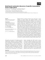
Figure 1 Histopathological Evaluation with Immunohistochemical Staining. A: Hematoxylin-eosin stain (original magnification ×250). The star
(*) indicates the malignant heterologous component of uterine MMMT. B: Hematoxylin-eosin stain (original magnification ×250). The arrows indicate
the malignant epithelial component of uterine MMMT. C: Staining with Bax antibody (original magnification ×250). The expression of Bax antibody is
diffuse with the thin arrowhead indicating weak staining, the thick arrowhead indicating medium staining, and the tailed arrow indicating strong
staining. D: Staining with p53 antibody(original magnification ×250). The star (*) indicates the negatively stained heterologous sarcomatous element,
and the arrow indicates positive staining in the epithelial component. E: Staining with p16 antibody (original magnification ×250). The star (*) indicates
the negatively stained region, and the arrow indicates the strong positive staining in the malignant epithelial glands. F: Staining with Cyclin D1 anti-
body (original magnification ×250). The star (*) indicates the negatively stained heterologous sarcomatous element, while the arrow indicates a focus
positive staining in the adjacent epithelial component.
Kanthan et al. World Journal of Surgical Oncology 2010, 8:60
/>Page 4 of 9
patients). The remaining proteins had less than 50% posi-
tive expression (Figure 3).
Discussion
Uterine MMMT are malignant neoplasms that contain
atypical malignant endometrial glands admixed with het-
erologous or homologous sarcomatous elements [2,10-
14] with the dominant element often being the epithelial
component yet distinct from endometrial carcinoma[13].
Occurring predominantly in postmenopausal women
[24,25], the prognosis of MMMT is generally worse than
endometrial carcinoma. These are rare tumors with an
annual incidence of 2/100 000 women, comprising 2-5%
of all gynecologic tumors [26,27]. Five-year survival rates
are reported between 18-39%. Many cases (70%) present
with advanced disease (Stage III/IV), contributing to
poor survival rates [21]. This tumor spreads locally
within the pelvic cavity and distally to the regional lymph
nodes, lungs and liver. DiSaia et al. [28] reported a 2 year
survival rate of 53% in patients with tumors confined to
the uterine corpus (Stage I), which dropped to 8.5% if the
disease had extended into the cervix, vagina or parame-
trium (Stages II/IIII). Less than two year survival was
seen in Stage IV disease [4], similar to other studies, with
5-year disease-free survival rates being: Stage 1 56%,
Stage II 31%, Stage III 13%, Stage IV 0% [29]. Our study
revealed similar trends. Three cases diagnosed as Stage
III did not survive beyond two years. 44.4% of seven cases
diagnosed as Stage IV did not survive beyond two years.
However, three stage IV patients had longer survival out-
comes in contrast to published literature [5]. This finding
may be related to small sample size.
The stage of the disease and the depth of myometrial
invasion were statistically significant prognostic factors
in our study, similar to reports by other authors [5,7].
Other demographic and clinico-pathological data includ-
ing age, postmenopausal bleeding, histological type,
metastasis, and treatment modalities were not found to
be statistically significant in our study.
Uterine MMMT metastasizes similar to endometrial
carcinoma of the uterus, with recurrence occurring com-
monly in the upper abdomen with occasional distant
spread [28]. In our study, 43.5% developed some form of
metastasis. Metastases occurred in 70.0% of subjects who
did not survive 2 years while longer survival time was
associated with lowered metastases (23.1%). This is sta-
tistically significant, and indicates that the presence of
metastasis at presentation is a strong prognostic indicator
for overall survival outcomes. The exact nature of
whether the carcinomatous or the sarcomatous element
is the more aggressive component and therefore has
greater propensity for metastases remains an unresolved
and controversial issue [13].
Uterine MMMTs consist of carcinomatous (CA) and
sarcomatous components (SA). Histopathological evalua-
tion of which component is responsible for biological
Table 1: Clone, dilution, and source of antibodies used for the immunohistochemical analysis in this study
ANTIBODY CLONE DILUTION SOURCE
EMA Clone E29 1/800 Dako
Ki-67 Clone MIB-1 1/50 Immunotech
PCNA Clone PC-10 N/A Ventana
Bcl-2 Clone 124 1/20 Dako
P53 Clone DO-7 1/50 Dako
Bad 48 1/20 BD (BioSciences)
Mcl-1 38G3 1/500 Novacastra
Bcl-x NC1 1/20 Novacastra
Mdm2 IB10 1/40 Novacastra
Bak polyclonal 1/20 Dako
Cyclin D1 polyclonal 1/50 Dako
Bax polyclonal 1/50 Dako
Ki67 Clone MIB-1 1/50 Immunotech
P53 Clone DO-7 1/50 Dako
P16 F-12 1/100 Santa-Cruz
P21 EA10 1/10 Oncogene
P27 SXS3G8 1/20 Dako
Cyclin D1 P2D11F11 Predilute Ventana
Kanthan et al. World Journal of Surgical Oncology 2010, 8:60
/>Page 5 of 9
tumor aggressiveness has not been greatly explored.
Yoshida et al [8] reported a higher microvessel density in
the carcinomatous region and a higher apoptotic index in
the sarcomatous areas, from which they concluded that
the carcinomatous components likely played an impor-
tant role in the aggressive biological behavior of MMMT
[22]. This biological behavior is similar to endometrial
carcinoma with which they share common etiopathologi-
cal factors.
Cell proliferation, including initiation, promotion and
progression (invasion and metastasis), plays a central role
in the multistep process of carcinogenesis. Replication of
damaged DNA is necessary to fix base substitutions,
frame shift mutations, allelic deletion and induction of
chromosomal translocations. Cancer cells commonly
demonstrate errors in these pathways during cell cycle
proliferation. Proliferative markers such as Ki67 provide
an index of cells in the Go/G1 pool of cycling cells [30].
Higher fractions of these cells represent an increased
number of cells subjected to genetic instability. In our
study Ki67 was overexpressed (50-80% positive cells) in
10 cases with no statistical difference between the carci-
nomatous and the sarcomatous areas. This lack of differ-
ence in antigen expression between the epithelial and the
sarcomatous areas is consistent with other studies [2];
thus supporting that the histogenesis of this tumor is
probably from a single pluripotential malignant clone
with divergent histologic differentiation [2].
Mutations in the p53 gene (tumor suppresser and gate-
keeper) remain one of the commonest genetic lesions
found in human cancers. This occurs in both the carcino-
matous and sarcomatous elements of uterine MMMT
Table 2: Patient demographics with clinico-pathological
data
Category Number of cases Percentage (%)
Age group
50-60 years 5 21.7
61-70 years 6 26.1
71-80 years 9 39.1
81-90 years 3 13.0
Postmenopausal bleeding
Yes 18 78.3
No 5 21.7
Histological type
Homologous 11 47.8
Heterologous 10 43.5
Stage
I1043.5
II 2 8.7
III 3 13.0
IV 7 30.4
Depth of invasion
Superficial 10 43.5
Deep 7 30.4
Metastasis
Any 18 78.3
Lung 2 8.7
Pelvic 5 21.7
Cervical 2 8.7
Other 9 39.1
Treatment
Surgery 20 87.0
Chemotherapy 2 8.7
Radiation 14 60.9
Other 8 34.8
The number of cases and their percentage are listed with respect to
age group, postmenopausal bleeding, histological type, stage,
depth of invasion, metastases, and treatment modalities.
Figure 2 Demographic and Clinical Data in Relation to 2 Year Sur-
vival Data. X-axis displays: age, postmenopausal bleeding, homolo-
gous elements, stage III/IV, depth of invasion, and metastasis. Y-axis
displays: survival outcome data-including overall survival (purple bars),
two-year survival (maroon bars), and less than two-year survival (yellow
bars). * p < 0.05 based on Fisher's Exact Test.
0. 0
20.0
40.0
60.0
80.0
100.0
Age >70
years
Post
menopausal
bleeding
Homologous
Stage III or
IV
Deep
Invasion
Depth
Me tas t a si s
Proportio n (%)
Overall Survived 2 years Did not Survive 2 years
*
*
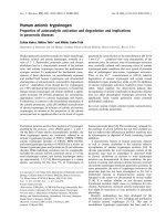
Figure 3 Cell Cycle and Apoptotic Regulatory Proteins in Relation
to 2 Year Survival Data. Statistical significance: *p < 0.05, †p < 0.10.
0.0
10.0
20.0
30.0
40.0
50.0
60.0
70.0
80.0
90.0
100.0
B
AD
MC
L
B
C
L2
B
CLX
BAK
MDM2
BAX
P16
P21
P53
KI
-67
PC
N
A
Propo rtion (%) posit ive
Ov eral l Survived 2 years Did not Survive 2 years
*
†
†
Kanthan et al. World Journal of Surgical Oncology 2010, 8:60
/>Page 6 of 9
[31-35]. Such mutations result in abnormal protein
expression, with increased intracellular accumulation due
to an increased half life which is easily detected by immu-
nohistochemical methods [36-38]. In our series, overex-
pression of p53 (70-95%) was negative in 8 cases and
positive in 15 cases, predominantly in the sarcomatous
regions as seen in Figure 1D. The average survival time in
p53 positive cases was 3.56 years as opposed to 8.94 years
in the negatively stained cases. The cohort of the positive
cases was also predominantly older (71-90 years).
Though p53 positive expression cases in our study were
not statistically significant in regard to survival beyond
two years, it was interesting to note that p53 negative
cases were associated with an improved clinical outcome.
Overexpression of p53 has been linked to decreased sur-
vival in several other malignancies. These include human
soft tissue sarcomas [39] and some cases of breast, lung
and colorectal carcinomas [40]. Such trends clearly sug-
gest that p53 may play a key role in the multistep evolu-
tion of disease progression in MMMT [41,42]; however, it
is postulated like in pulmonary carcinosarcoma to be a
late event in the disease progression with resultant better
survival outcomes in those cases that have not yet
acquired the defect [43]. In this context, further studies of
p53 mutation analysis by PCR-SSCP with sequencing will
help confirm these observed trends.
Central to the cell cycle regulatory protein machinery is
a family of serine-threonine kinases, the cyclin dependent
kinases (CDKs). These kinases are activated by cyclins D
and E and inactivated by CDK inhibitors (CDKIs) includ-
ing: p27, p16, and p21[44].
p16 specifically inhibits the cyclin D1-CDK4/6 complex
and, along with the main substrate, forms the retinoblas-
toma gene product (pRb), which is the most important
regulatory pathway involved in the G1/S transition
[45,46]. Frequent expression of p16 in primary tumors
suggests that the p16 protein is involved in the develop-
ment of these lesions [2]. Uncontrolled tumor cell prolif-
eration is frequent in tumor cells with the progression of
a normal cell to a transformed tumor cell involving many
genetic events that include the checkpoints of the cell
cycle machinery [44,47]. Overexpression of p16 is
believed to be the result of mutated p16 gene product
and/or an accumulation due to decreased turnover of the
protein [2]. Overexpression of p16 in the carcinomatous
regions of MMMT (Figure 1E) with inverse expression of
p21 in these regions denotes an upregulation of p16. The
latter is also supported by a failure to express cyclin D1 in
the majority of the tumor cells with focal expression only
in the carcinomatous elements (Figure 1F). This supports
the theory of a damaged regulatory pathway wherein p16
predominantly inhibits cyclin-D1 associated kinase activ-
ities [48]. P16 could also mediate contact inhibition of
growth and thus may be responsible for the invasive pow-
ers of the neoplasm. It is interesting to note that many of
the initial metastases in MMMT consist entirely of carci-
nomatous elements, thus supporting the theory that the
carcinomatous component is perhaps responsible for the
initial biological aggressiveness of the tumor. This change
over time is also reported in the literature as loss of p16 in
some cases of MMMTs when they recurred [2]. In our
study cases 46.2% of subjects who survived 2 years had
positive p16 overexpression in contrast to a lower expres-
sion in 10% of cases with less than two year survival.
Cell death plays an important role in normal tissue
homeostasis wherein the finite balance between new cell
productions caused by cell division is offset by cell loss in
tissues capable of cell renewal. Cells that succumb to this
mechanism of cell death undergo characteristic morpho-
logical and biochemical changes that are termed apopto-
sis. Apoptosis is one aspect of mammalian cell behavior,
which is of central importance in growth and develop-
ment and plays a key role in tumor-oncogenesis. The
three key features of apoptosis and cell survival are
related to inciting the signal transduction pathways of the
bcl-2 family of genes and the ICE family of proteases.
These components interact with other cell cycle related
genes such as p53. The central role of the Bcl-2 family of
genes in the regulation of apoptosis has been convinc-
ingly demonstrated [49-56]. The interaction of Bcl-2 fam-
ily of proteins is viewed in terms of two mechanisms: a) at
least two rheostats - the Bcl-2/Bax ratio and the Bcl-x
L
/
Table 3: Immunohistochemical expression of cell cycle and
apoptotic regulatory protein antibodies
Antibody Used Number of Cases % Positive
BAD 19 82.6
Mcl-1 8 34.8
Bcl-2 6 26.1
Bcl-x 3 13.0
Bak 17 73.9
Mdm-2 4 17.4
Bax 14 60.9
p16 7 30.4
p21 5 21.7
p27 0 0
p53 15 65.2
Cyc-D1 0 0
Ki-67 10 43.5
PCNA 11 47.8
The number of cases and their percentage of expression are listed
with respect to the oncoproteins used in this study: Bad, Mcl-1,
Bcl-2, Bcl-x, Bak, Mdm-2, Bax, p16, p21, p27, p53, Cyc-D1, Ki-67,
PCNA.
Kanthan et al. World Journal of Surgical Oncology 2010, 8:60
/>Page 7 of 9
Bcl-x
s
ratio and b) a quaternary complex involving anti-
apoptotic protein, pro-apoptotic protein, caspase and
Apf-1 equivalent protein. The susceptibility to apoptosis
is likely to be determined by the ratio of the positive regu-
lators (Bak, Bax, Bcl-x
s
) to negative regulators (Bcl-2, Bcl-
x
L
, Mcl-1 and A1) [57]. The role and contribution of each
of these factors is likely to be specific for different cells
and tissues. The function of Bcl-2 protein is dependent
on post-translational modification, specifically phospho-
rylation of serine/threonine residues.
Therefore, mere overexpression of the protein does not
provide complete information. Also, the finding that Bcl-
2 is not expressed in a variety of tumors indicates that
other apoptosis-modulating factors, especially Bcl-x
L
/
Bcl-x
s
, may play a role [58-63]. In view of the dimeric
interactions of Bcl-2 family proteins and interaction with
other apoptosis regulators, assessment of one protein
alone is unlikely to provide an understanding of apoptosis
regulation. Deregulation of the biochemical pathways
that control physiological cell death can contribute to
neoplastic cell expansion by preventing or delaying nor-
mal cell death. One of the critical regulators of apoptosis
is the protein encoded by the Bcl-2 gene [64,65].
Although the exact biochemical mechanism of Bcl-2
remains enigmatic, the Bcl-2 protein appears to control a
distal step in the final common pathway for apoptotic cell
death. Recently, a family of genes have been identified
whose encoded proteins share amino acid sequence
homology with Bcl-2. Some of these genes function as
blockers of cell death while others promote apoptosis
[56,57,66]. Among this multigene family, the protein
encoded by the Bax gene has emerged as a central regula-
tor [65,67,68]. The Bax protein is a promoter of cell death,
while others such as Bcl-x and Mcl-1 function as suppres-
sors of cell death. Further, it has been proposed that the
relative sensitivity of cells to apoptotic stimuli is governed
by the ratio of Bax: Bcl-2 and other antiapoptotic Bcl-2
family proteins [58]. Gene transfer experiments indicate
that Bax is a regulator, not an effector of the programmed
cell death pathway. As a result, it should be possible to
induce apoptosis even in the absence of Bax provided that
the apoptosis stimulus is of sufficient strength. Since Bcl-
2 can abrogate apoptosis promoted by Bax, it is possible
that it is Bax that mainly regulates the threshold of loss of
apoptosis. P53 is known to regulate Bax expression, with
inactivation of p53 leading to reduced Bax protein levels
[69,70]. Bax mutations and resistance to apoptosis have
been described in stomach, pancreas, endometrium,
hemopoietic malignancies, and a subset of colon and lung
cancers [71,72] indicating that inactivating Bax mutations
may play an important role in tumor progression in these
cancers.
In our study, all cases demonstrated diffuse expression
of Bax, Bad and Bak proteins in contrast to weak or nega-
tive expression of Mcl-1, MDM2 and Bcl-x. This supports
the existence of apoptosis protein dysregulation in these
lesions. The exact biochemical mechanism of such dys-
regulatory pathways remains unclear. Expression of Mcl-
1 was not statistically significant in regard to the two year
survival data; though, Mcl-1 was expressed in a higher
proportion of cases that survived 2 years. This finding
needs further investigation in larger samples.
Currently, there is no consensus treatment guidelines
related to improved survival outcomes. The rarity of this
tumor limits the potential for large clinical trials [73].
Nevertheless, the persistent high mortality rate and high
recurrence rate [73] with no significant improvement in
patient survival during the past 40 years [24] demands the
attention and time of researchers in a fight to improve
treatment modalities and widen the understanding of
uterine MMMT.
Conclusions
Our study supports that cell cycle and apoptotic regula-
tory protein dysregulation is an important pathway for
tumorigenesis. Apoptotic protein dysregulation may
result in epigenetic silencing of cell cycle pathways result-
ing in disarrayed/differential growth patterns. Future
genetic analysis of Bad/bax/bak pathway may provide fur-
ther insight in elucidating this mechanism.
In uterine MMMT p53+ve tumors occur in older
women with a short mean survival while p53-ve tumors
occur in younger women with longer survival. Such
trends suggest that p53 may be an important immuno-
prognostic marker in this neoplasm that warrants further
exploration.
Both p16 and Mcl-1 expression were associated with
longer survival (>2 years). Further research regarding
these cell cycle regulatory proteins will shed light into
their possibility as future predictive/prognostic markers.
Competing interests
The authors declare that they have no competing interests.
Authors' contributions
RK is the corresponding, and first author of this manuscript. JLBS contributed
to the acquisition, analysis, and interpretation of data. DD provided overall
expertise. All authors have read and approved the final manuscript.
Acknowledgements
This study was partly funded by The Scientific Teaching and Research Grant of
the College of Medicine, University of Saskatchewan.
Author Details
Department of Pathology and Laboratory Medicine, University of
Saskatchewan, Saskatoon, SK, Canada
References
1. Nguyen CP, Levi AW, Montz FJ, Bristow RE: Coexistent Choriocarcinoma
and Malignant Mixed Mesodermal Tumor of the Uterus. Gynecol Oncol
2000, 79(3):499-503.
Received: 15 October 2009 Accepted: 19 July 2010
Published: 19 July 2010
This article is available from: 2010 Kanthan et al; licensee BioMed Central Ltd. This is an Open Access article distributed under the terms of the Creative Commons Attribution License ( which permits unrestricted use, distribution, and reproduction in any medium, provided the original work is properly cited.World Journal of Surgical Oncology 2010, 8:60
Kanthan et al. World Journal of Surgical Oncology 2010, 8:60
/>Page 8 of 9
2. Robinson-Bennett B, Belch RZ, Han AC: Loss of p16 in Recurrent
Malignant Mixed Mullerian Tumors of the Uterus. International Journal
of Gynecological Cancer 2006, 16(3):1354-7.
3. De Brito PA, Silverberg SG, Orenstein JM: Carcinosarcoma (malignant
mixed mullerian (mesodermal) tumor) of the female genital tract:
immunohistochemical and ultrastructural analysis of 28 cases. Hum
Pathol 1993, 24(2):132-142.
4. Silverberg SG, Major FJ, Blessing JA, Fetter B, Askin FB, Liao SY, Miller A:
Carcinosarcoma (malignant mixed mesodermal tumor) of the uterus. A
Gynecologic Oncology group pathologic study of 203 cases. Int
Gynaecol Pathol 1990, 9(1):1-19.
5. Iwasa Y, Haga H, Konishi I, Kobashi Y, Higuchi K, Katsuyama E,
Minamiguchi S, Yamabe H: Prognostic factors in uterine
carcinosarcoma: a clinicopathologic study of 25 patients. Cancer 1998,
82(3):512-9.
6. Clement PB, Scully RE: Mullerian Adenosarcoma of the Uterus: A
Clinicopathologic Analysis of 100 Cases With a Review of the
Literature. Hum Pathol 1990, 21(4):363-381.
7. Blom R, Guerrieri C, Stal O, Malmstrom H, Sullivan S, Simonsen E:
Malignant mixed Mullerian tumors of the uterus: a clinicopathologic
DNA flow cytometric p53, and mdm-2 analysis of 44 cases. Gynecol
Oncol 1998, 68(1):18-24.
8. Yoshida Y, Kurokawa T, Fukuno N, Nishikawa Y, Kamitani N, Kotsuji F:
Markers of Apoptosis and Angiogenesis Indicate That Carcinomatous
Components Play an Important Role in the Malignant Behavior of
Uterine Carcinosarcoma. Hum Pathol 2000, 31(12):1448-1454.
9. Navarini R, Pineda RL: Malignant mixed mullerian tumors of the ovary.
Current Opinion in Obstetrics and Gynecology 2006, 18(1):20-23.
10. Yuan Y, Kim WH, Han HS, Lee JH, Park HS, Chung JK, Kang SB, Park JG:
Establishment and Characterization of Cell Lines Derived from Uterine
Malignant Mixed Mullerian Tumor. Gynecol Oncol 1997, 66(3):464-474.
11. Nicotina PA, Ferlazzo G, Vincelli AM: Proliferation indices and p53-
immunocytochemistry in uterine mixed mullerian tumors. Histol
Histopathol 1997, 12(4):967-72.
12. Pushkar I, Rao U: Final Diagnosis - Mixed Mullerian Tumors. [http://
path.upmc.edu/cases/case227/dx.html]. Retrieved on June 1, 2009.
13. Sreenan JJ, Hart WR: Carcinosarcomas of the female genital tract. A
pathologic study of 29 metastatic tumors: further evidence for the
dominant role for the epithelial component and the conversion theory
of histogenesis. American Journal of Surgical Pathology 1995,
19(6):666-74.
14. Jin Z, Ogata S, Tamura G, Katayama Y, Fukase M, Yajima M, Motoyama T:
Carcinosarcomas (malignant mullerian mixed tumors) of the uterus
and ovary: a genetic study with special reference to histogenesis.
International Journal of Gynaecological Pathology 2003, 22(4):368-73.
15. Conner MG: Uncommon and Relatively Uncommon Lesions of the
Female Reproductive System. Advances in Experimental Medicine and
Biology 2005, 563:10-20.
16. Abeln EC, Smit VT, Wessels JW, de Leeuw WJ, Cornelisse CJ, Fleuren GJ:
Molecular genetic evidence for the conversion hypothesis of the origin
of malignant mixed mullerian tumors. J Pathol 1997, 183(4):424-31.
17. Sebenik M, Yan Z, Khalbuss WE, Mittal K: Malignant Mixed Mullerian
Tumor of the Vagina: Case Report with Review of the Literature
Immunohistochemical Study and Evaluation for Human Papilloma
Virus. Human Pathology 2007, 38(8):1282-1288.
18. Kawaguchi W, Itamochi H, Kigawa J, Kanamori Y, Oishi T, Shimada M, Sato
S, Sato S, Terakawa N: Chemotherapy Consisting of Paclitaxel and
Carboplatin Benefits a Patient with Malignant Mixed Mullerian Tumor
of the Fallopian Tube. International Journal of Clinical Oncology 2008,
13(5):461-3.
19. Handa Y, Kato H, Kaneuchi M, Saitoh Y, Yamashita K: High-grade Broad
Ligament Cancer of Mullerian Origin: Immunohistochemical Analysis
of a Case and Review of the Literature. International Journal of
Gynecological Cancer 2007, 17(3):705-734.
20. Maitra RN, Lee J, McConnell DT, Kenwright DN, Dady P: Malignant Mixed
Mullerian Tumour of the Fallopian Tube Occuring in a Patient with
Peutz-Jegher's Syndrome. Australian and New Zealand Journal of
Obstetrics and Gynaecology 2004, 44(1):77-79.
21. Mok JE, Kim YM, Jung MH, Kim KR, Kim DY, Kim JH, Kim YT, Nam JH: Mixed
Mullerian Tumors of the Ovary: Experience with Cytoreductive Surgery
and Platinum-Based Combination Chemotherapy. International Journal
of Gynarcological Cancer 2006, 16(1):101-5.
22. Moe MM, El-Sharkawi S: Is There Any Association Between Uterine
Malignant Mixed Mullerian Tumor Breast Cancer and Prolonged
Tamoxifen Treatment? Journal of Obstetrics and Gynaecology 2003,
23(3):301-3.
23. Wang X, Tangjitgamol S, Liu J, Kavanagh JJ: Response of Recurrent
Uterine High-Grade Malignant Mixed Mullerian Tumor to Letrozole.
International Journal of Gynaecological Cancer 2005, 15(5):1243-8.
24. Callister M, Ramondetta LM, Jhingran A, Burke TW, Eifel PJ: Malignant
Mixed Mullerian Tumors of the Uterus: Analysis of Patterns of Failure
Prognostic Factors and Treatment Outcome. International Journal of
Radiation Oncology Biology, Physics 2004, 58(3):786-96.
25. Maheshwari A, Gupta S, Shet T, Wuntkal R, Tongaonkar HB: Diagnostic
dilemma in a case of malignant mixed mullerian tumor of the cervix.
World Journal of Surgical Oncology 2006, 4:36.
26. Vaidya AP, Horowitz NS, Oliva E, Halpern EF, Duska LR: Uterine Malignant
Mixed Mullerian Tumors Should not be Included in Studies of
Endometrial Carcinoma. Gynecologic Oncology 2006, 103(2):684-7.
27. Mikami M, Kuwabara Y, Tanaka K, Komiyama S, Ishikawa M, Hirose T:
Malignant Mixed Mullerian Tumor of Primary Mesenteric Origin.
International Journal of Gynarcological Cancer 2005, 15(6):1249-1253.
28. DiSaia PJ, Castro JR, Rutledge FN: Mixed mesodermal sarcoma of the
uterus. Am J Roentgenol 1973, 117(3):632-636.
29. Hoskins PJ, Le N, Ellard S, Lee U, Martin LA, Swenerton KD, Tinker AV, British
Columbia Cancer Agency: Carboplatin plus Paclitaxel for Advanced or
Recurrent Uterine Malignant Mixed Mullerian Tumors. The British
Columbia Cancer Agency Experience. Gynecologic Oncology 2008,
108(1):58-62.
30. Swisher EM, Gown AM, Skelly M, Ek M, Tamimi HK, Cain JM, Greer BE,
Muntz HG, Goff BA: The Expression of Epidermal Growth Factor
Receptor HER-2/Neu, p53, and Ki-67 Antigen in Uterine Malignant
Mixed Mesodermal Tumors and Adenosarcoma. Gynecol Oncol 1996,
60(1):81-88.
31. Kounelis S, Jones MW, Papadaki H, Bakker A, Swalsky P, Finkelstein SD:
Carcinosarcomas (malignant mixed mullerian tumors) of the female
genital tract: comparative molecular analysis of epithelial and
mesenchymal components. Hum Pathol 1998, 29(1):82-7.
32. Bur ME, Perlman C, Edelmann L, Fev E, Rose PG: p53 expression in
neoplasms of the uterine corpus. Am J Clin Pathol 1992, 98(1):81-7.
33. Porter PL, Gown AM, Kramp SG, Coltrera MD: Widespread p53
overexpression in human malignant tumors. An immunohistochemical
study using methacarn-fixed, embedded tissue. Am J Pathol 1992,
140(1):145-153.
34. Gagner JP, Mittal K: Malignant Mixed Mullerian Tumor of the Fimbriated
End of the Fallopian Tube: Origin as an Intraepithelial Carcinoma.
Gynecologic Oncology 2005, 97(1):219-222.
35. Geisler JP, Geisler HE, Wiemann MC, Zhou Z, Miller GA, Crabtree W: p53
Expression as a Prognostic Indicator of 5-Year Survival in Endometrial
Cancer. Gynecol Oncol 1999, 74(3):468-471.
36. Liu FS, Kohler MF, Marks JR, Bast RC Jr, Boyd J, Berchuck A: Mutation and
Overexpression of the p53 Tumor Suppressor Gene Frequently Occurs
in Uterine and Ovarian Sarcomas. Obs and Gynecol 1994, 83(1):118-123.
37. Kohler MF, Marks JR, Wiseman RW, Jacobs IJ, Davidoff AM, Clarke-Pearson
DL, Soper JT, Bast RC Jr, Berchuck A: Spectrum of mutation and
frequency of allelic deletion of the p53 gene in ovarian cancer. J Natl
Cancer Inst 1993, 85(18):1513-9.
38. Costa MJ, Vogelsan J, Young LJ: p53 gene mutation in female genital
tract carcinosarcomas (malignant mixed mullerian tumors): a
clinicopathologic study of 74 cases. Mod Pathol 1994, 7(6):619-27.
39. Toffoli G, Doglioni C, Cernigoi C, Frustaci S, Perin T, Canal B, Boiocchi M:
p53 overexpression in human soft tissue sarcomas: Relation to
biological aggressiveness. Ann Oncol 1994, 5(2):167-172.
40. Berchuck A, Kohler MF, Marks JR, Wiseman R, Boyd J, Bast RC Jr: The p53
tumor suppressor gene frequently is altered in gynecologic cancers.
Am J Obstet Gynecol 1994, 170(1 Pt 1):246-252.
41. Huang LW, Chou YY, Chao SL, Chen TJ, Lee TT: p53 and p21 Expression in
Precancerous Lesions and Carcinomas of the Uterine Cervix:
Overexpression of p53 Predicts Poor Disease Outcome. Gynecol Oncol
2001, 83(2):348-354.
42. Soong R, Knowles S, Hammond IG, Michael C, Lacopetta BJ: P53 protein
overexpression and gene mutation in mixed Mullerian tumors of the
uterus. Cancer Detect Prev 1999, 23(1):8-12.
Kanthan et al. World Journal of Surgical Oncology 2010, 8:60
/>Page 9 of 9
43. Holst VA, Finkelstein S, Colby TV, Myers JL, Yousem SA: p53 and K-ras
mutational genotyping in pulmonary carcinosarcoma spindle cell
carcinoma and pulmonary blastoma: implications for histogenesis.
Am J Surg Pathol 1997, 21(7):801-11.
44. Palmqvist R, Rutegard JN, Bozoky B, Landberg G, Stenling R: Human
Colorectal Cancers with an Intact p16/Cyclin D1/pRb Pathway Have
Up-Regulated p16 Expression and Decreased Proliferation in Small
Invasive Tumor Clusters. Am J Path 2000, 157(6):1947-53.
45. Milde-Langosch K, Riethdorf L, Bamberger AM, Loning T: P16/MTS1 and
pRB expression in endometrial carcinomas. Virchows Arch 1999,
434(1):23-8.
46. Ramirez PT, Gershenson DM, Tortolero-Luna G, Ramondetta LM,
Fightmaster D, Wharton JT, Wolf JK: Expression of Cell-Cycle Mediators in
Ovarian Cancer Cells after Transfection with p16, p21, and p53.
Gynecol Oncol 2001, 83(3):543-548.
47. Munirajan AK, Kannan K, Bhuvarahamurthy V, Ishida I, Fujinaga K, Tsuchida
N, Shanmugam G: The Status of Human Papillomavirus and Tumor
Suppressor Genes p53 and p16 in Carcinomas of Uterine Cervix from
India. Gynecol Oncol 1998, 69(3):205-209.
48. Shapiro GI, Edwards CD, Ewen ME, Rollins BJ: p16INK4A participates in a
G1 arrest checkpoint in response to DNA damage. Mol Cell Biol 1998,
18(1):378-387.
49. Dimitrakakis C, Kymionis G, Diakomanolis E, Papaspyrou I, Rodolakis A,
Arzimanoglou I, Leandros E, Michalas S: The Possible Role of p53 and bcl-
2 Expression in Cervical Carcinomas and Their Premalignant Lesions.
Gynecol Oncol 2000, 77(1):129-136.
50. Thompson C: Apoptosis in the Pathogenesis and Treatment of Disease.
Science 1995, 267(5203):1457-1461.
51. Soini Y, Paakko P, Lehto VP: Histopathological Evaluation of Apoptosis in
Cancer. Am J Pathol 1998, 153(4):1041-1051.
52. Kerr JF, Winterford CM, Harmon BV: Apoptosis - Its Significance in Cancer
and Cancer Therapy. Cancer 1994, 73(8):2013-2023.
53. Alnemri ES, Fernandes TF, Haldar S, Croce CM, Litwack G: Involvement of
bcl-2 in Glucocorticoid-Induced Apoptosis of Human Pre-B-Leukemias.
Cancer Res 1992, 52(2):491-5.
54. Johnstone RW, Ruefli AA, Lowe SW: Apoptosis: A Link between Cancer
Genetics and Chemotherapy. Cell 2002, 108(2):153-164.
55. Domen J, Gandy KL, Weissman IL: Systemic Overexpression of Bcl-2 in
the Hematopoietic System Protects Transgenic Mice from the
Consequences of Lethal Irradiation. Blood 1998, 91(7):2272-2282.
56. Adams JM, Cory S: The Bcl-2 Protein Family: Arbiters of Cell Survival.
Science 1998, 281(5381):1322-1326.
57. Suzuki M, Youle RJ, Tjandra N: Structure of Bax: Coregulation of Dimer
Formation and Intracellular Localization. Cell 2000, 103(4):645-653.
58. Geisler JP, Geisler HE, Wiemann MC, Zhou Z, Miller GA, Crabtree W: Lack of
Bcl-2 Persistence: an Independent Prognostic Indicator of Poor
Prognosis in Endometrial Carcinoma. Gynecol Oncol 1998, 71(2):305.
59. Crescenzi E, Palumbo G: Bcl-2 Exerts a pRb-Mediated Cell Cycle
Inhibitory Function in HEC1B Endometrial Carcinoma Cells. Gynecol
Oncol 2001, 81(2):184-192.
60. Nakamura T, Nomura S, Sakai T, Nariya S: Expression of bcl-2
Oncoprotein in Gastrointestinal and Uterine Carcinomas and their
Premalignant Lesions. Hum Pathol 1997, 28(3):309-15.
61. Yang HB, Chow NH, Sheu BS, Chan SH, Chien CH, Su IJ: The Role of Bcl-2
in the Progression of the Colorectal Adenoma-Carcinoma Sequence.
Anticancer Res 1999, 19(1B):727-30.
62. Krajewska M, Moss SF, Krajewski S, Song K, Holt PR, Reed JC: Elevated
Expression of Bcl-X and Reduced Bak in Primary Colorectal
Adenocarcinomas. Cancer Res 1996, 56(10):2422-7.
63. Muller W, Schneiders A, Hommel G, Gabbert HE: Prognostic Value of Bcl-
2 Expression in Gastric Cancer. Anticancer Res 1998, 18(6B):4699-704.
64. Crescenzi E, Criniti V, Pianese M, Tecce MF, Palumbo G: Differential
Expression of Antiapoptotic Genes in Human Endometrial Carcinoma:
bcl-XL Succeeds bcl-2 Function in Neoplastic Cells. Gynecol Oncol 2000,
77(3):419-428.
65. Oltvai ZN, Milliman CL, Korsmeyer SJ: Bcl-2 Heterodimerizes in vivo with
a Conserved Homologue Bax, that Accelerates Programmed Cell
Death. Cell 1993, 74:609-619.
66. Gross A, McDonnell JM, Korsmeyer SJ: Bcl-2 Family Members and the
Mitochondria in Apoptosis. Genes Dev 1999, 13(15):1899-1911.
67. Kokawa K, Shikone T, Otani T, Nishiyama R, Ishii Y, Yagi S, Yamoto M:
Apoptosis and the Expression of Bcl-2 and Bax in Patients with
Endometrioid Clear Cell and Serous Carcinomas of the Uterine
Endometrium. Gynecol Oncol 2001, 81(2):178-183.
68. Ouyang H, Furukawa T, Abe T, Kato Y, Horii A: The Bax Gene the Promoter
of Apoptosis is Mutated in Genetically Unstable Cancers of the
Colorectum Stomach, and Endometrium. Clin Cancer Res 1998,
4(4):1071-4.
69. Oda K, Arakawa H, Tanaka T, Matsuda K, Tanikawa C, Mori T, Nishimori H,
Tamai K, Tokino T, Nakamura Y, Taya Y: p53AIP1, a Potential Mediator of
p53-Dependent Apoptosis and Its Regulation by Ser-46-
Phosphorylated p53. Cell 2000, 102(6):849-862.
70. Miyashita T, Reed JC: Tumor suppressor p53 is a direct transcriptional
activator of the human bax gene. Cell 1995, 80(2):293-299.
71. Xu ZW, Friess H, Buchler M, Solioz M: Overexpression of Bax Sensitizes
Human Pancreatic Cancer Cells to Apoptosis Induced by
Chemotherapeutic Agents. Cancer Chemother Pharmacol 2002,
49(6):504-10.
72. Hanaoka T, Nakayama J, Haniuda M, Sato TA: Immunohistochemical
Demonstration of Apoptosis-Regulated Proteins Bcl-2 and Bax in
Resected Non-Small-Cell Lung Cancers. Int J Clin Oncol 2002, 7(3):152-8.
73. Wong L, See HT, Khoo-Tan HS, Low JS, Ng WT, Low JJ: Combined
Adjuvant Cisplating and Ifosfamide Chemotherapy and Radiotherapy
for Malignant Mixed Mullerian Tumors of the Uterus. International
Journal of Gynaecological Cancer 2006, 16(3):1364-9.
doi: 10.1186/1477-7819-8-60
Cite this article as: Kanthan et al., Malignant mixed Mullerian tumors of the
uterus: histopathological evaluation of cell cycle and apoptotic regulatory
proteins World Journal of Surgical Oncology 2010, 8:60