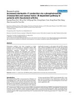
Báo cáo y học: "Increased interleukin-17 production via a phosphoinositide 3-kinase/Akt and nuclear factor κB-dependent pathway in patients with rheumatoid arthritis" ppt
Bạn đang xem bản rút gọn của tài liệu. Xem và tải ngay bản đầy đủ của tài liệu tại đây (529.8 KB, 10 trang )
Open Access
Available online />R139
Vol 7 No 1
Research article
Increased interleukin-17 production via a phosphoinositide
3-kinase/Akt and nuclear factor κB-dependent pathway in
patients with rheumatoid arthritis
Kyoung-Woon Kim*, Mi-La Cho*, Mi-Kyung Park, Chong-Hyeon Yoon, Sung-Hwan Park, Sang-
Heon Lee and Ho-Youn Kim
Department of Medicine, Division of Rheumatology, The Center for Rheumatic Diseases, and The Rheumatism Research Center (RhRC), Catholic
Research Institutes of Medical Sciences, Catholic University of Korea, Seoul, Korea
* Contributed equally
Corresponding author: Sang-Heon Lee,
Received: 27 Apr 2004 Revisions requested: 19 May 2004 Revisions received: 18 Oct 2004 Accepted: 3 Nov 2004 Published: 29 Nov 2004
Arthritis Res Ther 2005, 7:R139-R148 (DOI 10.1186/ar1470)
http://arthr itis-research.com/conte nt/7/1/R139
© 2004 Kim et al.; licensee BioMed Central Ltd.
This is an Open Access article distributed under the terms of the Creative Commons Attribution License ( />2.0), which permits unrestricted use, distribution, and reproduction in any medium, provided the original work is properly cited.
Abstract
Inflammatory mediators have been recognized as being
important in the pathogenesis of rheumatoid arthritis (RA).
Interleukin (IL)-17 is an important regulator of immune and
inflammatory responses, including the induction of
proinflammatory cytokines and osteoclastic bone resorption.
Evidence for the expression and proinflammatory activity of IL-
17 has been demonstrated in RA synovium and in animal
models of RA. Although some cytokines (IL-15 and IL-23) have
been reported to regulate IL-17 production, the intracellular
signaling pathways that regulate IL-17 production remain
unknown. In the present study, we investigated the role of the
phosphoinositide 3-kinase (PI3K)/Akt pathway in the regulation
of IL-17 production in RA. Peripheral blood mononuclear cells
(PBMC) from patients with RA (n = 24) were separated, then
stimulated with various agents including anti-CD3, anti-CD28,
phytohemagglutinin (PHA) and several inflammatory cytokines
and chemokines. IL-17 levels were determined by sandwich
enzyme-linked immunosorbent assay and reverse transcription–
polymerase chain reaction. The production of IL-17 was
significantly increased in cells treated with anti-CD3 antibody
with or without anti-CD28 and PHA (P < 0.05). Among tested
cytokines and chemokines, IL-15, monocyte chemoattractant
protein-1 and IL-6 upregulated IL-17 production (P < 0.05),
whereas tumor necrosis factor-α, IL-1β, IL-18 or transforming
growth factor-β did not. IL-17 was also detected in the PBMC
of patients with osteoarthritis, but their expression levels were
much lower than those of RA PBMC. Anti-CD3 antibody
activated the PI3K/Akt pathway; activation of this pathway
resulted in a pronounced augmentation of nuclear factor κB
(NF-κB) DNA-binding activity. IL-17 production by activated RA
PBMC is completely or partly blocked in the presence of the NF-
κB inhibitor pyrrolidine dithiocarbamate and the PI3K/Akt
inhibitor wortmannin and LY294002, respectively. However,
inhibition of activator protein-1 and extracellular signal-regulated
kinase 1/2 did not affect IL-17 production. These results
suggest that signal transduction pathways dependent on PI3K/
Akt and NF-κB are involved in the overproduction of the key
inflammatory cytokine IL-17 in RA.
Keywords: interleukin-17, nuclear factor κB, PI3K/Akt pathway, peripheral blood mononuclear cells, rheumatoid arthritis
Introduction
Rheumatoid arthritis (RA) is characterized by infiltrations of
macrophages and T cells into the joint, and synovial hyper-
plasia. Proinflammatory cytokines released from these cells
are known to be important in the destruction of joints in RA
[1]. The favorable clinical benefits obtained with inhibitors
of tumor necrosis factor (TNF)-α) and interleukin (IL)-1 sug-
gest that the blockade of key inflammatory cytokines has
been the important issue in the development of new thera-
peutic applications [2].
AP-1, activator protein-1; BSA = bovine serum albumin; EMSA = electrophoretic mobility-shift assay; GAPDH = glyceraldehyde-3-phosphate dehy-
drogenase; IL = interleukin; MAPK = mitogen-activated protein kinase; MCP-1 = monocyte chemoattractant protein-1; MIP = macrophage inflamma-
tory protein; NF-κB = nuclear factor κB; OA = osteoarthritis; PBMC = peripheral blood mononuclear cells; PDTC = pyrrolidine dithiocarbamate; PHA
= phytohemagglutinin; PI3K = phosphoinositide 3-kinase; RA = rheumatoid arthritis; TGF = transforming growth factor; Th = T helper; TNF = tumor
necrosis factor.
Arthritis Research & Therapy Vol 7 No 1 Kim et al.
R140
A little over a decade ago, the primacy of T cells in the
pathogenesis of autoimmune disease such as RA was
undisputed because they are the largest cell population
infiltrating the synovium. However, a series of studies dem-
onstrated paucity of T cell-derived cytokines such as IL-2
and interferon-γ in the joints of RA, whereas macrophage
and fibroblast cytokines including IL-1, IL-6, IL-15, IL-18
and TNF-α were abundant in rheumatoid synovium. This
paradox has questioned the role of T cells in the pathogen-
esis of RA [3]. Because we have already demonstrated the
enhanced proliferation of antigen specific T cells, espe-
cially to type II collagen, and the skewing of T helper type 1
(Th1) cytokines in RA [4], the role of T cells needs to be elu-
cidated in different aspects.
IL-17 is one of the inflammatory cytokines secreted mainly
by activated T cells, which can induce IL-6 and IL-8 by
fibroblasts [5]. This cytokine is of interest for two major rea-
sons: first, similarly to TNF-α and IL-1, IL-17 has proinflam-
matory properties; second, it is produced by T cells [6].
Recent observations demonstrated that IL-17 can also acti-
vate osteoclastic bone resorption by the induction of
RANKL (receptor activator of nuclear factor κB [NF-κB] lig-
and), which is involved in bony erosion in RA [7]. It also
stimulates the production of IL-6 and leukemia inhibitory
factor by synoviocytes, and of prostaglandin E
2
and nitric
oxide by chondrocytes, and has the ability to differentiate
and activate the dendritic cells [8-10]. Levels of IL-17 in
synovial fluids were significantly higher in patients with RA
than in patients with osteoarthritis (OA), and it was pro-
duced by CD4
+
T cells in the synovium [11,12].
IL-15, secreted from activated macrophages, has been
reported to be an important trigger of IL-17 production in
RA peripheral blood mononuclear cells (PBMC) by
cyclosporine and steroid sensitive pathways [13].
Recently, Happel and colleagues also showed that IL-23
could be an efficient trigger of IL-17 production from both
CD4
+
and CD8
+
T cells [14].
Although the contribution of IL-17 in joint inflammation in
RA has been documented in earlier studies [12,15,16], the
intracellular signal transduction pathway for IL-17 produc-
tion remains uncertain. In the present study we used vari-
ous stimuli to investigate IL-17 production in PBMC of
patients with RA and its signaling transduction pathway.
We found that the intracellular signaling pathway involving
phosphoinositide 3-kinase (PI3K)/Akt and NF-κB might be
involved in the overproduction of the key inflammatory
cytokine IL-17 in RA. These results might provide new
insights into the pathogenesis of RA and future directions
for new therapeutic strategies in RA.
Materials and methods
Patients
Informed consent was obtained from 24 patients (5 men
and 19 women) with RA who fulfilled the 1987 revised cri-
teria of the American College of Rheumatology (formerly
the American Rheumatism Association) [17]. The age of
the patients with RA was 50 ± 8 (mean ± SEM) years
(range 23–71 years). All medications were stopped 48
hours before entry to the study. Comparisons were made
with 14 patients with OA (3 men and 11 women) and with
14 healthy controls (3 men and 11 women) who had no
rheumatic diseases. The mean ages of the patients with OA
and the healthy controls were 50 ± 8 years (range 34–68
years) and 30 ± 6 years (range 24–57 years). Informed
consent was obtained, and the protocol was approved by
the Catholic University of Korea Human Research Ethics
Committee.
Reagents
Recombinant IL-17, IL-18, IL-15, monocyte chemoattract-
ant protein-1 (MCP-1), macrophage inflammatory protein
(MIP)-1α, MIP-1β, IL-6 and IL-8 were purchased from R &
D systems (Minneapolis, MN, USA). Recombinant trans-
forming growth factor (TGF)-β was purchased from Pepro-
tech (London, UK). Recombinant TNF-α and IL-1 were
purchased from Endogen Inc. (Cambridge, MA, USA).
Cyclosporin A was provided by Sandos Ltd. (Basel, Swit-
zerland). Phytohemagglutinin (PHA), pyrrolidine dithiocar-
bamate (PDTC), rapamycin, dexamethasone and curcumin
were all obtained from the Sigma Chemical Co. (St Louis,
MA, USA). Anti-CD3 monoclonal antibody and anti-CD28
monoclonal antibody were obtained from BD Biosciences
(San Diego, CA, USA). LY294002, SB203580, FK506,
wortmannin and PD98059 were obtained from Calbio-
chem (Schwalbach, Germany).
Production of IL-17 by T cell receptor activation,
cytokines or chemokines
PBMC were prepared from heparinized blood by Ficoll-
Hypaque (SG1077) density-gradient centrifugation. Cell
cultures were performed as described previously [18]. In
brief, the cell suspensions were adjusted to a concentra-
tion of 10
6
/ml in RPMI 1640 medium supplemented with
10% fetal calf serum, 100 U/ml penicillin, 100 mg/ml strep-
tomycin and 2 mM L-glutamine. Cell suspension (1 ml) was
dispensed into 24-well multi-well plates (Nunc, Roskilde,
Denmark), and incubated for 24 hours at 37°C in 5% CO
2
.
Subsequently, various concentrations of cyclosporin A
(10–500 ng/ml) were added to the medium and cells were
incubated for 24 hours. To each well was added FK506,
rapamycin, curcumin, PDTC, LY294002, SB203580,
PD98059, dexamethasone or wortmannin. After incubation
for 24 hours (unless otherwise stated), cell-free media were
collected and stored at -20°C until assayed. All cultures
Available online />R141
were set up in triplicate, and results are expressed as
means ± SEM.
CD4
+
T-cell isolation by MACS
Anti-CD4 microbeads were used essentially as recom-
mended by the manufacturer (Miltenyi) [19]. PBMC were
resuspended in 80 µl of FBS staining buffer. Anti-CD4
microbeads (20 µl) were added and incubated for 15 min
at 6–12°C. Saturating amounts of fluorochrome-conju-
gated antibodies were added for a further 10 min. Cells
were diluted in 2.5 ml of FBS staining buffer, pelleted,
resuspended in 500 µl and magnetically separated, usually
on an AutoMACS magnet fitted with a MACS MS column.
Flow-through and two 1 ml washes were collected as the
negative fraction. Enriched cells were collected in two 0.5
ml aliquots from the column after removal from the magnet.
Alternatively, cells stained with anti-CD4–phycoerythrin
were washed, magnetically labeled with anti-phycoerythrin
microbeads (20 µl added to 80 µl of cell suspension; 15
min, 6–12°C), and magnetically separated as described
above. The purity of cells was assessed by flow cytometric
analysis of stained cells on a FACS Vantage sorter. Most
(more than 97%) of the isolated cells had the CD4 T cell
marker.
Enzyme-linked immunosorbent assay of IL-17
IL-17 in culture supernatants was measured by sandwich
enzyme-linked immunosorbent assay as described previ-
ously [20]. In brief, a 96-well plate (Nunc) was coated with
4 µg/ml monoclonal antibodies against IL-17 (R & D Sys-
tems) at 4°C overnight. After blocking with phosphate-buff-
ered saline/1% bovine serum albumin (BSA)/0.05%
Tween 20 for 2 hours at room temperature (22–25°C), test
samples and the standard recombinant IL-17 (R & D Sys-
tems) were added to the 96-well plate and incubated at
room temperature for 2 hours. Plates were washed four
times with phosphate-buffered saline/Tween 20, and then
incubated with 500 ng/ml biotinylated mouse monoclonal
antibodies against IL-17 (R & D Systems) for 2 hours at
room temperature. After washing, streptavidin–alkaline
phosphate–horseradish peroxidase conjugate (Sigma) was
incubated for 2 hours, then washed again and incubated
with 1 mg/ml p-nitrophenyl phosphate (Sigma) dissolved in
diethanolamine (Sigma) to develop the color reaction. The
reaction was stopped by the addition of 1 M NaOH and the
optical density of each well was read at 405 nm. The lower
limit of IL-17 detection was 10 pg/ml. Recombinant human
IL-17 diluted in culture medium was used as a calibration
standard, ranging from 10 to 2000 pg/ml. A standard curve
was drawn by plotting optical density against the log of the
concentration of recombinant cytokines, and used for
determination of IL-17 in test samples.
Quantification of IL-17 mRNA by semiquantitative
reverse transcription–polymerase chain reaction
PBMC were incubated with various concentrations of anti-
CD3 in the presence or absence of inhibitors (LY294002,
PDTC). After 16 hours of incubation, mRNA was extracted
with RNAzol B (Biotex Laboratories, Houston, TX, USA) in
accordance with the manufacturer's instructions. Reverse
transcription of 2 µg of total mRNA was performed at 42°C
using the Superscript™ reverse transcription system
(Takara, Shiga, Japan). PCR amplification of cDNA aliquots
was performed by adding 2.5 mM dNTPs, 2.5 U of Taq
DNA polymerase (Takara) and 0.25 µM of sense and anti-
sense primers. The reaction was performed in PCR buffer
(1.5 mM MgCl
2
, 50 mM KCl, 10 mM Tris-HCl, pH 8.3) in a
total volume of 25 µl. The following sense and antisense
primers for each molecules were used: IL-17 sense, 5'-
ATG ACT CCT GGG AAG ACC TCA TTG-3'; IL-17 anti-
sense, 5'-TTA GGC CAC ATG GTG GAC AAT CGG-3';
glyceraldehyde-3-phosphate dehydrogenase (GAPDH)
sense, 5'-CGA TGC TGG GCG TGA GTA C-3'; GAPDH
antisense, 5'-CGT TCA GCT CAG GGA TGA CC-3'.
Reactions were processed in a DNA thermal cycler (Perkin-
Elmer Cetus, Norwalk, CT, USA) through cycles for 30 s of
denaturation at 94°C, 1 min of annealing at 56°C for
GAPDH and IL-17, followed by 1 min of elongation at
72°C. PCR rounds were repeated for 25 cycles each for
both GAPDH and IL-17; this was determined as falling
within the exponential phase of amplification for each mol-
ecule. The level of mRNA expression was presented as a
ratio of IL-17 PCR product over GAPDH product.
Figure 1
Levels of interleukin (IL)-17 production in peripheral blood mononuclear cells from patients with rheumatoid arthritis (RA; n = 24), patients with osteoarthritis (OA) (n = 14) and normal individuals (n = 14)Levels of interleukin (IL)-17 production in peripheral blood mononuclear
cells from patients with rheumatoid arthritis (RA; n = 24), patients with
osteoarthritis (OA) (n = 14) and normal individuals (n = 14). Each
peripheral blood mononuclear cell was stimulated for 24 hours with or
without phytohemagglutinin (PHA; 5 µg/ml). IL-17 was measured in cul-
ture supernatants by sandwich enzyme-linked immunosorbent assay.
Data are expressed as means and SEM. One representative result of
five independent experiments is shown. Student's t-test was used to
compare each group. *, P < 0.05; **, P < 0.001.
Arthritis Research & Therapy Vol 7 No 1 Kim et al.
R142
Western blot analysis of Akt, phosphorylated Akt and
IκB-α
PBMC were incubated with anti-CD3 (10 µg/ml) in the
presence or absence of LY294002 (20 µM). After incuba-
tion for 1 hour, whole cell lysates were prepared from about
10
7
cells by homogenization in the lysis buffer, and centri-
fuged at 14,000 r.p.m. (19,000 g) for 15 min. Protein con-
centrations in the supernatants were determined with the
Bradford method (Bio-Rad, Hercules, CA, USA). Protein
samples were separated by 10% SDS–PAGE and trans-
ferred to a nitrocellulose membrane (Amersham Pharmacia
Biotech, Uppsala, Sweden). For western hybridization,
membrane was preincubated with 0.1% skimmed milk in
TBS-T buffer (0.1% Tween 20 in Tris-buffered saline) at
room temperature for 2 hours, then primary antibodies
against Akt, phosphorylated Akt and IκB-α (Cell Signaling
Technology Inc., Beverly, MA, USA), diluted 1:1000 in 5%
BSA/TBS-T, were added and incubated overnight at 4°C.
After washing four times with TBS-T, horseradish peroxi-
dase-conjugated secondary antibodies were added and
allowed to incubate for 1 hour at room temperature. After
TBS-T washing, hybridized bands were detected with the
enhanced chemiluminescence (ECL) detection kit and
Hyperfilm-ECL reagents (Amersham Pharmacia).
Gel mobility-shift assay of NF-κB binding site
Nuclear proteins were extracted from about 5 × 10
6
PBMC. Oligonucleotide probes encompassing the NF-κB
binding site of the human IL-17 promoter (5'-ATG ACC
TGG AAA TAC CCA AAA TTC-3') were generated by 5'-
end labeling of the sense strand with [γ-
32
P]dATP (Amer-
sham Pharmacia) and T4 polynucleotide kinase (TaKaRa).
Unincorporated nucleotides were removed by NucTrap
probe purification columns (Stratagene, La Jolla, CA, USA).
Nuclear extracts (2 µg of protein) were incubated with radi-
olabeled DNA probes (10 ng; 100,000 c.p.m.) for 30 min
at room temperature in 20 µl of binding buffer consisting of
20 mM Tris-HCl, pH 7.9, 50 mM KCl, 1 mM dithiothreitol,
0.5 mM EDTA, 5% glycerol, 1 mg/ml BSA, 0.2% Nonidet
P40 and 50 ng/µl poly(dI-dC). Samples were subjected to
electrophoresis on nondenaturing 5% polyacrylamide gels
in 0.5 × Tris-borate-EDTA buffer (pH 8.0) at 100 V. Gels
were dried under vacuum and exposed to Kodak X-OMAT
film at -70°C with intensifying screens. Rabbit polyclonal
antibodies against NF-κB subunits p50, p65 and c-Rel
were from Santa Cruz Biotechnology (Santa Cruz, CA,
USA).
Cell viability (Trypan blue dye exclusion assay)
For cell viability assays, the trypan blue dye exclusion
method was used to evaluate the potential of direct cyto-
toxic effect of inhibitors on cells. After incubation for 24
hours, the cells were harvested and the percentage cell via-
bility was calculated with the formula 100 × (number of via-
ble cells/number of both viable and dead cells) [21].
Statistical analysis
Data are expressed as means ± SEM. Statistical analysis
was performed with Student's t-test for matched pairs. P
values less than 0.05 were considered significant.
Results
IL-17 production in PBMC from patients with RA,
patients with OA and normal individuals
PBMC were separated and cultured with PHA (5 µg/ml)
from patients with RA, patients with OA, and age-matched
normal controls; IL-17 levels were then determined in the
culture supernatants (Fig. 1). Although the amounts of
basal IL-17 secretion were not different between RA, OA
and normal controls (62 ± 31, 43 ± 19 and 43 ± 10 pg/ml,
respectively), the IL-17 production stimulated by PHA was
significantly higher in RA PBMC than in those from OA and
controls (768 ± 295 versus 463 ± 211 pg/ml [P < 0.05]
and 241 ± 29 pg/ml [P < 0.001]).
Increased IL-17 production in PBMC of patients with RA
by anti-CD3 and/or anti-CD28, and PHA
Because IL-17 was already known from earlier reports to
be produced mainly by activated T cells, we investigated
the effect of different concentrations of anti-CD3 (1, 5 and
10 µg/ml) as a T cell activation, which showed a dose-
dependent increase in IL-17 levels (data not shown). On
the basis of this, we chose 10 µg/ml as a stimulation con-
centration for anti-CD3. As shown in Table 1, anti-CD3 sig-
nificantly upregulated IL-17 production up to 3.7-fold, and
the combination of anti-CD28 and anti-CD3 produced
more IL-17 (approximately 1.3-1.5-fold) than anti-CD3
alone. Furthermore, when incubated with T cell mitogens
such as PHA, increased IL-17 production was more pro-
nounced than with anti-CD3 and anti-CD28 (588 ± 85 ver-
sus 211 ± 1 pg/ml; P < 0.05).
Regulation of IL-17 production in RA PBMC by
inflammatory cytokines and chemokines
Because RA PBMC include several cell types in addition to
T cells, some inflammatory cytokines released from macro-
phages and other lymphocytes might have affected the pro-
duction of IL-17 from T cells. To evaluate the effects of
inflammatory cytokines released by activated PBMC, we
tested the effects of several cytokines and chemokines on
IL-17 production. We detected an increase in IL-17 level
after stimulation with IL-15 (10 ng/ml), whereas with IL-1β
(10 ng/ml), TNF-α (10 ng/ml), IL-18 (10 ng/ml) or TGF-β
(10 ng/ml) the levels in IL-17 were unchanged (Fig. 2a).
When treated with MCP-1 (10 ng/ml) or IL-6 (10 ng/ml),
significant upregulations of IL-17 proteins were observed
(62 ± 42 and 50 ± 10 versus 31 ± 11 pg/ml, respectively;
P < 0.05), whereas none was observed with IL-8 (10 ng/
ml), MIP-1α (10 ng/ml) or MIP-1β (10 ng/ml) (Fig. 2b).
Available online />R143
Inhibition of IL-17 production by signal transduction
inhibitors and anti-rheumatic drugs
Having observed the increased IL-17 production in RA
PBMC, it was important to know which signal transduction
pathways were involved. As illustrated in Fig. 3, an signifi-
cant decrease in anti-CD3-induced IL-17 production was
observed when co-incubated with NF-κB inhibitor, PDTC
and dexamethasone in comparison with anti-CD3 alone
(38 ± 5 and 54 ± 11 versus 98 ± 19 pg/ml, respectively;
P < 0.05).
LY294002 and wortmannin, as an inhibitor of PI3K, also
markedly inhibited the anti-CD3-induced IL-17 production
in RA PBMC (98 ± 19 versus 38 ± 10 pg/ml [P < 0.005]
and 48 ± 4 pg/ml [P < 0.05], respectively).
The calcineurin inhibitors cyclosporin A and FK506 also
downregulated the IL-17 secretion as well as the mitogen-
activated protein kinase (MAPK) p38 inhibitor SB203580
did, whereas rapamycin and PD98059 had no effect on IL-
17 levels (Fig. 3). To evaluate the possibility of non-specific
inhibition by the drug at high concentrations, we observed
the dose response of PDTC and LY294002 for the inhibi-
tion of IL-17 production in PBMC. There were dose-
dependent inhibitions of IL-17 production with chemical
inhibitors (Fig. 4a). The other inhibitors in addition to PDTC
and LY294002 showed the same pattern of inhibition.
Cytotoxic effects on PBMC by the chemical inhibitors at
experimental concentrations were not observed (Fig. 4b).
IL-17 mRNA expression in RA PBMC
To see whether enhanced IL-17 production could be regu-
lated at a transcriptional level, semi-quantatitive reverse
transcription–polymerase chain reaction was performed.
Table 1
Production of interleukin-17 in response to anti-CD3 and mitogens by peripheral blood mononuclear cells and T cells from patients
with rheumatoid arthritis
RA cells Stimulation Interleukin-17 (pg/ml)
PBMC None 42 ± 11
Anti-CD3 155 ± 24
Anti-CD3 + anti-CD28 211 ± 1
PHA 588 ± 85
T cells None 30 ± 10
Anti-CD3 94 ± 41
PHA 122 ± 73
Rheumatoid arthritis (RA) peripheral blood mononuclear cells (PBMC) were stimulated for 24 hours with anti-CD3 (10 µg/ml) plus anti-CD28
antibody (1 µg/ml), phytohemagglutinin (PHA; 5 µg/ml), or none of these (medium only). RA T cells were stimulated for 24 hours with anti-CD3
(10 µg/ml) and PHA (5 µg/ml). The levels of interleukin-17 were measured in culture supernatants by enzyme-linked immunosorbent assay.
Results are means ± SEM of three independent experiments.
Figure 2
Production of interleukin (IL)-17 by peripheral blood mononuclear cells (PBMC) from patients with rheumatoid arthritis (RA)Production of interleukin (IL)-17 by peripheral blood mononuclear cells
(PBMC) from patients with rheumatoid arthritis (RA). (a) Production of
IL-17 by cytokine induction. PBMC from patients with RA were stimu-
lated for 24 hours with IL-15 (10 ng/ml), IL-1β (10 ng/ml), tumor necro-
sis factor-α (TNF-α; 10 ng/ml), IL-18 (10 ng/ml) and transforming
growth factor-β (TGF-β; 10 ng/ml). Levels of IL-17 were measured in
culture supernatants by enzyme-linked immunosorbent assay. Each
value represents the mean and SEM of three independent experiments.
(b) Production of IL-17 by chemokine induction. PBMC were cultured
in the presence of monocyte chemoattractant protein-1 (MCP-1; 10
ng/ml), macrophage inflammatory protein-1α (MIP-1α; 10 ng/ml), MIP-
1β (10 ng/ml), IL-6 (10 ng/ml) and IL-8 (10 ng/ml). *, P < 0.05.
Arthritis Research & Therapy Vol 7 No 1 Kim et al.
R144
We observed a dose-dependent increase in IL-17 mRNA
transcripts after stimulation with anti-CD3; this was
inhibited by the PI3K inhibitor LY294002 and by the NF-κB
inhibitor PDTC (Fig. 5).
Activation of PI3K/Akt signal transduction pathway on
IL-17 production by anti-CD3
To determine downstream effector molecules of the PI3K
pathway, we evaluated the activation of Akt by western
blotting. As shown in Fig. 6, at 10 min of incubation with
anti-CD3 (10 µg/ml) or LY294002 (20 µM), no difference
in the amounts of phosphorylated Akt was observed. How-
ever, after 30 min of incubation, phosphorylated Akt
increased (lane 2), and the effect of inhibition by
LY294002 (lane 3) reached a peak at 60 min, lasting to
120–240 min. In contrast, non-phosphorylated Akt and β-
actin remained unchanged regardless of incubation time.
PHA, concanavalin A and IL-15 also demonstrated the
same effect on phosphorylated Akt as shown with anti-
CD3, which was an inhibition by wortmannin and PDTC as
well as by LY294002 (data not shown).
Activation of the NF-κB and activator protein-1 (AP-1)
pathway in the IL-17 promoter region
To investigate further the intracellular signaling pathway
activated by anti-CD3 plus anti-CD28, concanavalin A,
PHA and IL-15, and responsible for inducing IL-17 expres-
sion, we performed an electrophoretic mobility-shift assay
(EMSA) of NF-κB recognition sites in the promoters of IL-
17. As shown in Fig. 7a, nuclear extracts from RA PBMC
stimulated with anti-CD3 plus anti-CD28 (lane 2) demon-
strated increased binding of NF-κB to IL-17 promoters in
comparison with that of controls (lane 1). A supershift
assay demonstrated shifted bands in p65 and p50 (lanes 3
and 4) not in c-Rel (lane 5). In normal PBMC the same pat-
tern was observed, but the degree of NF-κB activation by
anti-CD3 plus anti-CD28 was less intense than that in RA
PBMC (Fig. 7b). To confirm the link between PI3K activity
and NF-κB, we performed EMSA to determine the NF-κB
binding activity after treatment with both LY294002 and
PDTC. Both agents block NF-κB DNA-binding activity in
the IL-17 promoter (Fig. 7c). Western blotting for IκB-α
showed inhibition of degradation of IκB-α by LY294002
and PDTC at the same time (Fig. 7c). In contrast, the AP-1
pathway was not activated by stimulation with anti-CD3
plus anti-CD28 (data not shown), demonstrating that NF-
κB is the main intracellular signaling pathway in IL-17 pro-
duction by activated PBMC from patients with RA.
Discussion
IL-17 was first described as a T cell product with proinflam-
matory properties [5,22]. RA is characterized by hyperpla-
sia of synovial lining cells and an intense infiltration by
mononuclear cells [23]. Proinflammatory cytokines such as
IL-1 and TNF-α are abundant in rheumatoid synovium,
whereas the T cell-derived cytokines, especially IL-4 and
interferon-γ, have often proved difficult to detect in RA syn-
ovium [24]. Although T cells may have a role in the augmen-
tation of rheumatoid synovial inflammation, the lack of T
cell-derived cytokines has limited its importance. In this
respect, IL-17 is appealing because it has been described
as a T cell-derived cytokine with proinflammatory
properties.
In our studies, we tried to evaluate how IL-17 production is
regulated in RA PBMC, and which signaling pathway it
used. Levels of IL-17 were found to be higher in RA synovial
fluid than in OA synovial fluid [15]. However, there are few
data available on the agents that stimulate IL-17 production
in RA, although the highest level of IL-17 production can be
achieved by anti-CD3/anti-CD28 stimulation in healthy indi-
viduals [25]. In our experiments, PHA as mitogens, as well
as anti-CD3/anti-CD28 for signaling through the T cell
receptor, increased IL-17 production from RA PBMC in a
dose-dependent manner. We found, by a cell proliferation
assay (data not shown), that this upregulation of IL-17
might be due to increased cellular activity rather than to cel-
lular proliferation.
IL-17 is produced mainly by activated CD4
+
T cells, espe-
cially for Th1/Th0 cells, not the Th2 phenotype [26]. How-
ever, it can also be produced by CD8
+
T cells via an IL-23
triggering mechanism in Gram-negative pulmonary infec-
tion [14]. In addition, IL-17 production was significantly
Figure 3
Effects of protein kinase inhibitors and anti-rheumatic drug on anti-CD3 triggered interleukin (IL)-17 production by peripheral blood mononu-clear cells (PBMC) from patients with rheumatoid arthritisEffects of protein kinase inhibitors and anti-rheumatic drug on anti-CD3
triggered interleukin (IL)-17 production by peripheral blood mononu-
clear cells (PBMC) from patients with rheumatoid arthritis. PBMC pre-
treated for 1 hour with pyrrolidine dithiocarbamate (PDTC; 300 µM),
curcumin (10 µM), LY294002 (20 µM), wortmannin (200 nM),
Cyclosporin A (500 ng/ml), dexamethasone (DEX; 100 nM), FK506
(100 ng/ml), rapamycin (10 ng/ml), SB203580 (10 nM) or PD98059
(20 µM) in combination with anti-CD3 antibody (5 µg/ml). Culture
supernatant was assayed for IL-17 as described in the Materials and
methods section. Each value represents the mean and SEM of three
independent experiments. *, P < 0.05; **, P < 0.005.
Available online />R145
augmented by T cells recognizing type II collagen in a
collagen-induced arthritis model [27]. A complex interac-
tion between cells in inflamed RA joints might produce a
variety of proinflammatory cytokines and chemokines,
which also activate other cells in the joints. For example, IL-
17 stimulates rheumatoid synoviocytes to secrete several
cytokines such as IL-6, IL-8 and tumor necrosis factor-stim-
ulated gene 6 as well as prostaglandin E
2
in vitro
[12,28,29]. There are as yet few data available on the
agents that stimulate IL-17 production in RA, although
some cytokines (IL-15 and IL-23) have been known to reg-
ulate IL-17 production [13,14]. We therefore investigated
the in vitro production of IL-17 in RA PBMC responding to
a variety of cytokines/chemokines and mitogens as well as
T cell receptor (TCR) ligation using anti-CD3/anti-CD28.
Our studies demonstrated that IL-15 and MCP-1 as well as
TCR ligation significantly increased the production of IL-17
in RA PBMC. Adding IL-15 or MCP-1 to TCR ligation aug-
mented IL-17 production more markedly. In contrast, IL-1
and TNF-α, which are known to have proinflammatory prop-
erties and to be increased in RA joints, did not affect IL-17
production. Our data were consistent with a recent report
that IL-15 triggered in vitro IL-17 production in PBMC, but
TNF-α did not do so [13]. Although there were no data that
MCP-1 directly induces T cell activation, it might exert
effects indirectly on T cells through the activation of
monocytes/macrophages in PBMC cultures. As reported
for normal individuals [25], T cell activation through anti-
CD3/anti-CD28 also increases IL-17 induction in RA
PBMC.
Although the signaling pathway for the induction of
cytokines/chemokines by IL-17 has been documented
widely [8,30,31], no data have been available on how IL-17
production can be regulated by certain signaling pathways.
By using signal transduction inhibitors, we therefore
Figure 4
Dose-dependent effects of LY294002 or pyrrolidine dithiocarbamate (PDTC) in peripheral blood mononuclear cells (PBMC) from patients with rheumatoid arthritis (RA)Dose-dependent effects of LY294002 or pyrrolidine dithiocarbamate (PDTC) in peripheral blood mononuclear cells (PBMC) from patients with
rheumatoid arthritis (RA). (a) Effect of inhibitors on interleukin (IL)-17 release by anti-CD3-stimulated PBMC from patients with RA. (b) Effects of
LY294002 or PDTC on PBMC viability. PBMC were cultured at a concentration of 2 × 10
5
cells per well with medium, anti-CD3, anti-CD3 and
LY294002 or PDTC under the conditions described in the Materials and methods section. After 24 hours of treatment, cell viability was assessed by
the trypan blue dye exclusion method and expressed as a percentage with the formula 100 × (number of viable cells/number of both viable and dead
cells).
Arthritis Research & Therapy Vol 7 No 1 Kim et al.
R146
examined which signaling pathway was mainly involved in
the induction of IL-17 in RA PBMC.
We identified that anti-CD3-induced IL-17 production in
RA PBMC was significantly hampered by the PI3K inhibitor
LY294002 and the NF-κB inhibitor PDTC to comparable
levels of basal production without stimulation. We also
found that anti-CD3-induced IL-17 production was down-
regulated by the addition of SB203580, a p38 MAPK
inhibitor. It is interesting that a series of evidence supports
crosstalk between NF-κB and p38. In myocytes, IκB
kinase-β is activated by p38 [32], and the activated p38
can stimulate NF-κB by a mechanism involving histone
acetylase p300/CREB-binding protein [33]. Our results
revealed that p38 MAPK activation was not affected by
LY294002, whereas NF-κB binding activity was
decreased by LY294002, which provided the evidence for
a p38 MAPK pathway independent of PI3K activation. The
direct relationship between p38 and NF-κB for IL-17 pro-
duction needs to be studied in future experiments.
The search for a downstream pathway of PI3K seemed to
have a maximal response of Akt activation at 1 hour and a
gradual loss of activity at 2 hours. The fact that Akt is phos-
phorylated upon anti-CD3 stimulation suggests the possi-
ble involvement of PI3K in the induction of IL-17 in RA. In
view of the fact that NF-κB was also activated by anti-CD3/
anti-CD28, IL-15 or mitogens in our experiments, it is most
likely that the NF-κB pathway is also actively involved in the
induction of IL-17 in RA PBMC. In contrast, the AP-1 signal
transduction pathway, another important signaling pathway
for cytokines/chemokines, was not activated in our experi-
ments (data not shown). Although PI3K and its
downstream kinase Akt in association with NF-κB have
been reported to deliver activating signals in many cell
types, the data on the signal inducing IL-17 are lacking. Our
data clearly demonstrated that PI3K/Akt and resultant NF-
κB activation could be an important arbitrator of the upreg-
ulation of IL-17 in RA, on the basis of our experiments
showing simultaneous blocking of NF-κB binding activity in
the IL-17 promoter by PDTC and LY294002. Considering
its proinflammatory activities and successful induction of
anti-IL-17 for ameliorating arthritis in animal models
[2,6,34-36], understanding the IL-17 signaling pathway is
an important element of developing new targeted therapies
in RA.
Conclusions
We have detected a more pronounced production of IL-17
from RA PBMC in response to IL-15 and MCP-1 as well as
stimulation by anti-CD3/anti-CD28. We have also shown
that upregulation of IL-17 by activated T cells in patients
with RA could be the result of activation via the PI3K/Akt
pathway with resultant NF-κB activation. Our data provide
insights into cellular mechanisms of the regulation of IL-17
production in RA, and highlight the role of T cells, which
has hitherto been neglected in RA pathogenesis. Together
with recent data on the successful introduction of anti-IL-
17 in RA, our results have added information for the future
molecular targeting of new therapeutic applications in RA.
Competing interests
The author(s) declare that they have no competing
interests.
Figure 5
Effects of LY294002 or pyrrolidine dithiocarbamate (PDTC) on anti-CD3 antibody-triggered interleukin (IL)-17 mRNA expression by periph-eral blood mononuclear cells (PBMC) from patients with rheumatoid arthritisEffects of LY294002 or pyrrolidine dithiocarbamate (PDTC) on anti-
CD3 antibody-triggered interleukin (IL)-17 mRNA expression by periph-
eral blood mononuclear cells (PBMC) from patients with rheumatoid
arthritis. PBMC were cultured with medium only (lane 1), anti-CD3 anti-
body (1 µg/ml; lane 2), anti-CD3 antibody (10 µg/ml; lane 3), anti-CD3
antibody (10 µg/ml) plus LY294002 (20 µM; lane 4) or anti-CD3 anti-
body (10 µg/ml) plus PDTC (300 µM; lane 5) for 12 hours; lane 6
shows a negative control. Total RNA (2 µg) was used for cDNA synthe-
sis in a volume of 20 µl; 1 µl of the synthesized cDNA was used for
reverse transcription–polymerase chain reaction as described. PCR
reaction product (25 µl) was separated on an agarose gel containing
ethidium bromide. The relative intensities of the bands were revealed
under UV radiation.
Figure 6
Activation of phosphorylated Akt after interleukin (IL)-17 induction by anti-CD3 antibody, and its inhibition by LY294002Activation of phosphorylated Akt after interleukin (IL)-17 induction by
anti-CD3 antibody, and its inhibition by LY294002. Peripheral blood
mononuclear cells were cultured with medium only (lane 1), anti-CD3
antibody (10 µg/ml; lane 2) or anti-CD3 antibody (10 µg/ml) plus
LY294002 (20 µM; lane 3) for 10–120 min. Cell lysates were analyzed
for Akt activation by western blot analysis of total and Ser473-phospho-
rylated Akt (P-Akt) using specific antibodies. Levels of phosphorylated
Akt were compared at each time point, after normalization to Akt and β-
actin in the same sample. A representative example of three separate
experiments is shown.
Available online />R147
Authors' contributions
KWK performed the cellular immune response studies and
participated in the immunoassays. MLC participated in the
design of the study and performed the statistical analysis.
MKP participated in the isolation of the cells. CHY drafted
the manuscript. SHP participated in the molecular biology
and in the PCR. SHL conceived the study, participated in
its design and coordination and helped to draft the manu-
script. HYK helped to draft the manuscript. All authors read
and approved the final manuscript.
Acknowledgements
This study was supported by SRC grant R11-2002-098-04002-0 from
the Korea Science and Engineering Foundation (KOSEF) to the Rheu-
matism Research Center at the Catholic University of Korea, Seoul.
References
1. Smeets TJ, Barg EC, Kraan MC, Smith MD, Breedveld FC, Tak PP:
Analysis of the cell infiltrate and expression of proinflamma-
tory cytokines and matrix metalloproteinases in arthroscopic
synovial biopsies: comparison with synovial samples from
patients with end stage, destructive rheumatoid arthritis. Ann
Rheum Dis 2003, 62:635-638.
2. Taylor PC: Antibody therapy for rheumatoid arthritis. Curr Opin
Pharmacol 2003, 3:323-328.
3. Smeets TJ, Dolhain R, Miltenburg AM, de Kuiper R, Breedveld FC,
Tak PP: Poor expression of T cell-derived cytokines and activa-
tion and proliferation markers in early rheumatoid synovial
tissue. Clin Immunol Immunopathol 1998, 88:84-90.
4. Kim HY, Kim WU, Cho ML, Lee SK, Youn J, Kim SI, Yoo WH, Park
JH, Min JK, Lee SH, et al.: Enhanced T cell proliferative
response to type II collagen and synthetic peptide CII (255–
274) in patients with rheumatoid arthritis. Arthritis Rheum
1999, 42:2085-2093.
5. Fossiez F, Djossou O, Chomarat P, Flores-Romo L, Ait-Yahia S,
Maat C, Pin JJ, Garrone P, Garcia E, Saeland S, et al.: T cell inter-
leukin-17 induces stromal cells to produce proinflammatory
and hematopoietic cytokines. J Exp Med 1996, 183:2593-2603.
6. Miossec P: Interleukin-17 in rheumatoid arthritis: if T cells were
to contribute to inflammation and destruction through
synergy. Arthritis Rheum 2003, 48:594-601.
7. Lubberts E, van den Bersselaar L, Oppers-Walgreen B,
Schwarzenberger P, Coenen-de Roo CJ, Kolls JK, Joosten LA, van
den Berg WB: IL-17 promotes bone erosion in murine colla-
gen-induced arthritis through loss of the receptor activator of
NF-κB ligand/osteoprotegerin balance. J Immunol 2003,
170:2655-2662.
8. Attur MG, Patel RN, Abramson SB, Amin AR: Interleukin-17 up-
regulation of nitric oxide production in human osteoarthritis
cartilage. Arthritis Rheum 1997, 40:1050-1053.
9. Cai L, Yin JP, Starovasnik MA, Hogue DA, Hillan KJ, Mort JS, Filvar-
off EH: Pathways by which interleukin 17 induces articular car-
tilage breakdown in vitro and in vivo. Cytokine 2001, 16:10-21.
10. LeGrand A, Fermor B, Fink C, Pisetsky DS, Weinberg JB, Vail TP,
Guilak F: Interleukin-1, tumor necrosis factor alpha, and inter-
leukin-17 synergistically up-regulate nitric oxide and prostag-
landin E2 production in explants of human osteoarthritic knee
menisci. Arthritis Rheum 2001, 44:2078-2083.
11. Kotake S, Udagawa N, Takahashi N, Matsuzaki K, Itoh K, Ishiyama
S, Saito S, Inoue K, Kamatani N, Gillespie MT, et al.: IL-17 in syn-
ovial fluids from patients with rheumatoid arthritis is a potent
stimulator of osteoclastogenesis. J Clin Invest 1999,
103:1345-1352.
12. Chabaud M, Durand JM, Buchs N, Fossiez F, Page G, Frappart L,
Miossec P: Human interleukin-17: a T cell-derived proinflam-
matory cytokine produced by the rheumatoid synovium. Arthri-
tis Rheum 1999, 42:963-970.
13. Ziolkowska M, Koc A, Luszczykiewicz G, Ksiezopolska-Pietrzak K,
Klimczak E, Chwalinska-Sadowska H, Maslinski W: High levels of
IL-17 in rheumatoid arthritis patients: IL-15 triggers in vitro IL-
Figure 7
Effects of anti-CD3 plus anti-CD28 on NF-κB complex in nuclear extracts of rheumatoid arthritis (RA) peripheral blood mononuclear cells (PBMC) and normal PBMCEffects of anti-CD3 plus anti-CD28 on NF-κB complex in nuclear extracts of rheumatoid arthritis (RA) peripheral blood mononuclear cells (PBMC)
and normal PBMC. (a) NF-κB activity in the absence (lane 1) or presence (lane 2) of anti-CD3 plus anti-CD28 antibody; supershift assay of NF-κB
site with antibodies against p65 (lane 3), p50 (lane 4) and c-Rel (lane 5). PBMC from patients with RA were stimulated with anti-CD3 plus anti-
CD28 and were used for the supershift assay. (b) NF-κB activity in the absence (lane 1) or presence (lane 2) of anti-CD3 antibody plus anti-CD28
in normal PBMC. (c) NF-κB activity in the absence (lane 1) or presence (lane 2) of anti-CD3, anti-CD3 plus LY294002 (lane 3) or anti-CD3 plus pyr-
rolidine dithiocarbamate (PDTC; lane 4). Arrows denote a labeled oligonucleotide band shifted after NF-κB binding. The lower panel shows an
immunoblot for IκB-α and actin at the same time.
Arthritis Research & Therapy Vol 7 No 1 Kim et al.
R148
17 production via cyclosporin A-sensitive mechanism. J
Immunol 2000, 164:2832-2838.
14. Happel KI, Zheng M, Young E, Quinton LJ, Lockhart E, Ramsay AJ,
Shellito JE, Schurr JR, Bagby GJ, Nelson S, et al.: Cutting edge:
roles of Toll-like receptor 4 and IL-23 in IL-17 expression in
response to Klebsiella pneumoniae infection. J Immunol 2003,
170:4432-4436.
15. Kotake S, Kamatani N: The role of IL-17 in joint destruction.
Drug News Perspect 2002, 15:17-23.
16. Cho ML, Yoon CH, Hwang SY, Park MK, Min SY, Lee SH, Park
SH, Kim HY: Effector function of type II collagen-stimulated T
cells from rheumatoid arthritis patients: cross-talk between T
cells and synovial fibroblasts. Arthritis Rheum 2004,
50:776-784.
17. Arnett FC, Edworthy SM, Bloch DA, McShane DJ, Fries JF, Cooper
NS, Healey LA, Kaplan SR, Liang MH, Luthra HS, et al.: The Amer-
ican Rheumatism Association 1987 revised criteria for the
classification of rheumatoid arthritis. Arthritis Rheum 1988,
31:315-324.
18. Hirokawa M, Gray JD, Takahashi T, Horwitz DA: Human resting B
lymphocytes can serve as accessory cells for anti-CD2-
induced T cell activation. J Immunol 1992, 149:1859-1866.
19. Stanciu LA, Shute J, Holgate ST, Djukanovic R: Production of IL-
8 and IL-4 by positively and negatively selected CD4
+
and
CD8
+
human T cells following a four-step cell separation
method including magnetic cell sorting (MACS). J Immunol
Methods 1996, 189:107-115.
20. Asturias JA, Arilla MC, Aguirre M, Gomez-Bayon N, Martinez A, Pal-
acios R, Sanchez-Gascon F, Martinez J: Quantification of profi-
lins by a monoclonal antibody-based sandwich ELISA. J
Immunol Methods 1999, 229:61-71.
21. Perry SW, Epstein HA, Gellbard HA: In situ trypan blue staining
of monolayer cell cultures for permanent fixation and
mounting. BioTechniques 1997, 22:1020-1024.
22. Yao Z, Painter SL, Fanslow WC, Ulrich D, Macduff BM, Spriggs
MK, Armitage RJ: Human IL-17: a novel cytokine derived from T
cells. J Immunol 1995, 155:5483-5486.
23. Harris ED Jr: Rheumatoid arthritis. Pathophysiology and impli-
cations for therapy. N Engl J Med 1990, 322:1277-1289.
24. Firestein GS, Alvaro-Gracia JM, Maki R, Alvaro-Garcia JM: Quan-
titative analysis of cytokine gene expression in rheumatoid
arthritis. J Immunol 1990, 144:3347-3353.
25. Lenarczyk A, Helsloot J, Farmer K, Peters L, Sturgess A, Kirkham
B: Antigen-induced IL-17 response in the peripheral blood
mononuclear cells (PBMC) of healthy controls. Clin Exp
Immunol 2000, 122:41-48.
26. Aarvak T, Chabaud M, Miossec P, Natvig JB: IL-17 is produced by
some proinflammatory Th1/Th0 cells but not by Th2 cells. J
Immunol 1999, 162:1246-1251.
27. Lubberts E, Joosten LA, Oppers B, van den Bersselaar L, Coenen-
de Roo CJ, Kolls JK, Schwarzenberger P, van de Loo FA, van den
Berg WB: IL-1-independent role of IL-17 in synovial inflamma-
tion and joint destruction during collagen-induced arthritis. J
Immunol 2001, 167:1004-1013.
28. Yamamura Y, Gupta R, Morita Y, He X, Pai R, Endres J, Freiberg A,
Chung K, Fox DA: Effector function of resting T cells: activation
of synovial fibroblasts. J Immunol 2001, 166:2270-2275.
29. Kehlen A, Pachnio A, Thiele K, Langner J: Gene expression
induced by interleukin-17 in fibroblast-like synoviocytes of
patients with rheumatoid arthritis: upregulation of hyaluronan-
binding protein TSG-6. Arthritis Res Ther 2003, 5:R186-R192.
30. Kehlen A, Thiele K, Riemann D, Langner J: Expression, modula-
tion and signalling of IL-17 receptor in fibroblast-like synovio-
cytes of patients with rheumatoid arthritis. Clin Exp Immunol
2002, 127:539-546.
31. Jovanovic DV, Martel-Pelletier J, Di Battista JA, Mineau F, Jolicoeur
FC, Benderdour M, Pelletier JP: Stimulation of 92-kd gelatinase
(matrix metalloproteinase 9) production by interleukin-17 in
human monocyte/macrophages: a possible role in rheuma-
toid arthritis. Arthritis Rheum 2000, 43:1134-1144.
32. Craig R, Larkin A, Mingo AM, Thuerauf DJ, Andrews C,
McDonough PM, Glembotski CC: p38 MAPK and NF-kappa B
collaborate to induce interleukin-6 gene expression and
release. Evidence for a cytoprotective autocrine signaling
pathway in a cardiac myocyte model system. J Biol Chem
2000, 275:23814-23824.
33. Madrid LV, Mayo MW, Reuther JY, Baldwin AS Jr: Akt stimulates
the transactivation potential of the RelA/p65 subunit of NF-κB
through utilization of the IκB kinase and activation of the
mitogen-activated protein kinase p38. J Biol Chem 2001,
276:18934-18940.
34. Chabaud M, Garnero P, Dayer JM, Guerne PA, Fossiez F, Miossec
P: Contribution of interleukin 17 to synovium matrix destruc-
tion in rheumatoid arthritis. Cytokine 2000, 12:1092-1099.
35. Lubberts E, Koenders MI, Oppers-Walgreen B, van den Bersselaar
L, Coenen-de Roo CJ, Joosten LA, van den Berg WB: Treatment
with a neutralizing anti-murine interleukin-17 antibody after
the onset of collagen-induced arthritis reduces joint inflamma-
tion, cartilage destruction, and bone erosion. Arthritis Rheum
2004, 50:650-659.
36. Bush KA, Farmer KM, Walker JS, Kirkham BW: Reduction of joint
inflammation and bone erosion in rat adjuvant arthritis by
treatment with interleukin-17 receptor IgG1 Fc fusion protein.
Arthritis Rheum 2002, 46:802-805.