Báo cáo y học: "Osteoporosis in experimental postmenopausal polyarthritis: the relative contributions of estrogen deficiency and inflammation" potx
Bạn đang xem bản rút gọn của tài liệu. Xem và tải ngay bản đầy đủ của tài liệu tại đây (507.54 KB, 7 trang )
Open Access
Available online />R837
Vol 7 No 4
Research article
Osteoporosis in experimental postmenopausal polyarthritis: the
relative contributions of estrogen deficiency and inflammation
Caroline Jochems
1
, Ulrika Islander
1
, Malin Erlandsson
1
, Margareta Verdrengh
1
, Claes Ohlsson
2
and Hans Carlsten
1
1
Department of Rheumatology and Inflammation Research at the Sahlgrenska Academy, Göteborg, Sweden
2
Center for Bone Research at the Sahlgrenska Academy (CBS), Göteborg, Sweden
Corresponding author: Caroline Jochems,
Received: 18 Feb 2005 Revisions requested: 18 Mar 2005 Revisions received: 1 Apr 2005 Accepted: 12 Apr 2005 Published: 27 Apr 2005
Arthritis Research & Therapy 2005, 7:R837-R843 (DOI 10.1186/ar1753)
This article is online at: />© 2005 Jochems et al.; licensee BioMed Central Ltd.
This is an Open Access article distributed under the terms of the Creative Commons Attribution License ( />2.0), which permits unrestricted use, distribution, and reproduction in any medium, provided the original work is properly cited.
Abstract
Generalized osteoporosis in postmenopausal rheumatoid
arthritis (RA) is caused both by estrogen deficiency and by the
inflammatory disease. The relative importance of each of these
factors is unknown. The aim of this study was to establish a
murine model of osteoporosis in postmenopausal RA, and to
evaluate the relative importance and mechanisms of menopause
and arthritis-related osteoporosis. To mimic postmenopausal
RA, DBA/1 mice were ovariectomized, followed by the induction
of type II collagen-induced arthritis. After the mice had been
killed, paws were collected for histology, one femur for bone
mineral density (BMD) and sera for analyses of markers of bone
resorption (RatLaps; type I collagen cross-links, bone formation
(osteocalcin) and cartilage destruction (cartilage oligomeric
matrix protein), and for the evaluation of antigen-specific and
innate immune responsiveness. Ovariectomized mice displayed
more severe arthritis than sham-operated controls. At
termination of the experiment, arthritic control mice and non-
arthritic ovariectomized mice displayed trabecular bone losses
of 26% and 22%, respectively. Ovariectomized mice with
arthritis had as much as 58% decrease in trabecular BMD.
Interestingly, cortical BMD was decreased by arthritis but was
not affected by hormonal status. In addition, markers of bone
resorption and cartilage destruction were increased in arthritic
mice, whereas markers of bone formation were increased in
ovariectomized mice. This study demonstrates that the loss of
endogenous estrogen and inflammation contribute additively
and equally to osteoporosis in experimental postmenopausal
polyarthritis. Markers of bone remodeling and bone marrow
lymphocyte phenotypes indicate different mechanisms for the
development of osteoporosis caused by ovariectomy and
arthritis in this model.
Introduction
Rheumatoid arthritis (RA) is a common inflammatory joint dis-
ease with a prevalence of 0.5 to 1% [1]. RA is more common
in women than in men, and the peak incidence in women coin-
cides with the time of menopause [2]. There is evidence that
the female sex hormone estrogen can influence both the inci-
dence and the progression of RA. Exposure to oral contracep-
tives has been shown to reduce the risk of developing RA [3],
and disease activity often decreases during pregnancy [4],
when levels of female sex hormones are elevated. Recently, we
reported beneficial effects of hormone replacement therapy in
women with postmenopausal RA. Patients treated with hor-
mone replacement therapy displayed increased bone mineral
density (BMD), better clinical outcome, decreased erythrocyte
sedimentation rate and elevated levels of serum hemoglobin
as well as retarded progression of joint erosion [5].
RA is characterized by different skeletal manifestations includ-
ing periarticular osteoporosis, bone erosions and generalized
osteoporosis. The frequency of generalized osteoporosis in
postmenopausal RA has been shown to be almost 50% [6,7],
and these patients are at high risk for fractures. The bone loss
in postmenopausal RA is believed to be caused by the com-
bined effects of estrogen deficiency [8] and the inflammatory
BMD = bone mineral density; CIA = collagen-induced arthritis; CII = type II collagen; COMP = cartilage oligomeric matrix protein; ELISA = enzyme-
linked immunosorbent assay; FACS = fluorescence-activated cell sorting; IL = interleukin; OVX = ovariectomy; pQCT = peripheral quantitative com-
puted tomography; RA = rheumatoid arthritis.
Arthritis Research & Therapy Vol 7 No 4 Jochems et al.
R838
disease [9]. The relative importance of each of these two fac-
tors is not yet known.
Collagen-induced arthritis (CIA) is a well established murine
model for human RA [10]. It has been shown that treatment
with physiological doses of estradiol suppresses the disease
progression in this model [11], whereas loss of endogenous
estrogen by ovariectomy (OVX) leads to a more severe dis-
ease. OVX of mice leads to significant bone loss and is used
as a model of postmenopausal osteopenia [12]. It has been
demonstrated that OVX enhances the severity of arthritis and
bone loss in CIA in rats, whereas exposure to estrogen sup-
presses it [13].
The aim of this study was to establish a murine model for stud-
ies of osteoporosis in postmenopausal RA, and to evaluate the
relative importance and possible different mechanisms of
estrogen deficiency versus joint inflammation for the induction
of bone loss.
Materials and methods
Mice
The ethical committee for animal experiments at the University
of Göteborg approved this study. Female DBA/1 mice
(Taconic M&B A/S, Ry, Denmark) were kept, 5 to 10 animals
to a cage, under standard environmental conditions and were
fed with standard laboratory chow and tap water ad libitum.
Castration
OVX or sham operation was performed at 10 weeks of age.
Ovaries were removed by using a midline incision of the skin,
and flank incisions of the peritoneum. The skin incision was
closed with metallic clips. Sham-operated animals had their
ovaries exposed but not removed. Surgery was performed
under Ketalar
®
(Pfizer AB, Täby, Sweden) and Domitor
®
(Orion Pharma, Espoo, Finland) anesthesia.
Induction and evaluation of arthritis
Nine days after surgery the mice were immunized with 100 µg
of chicken type II collagen (CII; Sigma, St Louis, MO) dis-
solved in 0.1M acetic acid and emulsified with an equal vol-
ume of incomplete Freund's adjuvant (Sigma) supplemented
with 0.5 mg/ml Mycobacterium tuberculosis (Sigma). A total
volume of 100 µl was injected intradermally at the base of the
tail (50 µl on each side). After 21 days mice received a booster
injection in the same way using CII emulsified in incomplete
Freund's adjuvant.
The animals were observed twice weekly for frequency and
severity of arthritis. Severity was graded as described previ-
ously [14], scoring 1 to 3 in each paw (maximum of 12 points
per mouse) as follows: 1, swelling or erythema in one joint; 2,
swelling or erythema in two joints; 3, severe swelling of the
entire paw or ankylosis.
Tissue collection and histological examination
At 45 days after immunization mice were anaesthetized with
Ketalar
®
/Domitor
®
, bled, and killed by cervical dislocation.
Sera were individually stored at -20°C until use. Paws and
femurs were collected.
Paws were placed in 4% paraformaldehyde dissolved in
water, decalcified, and embedded in paraffin. Sections were
stained with eosin/hematoxylin and encoded before examina-
tion. In each animal the front and back of all four paws were
graded separately on a scale 0 to 4 and divided by 2, with a
maximum of 16 points per mouse, as follows: 1, synovial
hypertrophy; 2, pannus, erosions of cartilage; 3, erosions of
bone; 4, complete ankylosis.
Bone mineral density
One femur was subjected to a peripheral quantitative com-
puted tomography (pQCT) scan with a Stratec pQCT XCT
Research M, software version 5.4 B (Norland, Fort Atkinson,
WI) at a resolution of 70 µm, as described previously [15].
Trabecular BMD was determined with a metaphyseal scan at
a point 3% of the length of the femur from the growth plate.
The inner 45% of the area was defined as the trabecular bone
compartment. Cortical BMD was determined with a mid-dia-
physeal scan, which contains only cortical bone.
Serological markers of bone and cartilage remodeling
As a marker of bone resorption, serum levels of fragments of
type I collagen were assessed using a RatLaps ELISA kit (Nor-
dic Bioscience Diagnostics A/S, Herlev, Denmark). Serum lev-
els of osteocalcin, a marker of bone formation, were
determined with a Mouse Osteocalcin IRMA kit (Immutopics,
Inc., San Clemente, CA).
As a marker of cartilage destruction, serum levels of COMP
(cartilage oligomeric matrix protein) were determined with an
Animal COMP
®
ELISA kit (provided by AnaMar Medical AB,
Uppsala, Sweden).
Quantification of serum IgG and CII-specific antibodies
Serum levels of IgG were measured by single radial immunod-
iffusion as described previously [16]. By use of a previously
described ELISA, serum levels of anti-CII antibodies were
determined [17].
Interleukin-6 bioassay
A bioassay [18] with cell line B13.29, subclone B9 (which is
dependent on interleukin (IL)-6 for growth), was used to meas-
ure levels of IL-6 in serum. B9 cells were seeded with 5,000
cells per well into flat-bottomed 96-well plates (Nunc,
Roskilde, Denmark) and cultured in Iscove's medium (Sigma)
enriched with 50 µg/ml gentamicin (Sigma), 4 mM L-glutamine
(Sigma), 50 µM mercaptoethanol (Sigma) and 10% fetal calf
serum (Biological Ind., Beit Haemek, Israel). Sera were diluted
1:50 and added in triplicates. After 68 hours of culture, 1 µCi
Available online />R839
of
3
H-thymidine (Amersham Pharmacia Biotech, Uppsala,
Sweden) was added; the cells were harvested 4 hours later.
Recombinant mouse IL-6 (National Institute for Biological
Standards and Control, Potters Bar, Hertfordshire, UK) was
used as a standard.
Analysis of bone marrow cells
One femur was flushed with 2 ml of phosphate-buffered saline
through the bone cavity to harvest bone marrow cells. After
centrifugation at 515 g for 5 min, the pellet was resuspended
in Tris-buffered 0.83% NH
4
Cl solution, pH 7.29, for 5 min to
lyse erythrocytes, and then washed in phosphate-buffered
saline. The cells were kept in complete Iscove's medium
(described above) until use. Leukocytes were counted with an
automated cell counter (Sysmex, Kobe, Japan).
The cells were stained with anti-CD45R/B220 conjugated
with fluorescein isothiocyanate (clone RA3-6B2; BD) for B-
lymphocytes and anti-CD3-conjugated with phycoerythrin
(PE) (clone 145-2C11; BD), anti-CD4-biotin (clone RM4-5;
BD), anti-CD8-biotin (clone 53-6.7; BD), anti-CD69-PE (clone
H1.2F3; BD) and anti-CD25-PE (clone 7D4; BD) for T-lym-
phocytes. Cells were then subjected to fluorescence-acti-
vated cell sorting (FACS) analysis with FACSCalibur (BD
Pharmingen, Franklin Lakes, NJ) and analyzed with Paint-A-
Gate software (BD). Results are expressed as the numbers of
positively stained cells per femur.
Statistical analysis
For statistical evaluation the non-parametric Kruskal–Wallis
test followed by a post hoc test was used between all four
groups. A Mann–Whitney test was used when two groups
were compared. P < 0.05 was considered statistically
significant.
Results
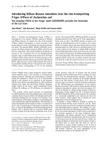
OVX results in more severe arthritis
Nine days after OVX/sham operation, mice were immunized
(day 0) with chicken CII, and 3 weeks later (day 21) they
received a booster injection. Arthritis developed from day 24,
and arthritic score was evaluated twice a week. Ovariect-
omized mice displayed a more severe disease (Fig. 1) than
sham-operated mice.
Arthritis and loss of endogenous estrogen lead to an
additive and similar degree of bone loss
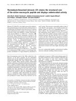
After termination of the experiment (day 45), BMD of the right
femur was measured by pQCT. Mice subjected to OVX dis-
played a trabecular bone loss of 22% compared with sham-
operated non-arthritic controls. Arthritic sham-operated mice
displayed a bone loss of 26% and, finally, ovariectomized mice
with arthritis had a 58% decrease in trabecular BMD (Figs 2a
and 3). (These values were obtained by dividing the difference
between the medians of each group and the sham-operated
control group by the median of the sham-operated control
group.) The cortical BMD was decreased by arthritis but was
unaffected by hormonal status (Fig. 2b).
Arthritis is associated with increased bone resorption,
and OVX with increased bone formation
At day 45, serum levels of osteocalcin were increased in ova-
riectomized mice compared with sham-operated mice (Fig.
4a). Immunization with CII did not affect the levels of osteocal-
cin. Serum levels of RatLaps (type I collagen cross-links) were
greatly enhanced in the CII-immunized mice, in comparison
with controls (Fig. 4b). In contrast, OVX did not increase the
levels of RatLaps.
Arthritis, but not estrogen deficiency, increases cartilage
destruction
Serum levels of COMP were increased in arthritic mice but
were not affected by hormonal status (Fig. 4c).
Figure 1
Mice after ovariectomy (OVX) displayed a significantly more severe dis-ease than sham-operated miceMice after ovariectomy (OVX) displayed a significantly more severe dis-
ease than sham-operated mice. (a) The mice were observed twice
weekly for frequency of arthritis. They were considered arthritic when
they displayed signs of arthritis in one joint for two consecutive assess-
ments, or arthritis in more than one joint. (b) Severity of arthritis was
evaluated twice weekly. Severity was graded 1 to 3 in each paw (maxi-
mum 12 points per mouse). Open circles, sham (n = 18); filled circles,
ovariectomy (n = 15). *P < 0.05; **P < 0.01; ***P < 0.001. CII, type II
collagen.
Arthritis Research & Therapy Vol 7 No 4 Jochems et al.
R840
Hormonal status does not affect arthritis-induced
increased levels of pro-inflammatory cytokines, IgG and
CII antibodies
As shown in Table 1, serum levels of the pro-inflammatory
cytokine IL-6 were low in non-arthritic mice in comparison with
the higher levels found in arthritic mice. All arthritic mice dis-
played high serum levels of IgG and anti-CII antibodies, but no
significant differences between the ovariectomized and sham-
operated mice were demonstrated.
Phenotypes of bone marrow lymphocytes are influenced
both by OVX and by arthritis
Flow cytometry analysis was performed to evaluate the effects
of OVX and arthritis on phenotypes of bone marrow
lymphocytes (Table 2). OVX was associated with an increased
number of B lymphocytes per femur, whereas CII immunization
led to a decreased number of B cells. The total numbers of T
lymphocytes (CD3
+
) and CD4
+
cells per femur were not
affected by either OVX or CII immunization. In contrast, the
number of CD8
+
cells was significantly decreased in both
sham-operated and ovariectomized arthritic mice compared
with controls. The CD69 expression, a marker of early activa-
tion, was increased on CD4
+
and CD8
+
cells in arthritic mice.
In contrast, T cell CD25 expression remained unchanged in all
groups (data not shown).
Histological findings
There was no significant difference in the degree of histologi-
cal destruction score between ovariectomized and sham-oper-
ated arthritic mice (Table 1).
Discussion
Osteoporosis is one of the major problems in postmenopausal
RA [7,19] and is a factor contributing to increased risk for frac-
tures [20]. The mechanisms and relative importance of estro-
gen deficiency versus inflammation for the bone loss in
postmenopausal RA are not fully understood. Our study is the
first to demonstrate equal contributions of estrogen deficiency
and polyarthritis to bone loss in a model of human postmeno-
pausal RA. In addition, serum markers of bone and cartilage
turnover and FACS analysis of bone marrow leukocyte pheno-
types indicate different mechanisms for the development of
osteoporosis.
OVX of the DBA/1 mice several weeks before the develop-
ment of arthritis enabled separate and concurrent analyses of
the effects of estrogen deficiency and the inflammatory
Figure 2
Ovariectomy decreased trabecular BMD whereas arthritis decreased both trabecular and cortical BMDOvariectomy decreased trabecular BMD whereas arthritis decreased
both trabecular and cortical BMD. Peripheral quantitative computer
tomography (pQCT) was performed to measure trabecular and cortical
bone mineral density (BMD). (a) Trabecular bone mineral density
(BMD) was determined with a metaphyseal scan at a point 3% of the
length of the femur from the growth plate and the inner 45% of the area
was defined as the trabecular bone compartment. (b) Cortical BMD of
the femur was determined with a mid-diaphyseal scan. Results are
shown as box plots (values are given as medians (horizontal lines),
interquartile ranges (box) and ranges (whiskers); circles represent out-
liers). For controls, n = 10 for sham (open boxes) and ovariectomy
(filled boxes); for immunized mice, n = 18 for sham and n = 14 for ova-
riectomy. **P < 0.01; ***P < 0.001. CII, type II collagen.
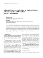
Figure 3
Peripheral quantitative computed tomography (pQCT) scans of one representative mouse in each groupPeripheral quantitative computed tomography (pQCT) scans of one
representative mouse in each group. Trabecular bone mineral density
(BMD) was determined with a metaphyseal scan at a point 3% of the
length of the femur from the growth plate and the inner 45% of the area
was defined as the trabecular bone compartment. (a) Sham-operated
control; (b) ovariectomy control; (c) sham-operated, arthritic mouse; (d)
ovariectomized, arthritic mouse. The bar shows the density of the bone,
from 0 (black) to 750 mg/cm
3
(white).
Available online />R841
disease on bone loss. Our results show that the loss of
endogenous estrogen and the ongoing arthritic disease cause
a similar degree of trabecular bone loss (22% and 26%,
respectively) and clearly have an additive effect, because ova-
riectomized mice with arthritis lost 58% of trabecular BMD.
Interestingly, arthritis also induced a significant decrease in
cortical BMD, whereas OVX, irrespective of inflammatory sta-
tus, did not affect this parameter.
It has previously been demonstrated in CIA in rats that OVX
enhances the severity of arthritis and bone loss, whereas expo-
sure to estrogen suppresses it [13]. A more detailed compar-
ison between the previous study and ours is not possible
because we ovariectomized the mice 2 weeks before initial
immunization (that is, 5 weeks before the development of
arthritis) to achieve an established postmenopausal state,
whereas Yamasaki and colleagues ovariectomized the rats 1
week after sensitization.
Systemic inflammation, impaired physical activity, low body
mass and treatment with corticosteroids are some important
factors associated with the development of osteoporosis in
RA. The pathophysiological mechanisms of bone loss in arthri-
tis have been shown to be mediated through the activation of
osteoclasts by the macrophage-derived proinflammatory
cytokines tumor necrosis factor-α and IL-1, and by the produc-
tion of RANKL by activated T-lymphocytes and fibroblasts.
Garnero and colleagues [21] found increased serum levels of
markers of bone resorption in patients with erosive RA, and
decreased markers of bone formation. The discrepancy
between bone formation and bone resorption results in the
enhanced bone loss in arthritis.
We showed that there was strongly increased bone resorption
measured by RatLaps in the arthritic mice but not in ovariect-
omized mice. This was expected, as we sought to study
changes in established menopause, and not the rapid phase
of bone loss that follows OVX. In contrast to what Garnero and
colleagues found in RA patients, we failed to demonstrate
decreased serum levels of osteocalcin associated with arthri-
tis. In accord with our results, Nishida and colleagues [22]
have previously suggested that reduced bone formation might
not be a substantial contributor to bone loss in DBA/1 mice,
so this difference might be species dependent.
The exact mechanism whereby OVX induces bone loss in
mice is not yet known. Several mechanisms are involved, and
recent studies have shown that OVX of mice was associated
with an increase in the number of activated, tumor necrosis
factor-producing, bone marrow T lymphocytes stimulating
monocytes to differentiate into osteoclasts [12,23,24]. We did
not show an increase in bone marrow T lymphocytes. The
explanation for this discrepancy could be either that we used
CD3 as a marker for T cells, whereas others have used anti-
CD90 (which is also expressed on natural killer cells,
Figure 4
Ovariectomy increased bone formation and arthritis increased bone resorption and cartilage destructionOvariectomy increased bone formation and arthritis increased bone
resorption and cartilage destruction. (a) Ovariectomy (OVX) increased
bone formation. Serum levels of osteocalcin were analyzed by immuno-
radiometric assay. (b) Arthritis increased bone resorption. Serum levels
of RatLaps were analyzed by ELISA. For controls, n = 10 for sham
(open boxes) and ovariectomy (filled boxes); for immunized mice, n =
18 for sham and n = 15 for ovariectomy. (c) Arthritis increased carti-
lage destruction. Serum levels of cartilage oligomeric matrix protein
(COMP) were analyzed by ELISA. For controls, n = 9 for sham (open
boxes) and n = 10 for ovariectomy (filled boxes); for immunized mice, n
= 17 for sham and n = 14 for ovariectomy. **P < 0.01, ***P < 0.001.
Results are shown as box plots (values are given as medians (horizontal
lines), interquartile ranges (box) and ranges (whiskers), circles repre-
sent outliers).
Arthritis Research & Therapy Vol 7 No 4 Jochems et al.
R842
monocytes and dendritic cells), or the very late time point (8
weeks after OVX) that we used for analysis of the bone mar-
row. Indeed, the finding that RatLaps, a serum marker of bone
resorption, was unaltered whereas osteocalcin, a serum
marker of bone formation, was increased in ovariectomized
mice indicates that the period of OVX-induced increased
activation of osteoclasts had already ended at this late time
point. As has been shown previously, the number of B lym-
phocytes in bone marrow was increased after OVX [25] and
decreased in the arthritic mice [26]. As increased B lym-
phopoiesis has been shown to be associated with bone loss
[27], our data suggest separate mechanisms for the bone loss
found in estrogen deficiency and in arthritis.
COMP is an extracellular matrix protein initially found in carti-
lage but recently also shown to be secreted by synovial fibrob-
lasts. Serum levels of COMP are used as a marker of cartilage
destruction and have previously been evaluated in CIA in rats
[28,29]. We found increased serum COMP levels in all
arthritic mice, irrespective of the estrogen level, indicating a
lack of cartilage protection by endogenous ovarian hormones.
Taken together, although the analyses in this study were all
performed on day 45, the differences in serum levels of Rat-
Laps, osteocalcin, COMP and frequencies and phenotypes of
bone marrow lymphocytes between mice subjected to OVX
and CIA suggest the possibility of different mechanisms for
the development of osteoporosis in estrogen deficiency and
arthritic disease.
The female sex hormone estradiol not only preserves bone but
also has a clear anti-arthritic effect both in human RA [4,5] as
well as in rat [13] and murine [11,30] CIA. Clinically, the
arthritic ovariectomized mice developed a more severe dis-
ease than the sham-operated mice. However, at termination of
the experiment all mice, irrespective of hormonal status, had
developed severe arthritic disease, with histological
destruction score, pro-inflammatory cytokines and CII antibod-
ies at similar levels.
Conclusion
We demonstrate that CIA in ovariectomized DBA/1 mice is a
relevant model for studies of osteoporosis in postmenopausal
RA. Furthermore, the loss of endogenous estrogen and the
inflammation contribute equally to bone loss in this model.
Markers of bone and cartilage turnover, as well as bone mar-
row lymphocyte phenotypes, indicate different mechanisms
for bone loss induced by estrogen deficiency and
inflammation, respectively. We suggest that this model is well
suited for future studies, both on anti-arthritic and anti-oste-
oporotic properties of new medications and on mechanisms
for bone loss in postmenopausal polyarthritis.
Competing interests
The author(s) declare that they have no competing interests.
Table 1
Serological markers of inflammation and histopathological findings were not significantly affected by ovariectomy
Arthritis OVX No. of mice IgG (mg/ml) CII antibody (ng/
ml)
Interleukin-6 (pg/
ml)
Frequency of arthritis,
day 45 (%)
Arthritic score,
day 45
Histopathology
(score)
- - 10 10 (10–12) n.d 62 (48–67) 0 0 0
+ 10 10 (7–15) n.d 80 (42–111) 0 0 0
+ - 18 18 (15–23) 4.6 (2.6–8.0) 343 (214–755) 100 8 (7–10)*** 9.5 (7.0–11.5)
+ 15 18 (15–18) 3.5 (2.3–4.6) 371 (280–602) 100 11 (10–12) 11.0 (8.9–13.0)
Values are medians and interquartile ranges for each group. The maximum arthritic score was 12 points per mouse. ***P < 0.001 between sham-
operated and ovariectomized arthritic mice. CII, type II collagen; n.d., not detectable; OVX, ovariectomy.
Table 2
Characteristics of bone marrow lymphocytes were influenced both by ovariectomy and by arthritis
Arthritis OVX n Bone marrow
cellularity (× 10
6
)
B cells per femur
(× 10
6
)
T cells per femur
(× 10
6
)
CD4
+
cells per
femur (× 10
6
)
CD69
+
/CD4
+
cells (%)
CD8
+
cells per femur (×
10
6
)
CD69
+
/CD8
+
cells (%)
- - 10 5.1 (4.0–6.4) 1.5 (1.0–1.7)*** 0.06 (0.05–0.09) 0.02 (0.02–0.04) 25 (18–30) 0.013 (0.008–0.018) 3 (1–3)
+ 10 6.0 (5.0–8.6) 2.4 (2.0–3.0) 0.05 (0.04–0.07) 0.02 (0.02–0.03) 29 (26–31) 0.008 (0.006–0.012) 4 (2–6)
+ - 18 5.2 (4.7–7.0) 1.0 (0.8–1.3)**
†
0.05 (0.04–0.06) 0.02 (0.01–0.02) 50 (43–58)***
†††
0.004 (0.002–0.007)
†††
9 (4–25)
†††
+ 15 6.1 (5.3–8.4) 1.5 (1.0–2.5)
†
0.05 (0.03–0.05) 0.02 (0.01–0.02) 35 (29–40) 0.005 (0.002–0.006)
†††
10 (5–16)
†
Values are medians and interquartile ranges for each group; n is the number of mice. Comparison between sham operation and ovariectomy (OVX):
**P < 0.01; ***P < 0.001. Comparison between arthritic mice and their controls:
†
P < 0.05;
†††
P < 0.001.
Available online />R843
Authors' contributions
HC and CO participated in study design, interpretation of data
and manuscript preparation. UI aided with analysis of data and
statistical analysis. ME and MV aided with acquisition of data.
The study was performed mainly by CJ. All authors read and
approved the final manuscript.
Acknowledgements
We thank Berit Eriksson, Anette Hansevi and Maud Petersson for excel-
lent technical assistance. This study was supported by grants from the
Göteborg Medical Society, King Gustav V's 80 years' foundation, the
Sahlgrenska Foundation, the Novo Nordic Foundation, the Börje Dahlin
foundation, the Association against Rheumatism, Reumaforskningsfond
Margareta, the Medical Faculty of Göteborg University (ALF) and the
Swedish Research Council.
References
1. Doran MF, Pond GR, Crowson CS, O'Fallon WM, Gabriel SE:
Trends in incidence and mortality in rheumatoid arthritis in
Rochester, Minnesota, over a forty-year period. Arthritis Rheum
2002, 46:625-631.
2. Goemaere S, Ackerman C, Goethals K, De Keyser F, Van der Stra-
eten C, Verbruggen G, Mielants H, Veys EM: Onset of symptoms
of rheumatoid arthritis in relation to age, sex and menopausal
transition. J Rheumatol 1990, 17:1620-1622.
3. Doran MF, Crowson CS, O'Fallon WM, Gabriel SE: The effect of
oral contraceptives and estrogen replacement therapy on the
risk of rheumatoid arthritis: a population based study. J
Rheumatol 2004, 31:207-213.
4. Ostensen M, Aune B, Husby G: Effect of pregnancy and hormo-
nal changes on the activity of rheumatoid arthritis. Scand J
Rheumatol 1983, 12:69-72.
5. Forsblad D'Elia H, Larsen A, Mattsson LA, Waltbrand E, Kvist G,
Mellstrom D, Saxne T, Ohlsson C, Nordborg E, Carlsten H: Influ-
ence of hormone replacement therapy on disease progres-
sion and bone mineral density in rheumatoid arthritis. J
Rheumatol 2003, 30:1456-1463.
6. Sinigaglia L, Nervetti A, Mela Q, Bianchi G, Del Puente A, Di
Munno O, Frediani B, Cantatore F, Pellerito R, Bartolone S, et al.:
A multicenter cross sectional study on bone mineral density in
rheumatoid arthritis. Italian Study Group on Bone Mass in
Rheumatoid Arthritis. J Rheumatol 2000, 27:2582-2589.
7. Forsblad D'Elia H, Larsen A, Waltbrand E, Kvist G, Mellstrom D,
Saxne T, Ohlsson C, Nordborg E, Carlsten H: Radiographic joint
destruction in postmenopausal rheumatoid arthritis is
strongly associated with generalised osteoporosis. Ann
Rheum Dis 2003, 62:617-623.
8. Riggs BL, Khosla S, Melton LJ 3rd: Sex steroids and the con-
struction and conservation of the adult skeleton. Endocr Rev
2002, 23:279-302.
9. Walsh NC, Gravallese EM: Bone loss in inflammatory arthritis:
mechanisms and treatment strategies. Curr Opin Rheumatol
2004, 16:419-427.
10. Holmdahl R, Bockermann R, Backlund J, Yamada H: The molecu-
lar pathogenesis of collagen-induced arthritis in mice – a
model for rheumatoid arthritis. Ageing Res Rev 2002,
1:135-147.
11. Holmdahl R, Jansson L, Andersson M: Female sex hormones
suppress development of collagen-induced arthritis in mice.
Arthritis Rheum 1986, 29:1501-1509.
12. Roggia C, Gao Y, Cenci S, Weitzmann MN, Toraldo G, Isaia G,
Pacifici R: Up-regulation of TNF-producing T cells in the bone
marrow: a key mechanism by which estrogen deficiency
induces bone loss in vivo. Proc Natl Acad Sci U S A 2001,
98:13960-13965.
13. Yamasaki D, Enokida M, Okano T, Hagino H, Teshima R: Effects
of ovariectomy and estrogen replacement therapy on arthritis
and bone mineral density in rats with collagen-induced
arthritis. Bone 2001, 28:634-640.
14. Holmdahl R, Jansson L, Larsson E, Rubin K, Klareskog L: Homol-
ogous type II collagen induces chronic and progressive arthri-
tis in mice. Arthritis Rheum 1986, 29:106-113.
15. Windahl SH, Vidal O, Andersson G, Gustafsson JA, Ohlsson C:
Increased cortical bone mineral content but unchanged
trabecular bone mineral density in female ERβ
-/-
mice. J Clin
Invest 1999, 104:895-901.
16. Mancini G, Carbonara AO, Heremans JF: Immunochemical
quantitation of antigens by single radial immunodiffusion.
Immunochemistry 1965, 2:235-254.
17. Verdrengh M, Jonsson IM, Holmdahl R, Tarkowski A: Genistein as
an anti-inflammatory agent. Inflamm Res 2003, 52:341-346.
18. Helle M, Boeije L, Aarden LA: Functional discrimination between
interleukin 6 and interleukin 1. Eur J Immunol 1988,
18:1535-1540.
19. Haugeberg G, Uhlig T, Falch JA, Halse JI, Kvien TK: Bone mineral
density and frequency of osteoporosis in female patients with
rheumatoid arthritis: results from 394 patients in the Oslo
County Rheumatoid Arthritis register. Arthritis Rheum 2000,
43:522-530.
20. Huusko TM, Korpela M, Karppi P, Avikainen V, Kautiainen H,
Sulkava R: Threefold increased risk of hip fractures with rheu-
matoid arthritis in Central Finland. Ann Rheum Dis 2001,
60:521-522.
21. Garnero P, Jouvenne P, Buchs N, Delmas PD, Miossec P: Uncou-
pling of bone metabolism in rheumatoid arthritis patients with
or without joint destruction: assessment with serum type I col-
lagen breakdown products. Bone 1999, 24:381-385.
22. Nishida S, Tsurukami H, Sakai A, Sakata T, Ikeda S, Tanaka M, Ito
M, Nakamura T: Stage-dependent changes in trabecular bone
turnover and osteogenic capacity of marrow cells during
development of type II collagen-induced arthritis in mice. Bone
2002, 30:872-879.
23. Cenci S, Toraldo G, Weitzmann MN, Roggia C, Gao Y, Qian WP,
Sierra O, Pacifici R: Estrogen deficiency induces bone loss by
increasing T cell proliferation and lifespan through IFN-
gamma-induced class II transactivator. Proc Natl Acad Sci U S
A 2003, 100:10405-10410.
24. Cenci S, Weitzmann MN, Roggia C, Namba N, Novack D,
Woodring J, Pacifici R: Estrogen deficiency induces bone loss
by enhancing T-cell production of TNF-alpha. J Clin Invest
2000, 106:1229-1237.
25. Erlandsson MC, Jonsson CA, Lindberg MK, Ohlsson C, Carlsten
H: Raloxifene- and estradiol-mediated effects on uterus, bone
and B lymphocytes in mice. J Endocrinol 2002, 175:319-327.
26. Inoue M, Wakabayashi K, Ogihara Y: Variation of lymphocytes in
peripheral blood and bone marrow in collagen-induced
arthritis. Chem Pharm Bull (Tokyo) 1994, 42:733-735.
27. Miyaura C, Onoe Y, Inada M, Maki K, Ikuta K, Ito M, Suda T:
Increased B-lymphopoiesis by interleukin 7 induces bone loss
in mice with intact ovarian function: similarity to estrogen
deficiency. Proc Natl Acad Sci U S A 1997, 94:9360-9365.
28. Larsson E, Erlandsson Harris H, Larsson A, Mansson B, Saxne T,
Klareskog L: Corticosteroid treatment of experimental arthritis
retards cartilage destruction as determined by histology and
serum COMP. Rheumatology (Oxford) 2004, 43:428-4234.
29. Larsson E, Erlandsson Harris H, Lorentzen JC, Larsson A, Mans-
son B, Klareskog L, Saxne T: Serum concentrations of cartilage
oligomeric matrix protein, fibrinogen and hyaluronan distin-
guish inflammation and cartilage destruction in experimental
arthritis in rats. Rheumatology (Oxford) 2002, 41:996-1000.
30. Jansson L, Holmdahl R: Oestrogen induced suppression of col-
lagen arthritis. IV: Progesterone alone does not affect the
course of arthritis but enhances the oestrogen-mediated ther-
apeutic effect. J Reprod Immunol 1989, 15:141-150.